Review
. 2024 Jul 10;5(7):e649.
doi: 10.1002/mco2.649. eCollection 2024 Jul.

Skeletal muscle: molecular structure, myogenesis, biological functions, and diseases

Affiliations
Review

Skeletal muscle: molecular structure, myogenesis, biological functions, and diseases

Lan-Ting Feng et al. MedComm (2020). .

Abstract

Skeletal muscle is an important motor organ with multinucleated myofibers as its smallest cellular units. Myofibers are formed after undergoing cell differentiation, cell-cell fusion, myonuclei migration, and myofibril crosslinking among other processes and undergo morphological and functional changes or lesions after being stimulated by internal or external factors. The above processes are collectively referred to as myogenesis. After myofibers mature, the function and behavior of skeletal muscle are closely related to the voluntary movement of the body. In this review, we systematically and comprehensively discuss the physiological and pathological processes associated with skeletal muscles from five perspectives: molecule basis, myogenesis, biological function, adaptive changes, and myopathy. In the molecular structure and myogenesis sections, we gave a brief overview, focusing on skeletal muscle-specific fusogens and nuclei-related behaviors including cell-cell fusion and myonuclei localization. Subsequently, we discussed the three biological functions of skeletal muscle (muscle contraction, thermogenesis, and myokines secretion) and its response to stimulation (atrophy, hypertrophy, and regeneration), and finally settled on myopathy. In general, the integration of these contents provides a holistic perspective, which helps to further elucidate the structure, characteristics, and functions of skeletal muscle.

Keywords: biological function; myogenesis; myonuclear; myopathy; skeletal muscle.


Conflict of interest statement

The authors declare that they have no conflict of interest.

Figures

FIGURE 1
FIGURE 1
The formation process and state change of myofibers. During the embryonic period, myoblasts differentiate from precursor cells and fuse to form nascent myotubes. Subsequently, the myoblasts continue to fuse with the myotubes to expand and increase the number of myonuclei. Subsequently, nuclear aggregates are newly added to the center under the traction of the cytoskeleton and form a cluster. Then, myonuclei become spatially distant from one another and are eventually transferred to the plasma membrane. The migrated myonuclei are fixed and uniformly distributed at appropriate distances to maximize their function. The skeletal muscle state changes under stimulation. At the cellular level, this primarily depends on the relationship between protein synthesis and degradation. When protein synthesis is less than degradation, the skeletal muscle shows an atrophied state; otherwise, it shows hypertrophy. Additionally, muscle hypertrophy is associated with MuSC fusion. Muscle regeneration mediated by MuSCs that localize between myofibers can partially or even completely restore atrophied and damaged muscle tissue, which is crucial for recovery from injury and disease (not shown in the figure).
FIGURE 2
FIGURE 2
Putative membrane fusion process mediated by myomakers and myomergers. After the fusogen is anchored on another membrane involved in fusion (not shown in the figure), the anchored membranes overcome the hydrophilic resistance and continue to approach each other with the allosteric action of the fusogen. Subsequently, outer layers of anchored membranes fuse to form the hemifusion state, promoting the convergence of inner membrane layers and facilitating their fusion. A fusion pore is formed and continuously expands, and irreversible membrane fusion is completed after cytoplasmic exchange. In myoblast–myoblast and myoblast–myotube fusion, myomakers mediate the processes preceding fusion pore formation, whereas myomergers induce the remaining steps.
FIGURE 3
FIGURE 3
Structure of myomakers and myomergers. (A) Myomakers contain seven transmembrane domains and a 25‐amino acid intracellular C‐terminal tail, as indicated by myomaker topology (modified from Ref. 23). (B) Myomergers have only one transmembrane domain, an N‐terminal hydrophobic domain, and a C‐terminal domain containing helix‐1 and helix‐2, with a total length of 84 amino acids (modified from Ref. 31).
FIGURE 4
FIGURE 4
Molecular mechanism of myonuclear migration. Myonuclear migration has three main stages: cluster formation, spreading, and dispersion. (A) Cluster formation is mainly mediated by the dynein/dynactin complex. Dynein/dynactin complex links MTs and myonuclei via Par6β, pulling the newly fused myonuclear towards the cluster. Amphiphysins maintain cluster aggregation. (B) Myonuclei spreading is related to kinesin‐1 and dynein function. Myonuclei emit MTs via the SYNE–sk‐CIP–MTOC protein. Adjacent myonuclei can move on these MTs through the antiparallel MT–Kif5b–MAP7–MT, nuclear–nesprin–KLC–MT, and myotube pole–Raps/Pins–dynein–MT modes. MTs are fixed on the myotube pole using CLIP‐190 in the myotube pole–Raps/Pins–dynein–MT mode. (C) Myonuclei dispersion is based on myofibril crosslinking. Myofibril crosslinking is initiated by fibronectin secreted by myofibroblasts, which interacts with α5‐integrin and phosphorylates FAK–Src complex and paxillin to recruit β‐Pix. Then, Cdc42, N‐Wasp, and Arp2/3 complexes are sequentially activated by the former to promote the function of γ‐actin and desmin, completing myofibril crosslinking. Subsequently, the myonuclei are squeezed to the periphery by myotube contraction. Par6, partitioning defective 6; LINC, linkers of the nucleoskeleton and cytoskeleton; sk‐CIP, skeletal muscle‐cardiac islet‐1 interaction protein; MTOC, microtubule organizing center; MAP7, microtubule‐associated protein 7; Raps, rapsynoid; Pins, partner of inscuteable; CLIP‐190, cytoplasmic linker protein 190; FAK, focal adhesion kinase; β‐Pix, PAK‐interacting exchange factor‐β; Cdc42, cell division cycle 42; N‐Wasp, neural Wiskott‐Aldrich syndrome protein; Amph2, Amphiphysin‐2; Bin1, bridging integrator 1; Arp, actin‐related protein.
FIGURE 5
FIGURE 5
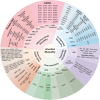
Classification and pathogenic genes of inherited myopathies. Inherited myopathies are divided into five categories: mitochondrial myopathies, congenital myopathies, metabolic myopathies, channelopathies and muscular dystrophies. Mitochindrial myopathies: Kearns–Sayre syndrome (KSS), chronic progressive external ophthalmoplegia (CPEO), leigh syndrome, mitochondrial DNA depletion syndrome (MDS), mitochondrial encephalomyopathy, lactic acidosis and stroke like episodes (MELAS), myoclonus epilepsy with ragged red fibers (MERRF), maternally inherited deafness and diabetes (MIDD), mitochondrial neurogastrointestinal encephalomyopathy (MNGIE), neuropathy, ataxia, and retinitis pigmentosa (NARP), and Pearson syndrome (PS). Congenital myopathies: core myopathy, nemaline myopathy (NM), centronuclear myopathy (CNM), congenital fiber type disproportion (CFTD), and myosin storage myopathy (MSM). Metabolic myopathies: glycogen storage disease (GSD) and lipid myopathy. Channelopathies: nondystrophic myotonias (NDM) including myotonia congenita (MC), paramyotonia congenita (PMC), and sodium channel myotonia (SCM); periodic paralysis (PP) including hypokalemic periodic paralysis (HypoPP), hyperkalemic periodic paralysis (HyperPP), and Andersen–Tawil syndrome (ATS). Muscular dystrophies: Duchenne muscular dystrophy (DMD), Becker muscular dystrophy (BMD), myotonic dystrophy (DM), facioscapulohumeral muscular dystrophy (FSHD), oculopharyngeal muscular dystrophy (OPMD), congenital muscular dystrophy (CMD), Limb‐Girdle muscular dystrophy (LGMD), and Emery–Dreifuss muscular dystrophy (EDMD).

References

    1. Wang X, Vannier J, Yang X, et al. Muscle systems and motility of early animals highlighted by cnidarians from the basal Cambrian. Elife. 2022:11. - PMC - PubMed
    1. Liu AG, Matthews JJ, Menon LR, McIlroy D, Brasier MD. Haootia quadriformis n. gen., n. sp., interpreted as a muscular cnidarian impression from the Late Ediacaran period (approx. 560 Ma). Proc Biol Sci. 2014;281(1793). - PMC - PubMed
    1. Murphy M, Kardon G. Origin of vertebrate limb muscle: the role of progenitor and myoblast populations. Curr Top Dev Biol. 2011;96:1‐32. - PMC - PubMed
    1. Chal J, Pourquié O. Making muscle: skeletal myogenesis in vivo and in vitro. Development. 2017;144(12):2104‐2122. - PubMed
    1. Leikina E, Gamage DG, Prasad V, et al. Myomaker and Myomerger work independently to control distinct steps of membrane remodeling during myoblast fusion. Dev Cell. 2018;46(6):767‐780. e7. - PMC - PubMed

LinkOut - more resources