

The role of ultra-widefield imaging with navigated central and peripheral cross-sectional and three-dimensional swept source optical coherence tomography in ophthalmology: Clinical applications
- PMID: 38988788
- PMCID: PMC11232741
- DOI: 10.4103/sjopt.sjopt_59_24
The role of ultra-widefield imaging with navigated central and peripheral cross-sectional and three-dimensional swept source optical coherence tomography in ophthalmology: Clinical applications
Abstract
Purpose: To assess central and peripheral retinal and choroidal diseases using ultra-widefield (UWF) fundus imaging in combination with navigated central and peripheral cross-sectional and three-dimensional (3D) swept source optical coherence tomography (SS-OCT) scans.
Methods: Retrospective study involving 332 consecutive patients, with a nearly equal distribution of males and females. The mean age of patients was 52 years (range 18-92 years). Average refractive error was -3.80 D (range +7.75 to -20.75 D).
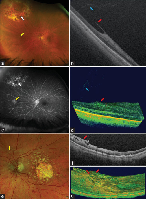
Results: The observations in this study demonstrate the efficacy of peripheral navigated SS-OCT in assessing various ocular conditions. The technology provides high-quality images of the peripheral vitreous, vitreoretinal interface, retina, and choroid, enabling visualization of vitreous floaters and opacities, retinal holes and tears, pigmented lesions, and peripheral retinal degenerations. 3D OCT scans enhance the visualization of these abnormalities and improve diagnostic and therapeutic decisions.
Conclusion: Navigated central and peripheral cross-sectional and 3D SS-OCT scans offer significant complementary benefits in the assessment and management of retinal diseases. Their addition to UWF imaging provides a comprehensive view of central and peripheral ocular structures, aiding in early detection, precise anatomical measurements, and objective monitoring of disease progression. In addition, this technology serves as a valuable tool for patient education, a teaching tool for trainees, and documentation for medico-legal purposes.
Keywords: Branch vein occlusion; diabetic retinopathy; inherited retinal diseases; lattice degeneration; navigated swept source optical coherence tomography; ora serrata; pigmented lesion; retinal detachment; retinal hole; retinal tear; retinal vasculopathies; retinitis pigmentosa; retinoschisis; sickle cell retinopathy; swept source optical coherence tomography; three-dimensional navigated optical coherence tomography; ultra widefield imaging; vitreoretinal tuft; vitreous; vitreous floaters and opacities; widefield imaging.
Copyright: © 2024 Saudi Journal of Ophthalmology.
Conflict of interest statement
There are no conflicts of interest.
Figures


Similar articles
-
Navigated single-capture 3D and cross-sectional wide-field OCT of the mid and peripheral retina and vitreoretinal interface.Eur J Ophthalmol. 2022 May;32(3):1642-1651. doi: 10.1177/11206721211026100. Epub 2021 Jul 3. Eur J Ophthalmol. 2022. PMID: 34218694
-
Silicone oil emulsification: A literature review and role of widefield imaging and ultra-widefield imaging with navigated central and peripheral optical coherence tomography technology.Saudi J Ophthalmol. 2024 Jan 6;38(2):112-122. doi: 10.4103/sjopt.sjopt_193_23. eCollection 2024 Apr-Jun. Saudi J Ophthalmol. 2024. PMID: 38988778 Free PMC article.
-
Single-capture ultra-widefield guided swept-source optical coherence tomography in the management of rhegmatogenous retinal detachment and associated peripheral vitreoretinal pathology.Br J Ophthalmol. 2023 Sep;107(9):1356-1362. doi: 10.1136/bjophthalmol-2021-320149. Epub 2022 May 26. Br J Ophthalmol. 2023. PMID: 35618409
-
Retinal applications of swept source optical coherence tomography (OCT) and optical coherence tomography angiography (OCTA).Prog Retin Eye Res. 2021 Sep;84:100951. doi: 10.1016/j.preteyeres.2021.100951. Epub 2021 Jan 28. Prog Retin Eye Res. 2021. PMID: 33516833 Review.
-
Impact of swept source optical coherence tomography on ophthalmology.Taiwan J Ophthalmol. 2016 Apr-Jun;6(2):58-68. doi: 10.1016/j.tjo.2015.09.002. Epub 2015 Dec 2. Taiwan J Ophthalmol. 2016. PMID: 29018713 Free PMC article. Review.
References
-
- Quinn N, Csincsik L, Flynn E, Curcio CA, Kiss S, Sadda SR, et al. The clinical relevance of visualising the peripheral retina. Prog Retin Eye Res. 2019;68:83–109. - PubMed
-
- Bravo FJ, Ayliffe W, Stanga SF, Reinstein UI, Moxham R, Tariq Z, et al. New imaging technology for simultaneous multiwavelength-UWF fundus fluorescein angiography and indocyanine green angiography with navigated central and peripheral SS-OCT. Ophthalmic Surg Lasers Imaging Retina. 2023;54:401–10. - PubMed
LinkOut - more resources
Full Text Sources