

Influence of periapical lesion volume on the radiodensity of surrounding bone: A CBCT study
- PMID: 38989501
- PMCID: PMC11232769
- DOI: 10.4103/JCDE.JCDE_178_24
Influence of periapical lesion volume on the radiodensity of surrounding bone: A CBCT study
Abstract
Aim: This study assesses if the size of periapical lesions has an effect on the bone immediately peripheral to an apical lesion.
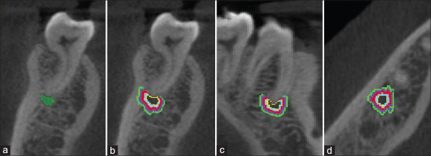
Methods: Cone-beam computed tomography (CBCT) images of 271 periapical lesions were analyzed using Mimics Research™ to determine the CBCT periapical lesion volume index (CBCTPAVI) score, along with the radiodensity of the lesion, lesion border, and surrounding bone in 0.5 mm increments up to 2.0 mm peripheral to the apical lesion. The one-way analysis of variance was used to assess for significant differences in the radiodensity of the lesion, border, and peripheral bone, as well as differences among CBCTPAVI scores.
Results: The radiodensity of bone peripheral to the apical lesion increased significantly up to 1.0 mm around the lesion's perimeter. In addition, lesions with higher CBCTPAVI scores showed a significantly greater difference in the radiodensity from the lesion to the lesion border and the peripheral bone, compared to lesions with smaller CBCTPAVI scores.
Conclusions: This study for the first time shows the influence of periapical lesion size on the radiodensity of bone peripheral to an apical lesion. Variations in radiodensity at the perimeter of a periapical lesion can be influenced by the size of the lesion, possibly indicating differences in defense response. Knowledge of these phenomena may provide information on bone healing and enhance our understanding of bone peripheral to a periapical lesion.
Keywords: Cone-beam computed tomography; endodontics; periapical lesion; radiodensity; volume.
Copyright: © 2024 Journal of Conservative Dentistry and Endodontics.
Conflict of interest statement
There are no conflicts of interest.
Figures

References
-
- Yusof Z, Nambiar P. Radiographic considerations in endodontics. Malays Dent J. 2007;28:51–8.
-
- Kaya S, Yavuz I, Uysal I, Akkuş Z. Measuring bone density in healing periapical lesions by using cone beam computed tomography: A clinical investigation. J Endod. 2012;38:28–31. - PubMed
-
- Shapurian T, Damoulis PD, Reiser GM, Griffin TJ, Rand WM. Quantitative evaluation of bone density using the Hounsfield index. Int J Oral Maxillofac Implants. 2006;21:290–7. - PubMed
-
- Clementino-Luedemann TN, Kunzelmann KH. Mineral concentration of natural human teeth by a commercial micro-CT. Dent Mater J. 2006;25:113–9. - PubMed
-
- Valiyaparambil JV, Yamany I, Ortiz D, Shafer DM, Pendrys D, Freilich M, et al. Bone quality evaluation: Comparison of cone beam computed tomography and subjective surgical assessment. Int J Oral Maxillofac Implants. 2012;27:1271–7. - PubMed
LinkOut - more resources
Full Text Sources