. 2024 Jun 7;6(14):3655-3667.
doi: 10.1039/d4na00204k. eCollection 2024 Jul 9.

Contrast enhanced photoacoustic detection of fibrillar collagen in the near infrared region-I

Affiliations

Contrast enhanced photoacoustic detection of fibrillar collagen in the near infrared region-I

Inna Solomonov et al. Nanoscale Adv. .

Abstract

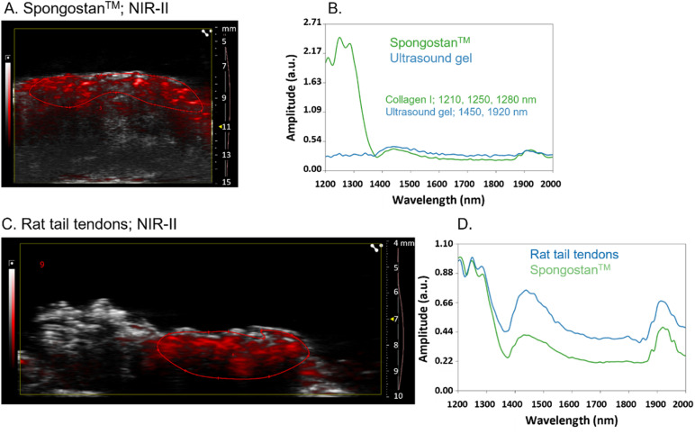
Fibrillar collagen accumulation emerges as a promising biomarker in several diseases, such as desmoplastic tumors and unstable atherosclerotic plaque. Gold nanorods (GNRs) hold great potential as contrast agents in high-resolution, biomedically safe, and non-invasive photoacoustic imaging (PAI). This study presents the design and characterization of a specialized imaging tool which exploits GNR assisted targeted photoacoustic imaging that is tailored for the identification of fibrillar collagen. In addition to the photoacoustic characterization of collagen in the NIR 1 and 2 regions, we demonstrate the detailed steps of conjugating a decoy to GNRs. This study serves as a proof of concept, that demonstrates that conjugated collagenase-1 (MMP-1) generates a distinct and collagen-specific photoacoustic signal, facilitating real-time visualization in the wavelength range of 700-970 nm (NIR I). As most of the reported studies utilized the endogenous contrast of collagen in the NIR II wavelength that has major limitations to perform in vivo deep tissue imaging, the approach that we are proposing is unique and it highlights the promise of MMP-1 decoy-functionalized GNRs as novel contrast agents for photoacoustic imaging of collagen in the NIR 1 region. To our knowledge this is the first time functionalized GNRs are optimized for the detection of fibrillar collagen and utilized in the field of non-invasive photoacoustic imaging that can facilitate a better prognosis of desmoplastic tumors and broken atherosclerotic plaques.


Conflict of interest statement

The authors declare no competing financial interests; GM and JJ are employees of FujiFilm VisualSonics.

Figures

Fig. 1
Fig. 1. Characterization of the MMP-1 decoy. (A) SDS-PAGE analysis of MMP-1 decoy purification. Lane labels: molecular mass standards; L, load; F-T, flow-through; His-Trap, protein eluted using imidazole from the Ni column; SEC, size exclusion chromatography using a Superdex 75. (B) MMP-1 decoy thermostability by DSF, showing the melting curve and derivative plot used to determine the Tm of the decoy. (C) BLAST search results; red rectangles show the alignment of collagen fragments from different origins with human collagen MMP interaction domains identified or supposed to be MMP interaction domains. (D) Direct specific binding of the MMP1-decoy to collagen type I from different species revealed by pull down assay. Left: the SDS-PAGE image of the decoy pulling down different collagens. lanes 1 and 2: negative control and beads immobilized with the MMP14 based decoy (MMP-14 SIA). Lanes 3 and 4: the MMP-1 decoy binds to reconstituted collagen type I from rats and humans, respectively. Right: the densitometry analysis of total pulled down collagen (all bands above 139 kDa). The bars represent the normalized integrated density of collagen compared to that of the decoy.
Fig. 2
Fig. 2. Coupling of the His-tagged decoy to chitosan-coated GNRs. First, the free amino groups of GNRs@Chit are reacted with the isothiocyanate terminus of the bifunctional linker. Then, the NTA terminus is exploited for Ni2+ complexation, thus forming a Ni-NTA complex able to bind His-tagged proteins.
Fig. 3
Fig. 3. Effectiveness of the conjugation approach and characterization of GNRs@Chit-Dec. (A) Fluorescence emission spectrum of GNRs@Chit-GFP compared to the wastewaters collected during its purification, revealing quantitative attachment of the His-tagged protein to the Ni-activated nanosystem. (B) Representative TEM image of GNRs@Chit-Dec. (C) Normalized Uv-vis spectrum of GNRs@Chit-Dec compared to GNRs@Chit. (D) Distribution of widths and lengths of n = 55 GNRs@Chit-Dec measured by TEM. The solid lines represent the Gaussian fit for both dimensions. Photoacoustic imaging of an agar drop containing GNR@Chit-NTA-Ni (6 nmol Au) (E) and its associated PA spectra in NIR-I (680–970 nm), followed by the overlay with the PA spectra of GNR@Chit-Dec (6 nmol Au) (F).
Fig. 4
Fig. 4. Images illustrating the progression of bladder carcinoma in rats and the interactions of GNRs with collagen fibrils enriched in ECM scaffolds derived from bladder carcinoma tissues. Representative H&E section bladder from an 8-month rat (a) and rat bladder carcinoma after 6 months of BBN treatment (b), used for the isolation of the ECM; the red asterisk shows a typical desmoplastic stroma, which applies to all tumors present in that bladder. Representative SEM images of the ECM isolated from healthy (c) and carcinoma rat bladder (d) tissues; ECM scaffolds from healthy tissues consist of tightly packed collagen fibers with similar diameters, creating a wavy pattern. In contrast, bladder carcinoma scaffolds exhibit disrupted collagen packing, characterized by a disorganized composition of fibrils (thinnest fibrils indicated by asterisks) and fibers with varying diameters. Linearization of thick collagen fibers, as expected in solid tumors, is also observed (indicated by the arrow). The images of at least 7 fields in different locations within the desmoplastic region of each decellularized bladder from three rats were captured. Topography of the tumor ECM captured in the secondary electron mode (e) and acquired in the backscattered electron mode (f) to highlight the binding of GNRs@Chit-Dec to collagen fibrils (indicated by white dots). Panels (g) and (h) refer to the same experiment shown in panels (e) and (f), but with the ECM incubated with the control nanoparticles GNRs@Chit-NTA-Ni.
Fig. 5
Fig. 5. PA spectra of collagen I in NIR-II. Photoacoustic imaging (overlay of B mode in grayscale and photoacoustic signal in red scale) of Spongostan™ (A) and its associated PA spectra in NIR-II (1200–2000 nm) followed by the overlay with the PA spectra of the ultrasound gel (B); the wavelength of the three peaks of the collagen spectra and of the two peaks of the ultrasound gel are reported. Photoacoustic imaging of rat tail tendons (C) and their associated PA spectra in NIR-II, followed by the overlay with the PA spectra of Spongostan™ (D); the PA spectra of rat tail tendons and Spongostan™ were normalized to highlight the similarities. The right axis of US imaging shows (i) the acquisition depth in millimeters (mm), (ii) the yellow arrow the focus of the US imaging, and (iii) the red line the time gain compensation.
Fig. 6
Fig. 6. GNRs@Chit-Dec allows photoacoustic detection of collagen in NIR-I. Reference spectra of GNRs@Chit-Dec and rat tail tendons in NIR-I (A). PA spectra or rat tail tendons alone, incubated with GNRs@Chit-NTA-Ni, or GNRs@Chit-Dec (B). PA imaging of rat tail tendons alone, incubated with GNRs@Chit-NTA-Ni (100 nmol Au) or GNRs@Chit-Dec (100 nmol Au), after spectral unmixing using the reference spectra of rat tail tendons and GNRs@Chit-Dec acquired in NIR-I (C); the right axis of US imaging shows (i) the acquisition depth in millimeters (mm), (ii) the yellow arrow the focus of the US imaging, and (iii) the red line the time gain compensation.

References

    1. Winkler J. Abisoye-Ogunniyan A. Metcalf K. J. Werb Z. Nat. Commun. 2020;11:5120. doi: 10.1038/s41467-020-18794-x. - DOI - PMC - PubMed
    1. Zhou Z. H. Ji C. D. Xiao H. L. Zhao H. B. Cui Y. H. Bian X. W. J. Cancer. 2017;8:1466–1476. doi: 10.7150/jca.18466. - DOI - PMC - PubMed
    1. Whatcott C. J. Diep C. H. Jiang P. Watanabe A. LoBello J. Sima C. Hostetter G. Shepard H. M. Von Hoff D. D. Han H. Clin. Cancer Res. 2015;21:3561–3568. doi: 10.1158/1078-0432.CCR-14-1051. - DOI - PMC - PubMed
    1. Iacobuzio-Donahue C. A. Argani P. Hempen P. M. Jones J. Kern S. E. Cancer Res. 2002;62:5351–5357. - PubMed
    1. Amin M. B. Mod. Pathol. 2009;22(Suppl 2):S96–S118. doi: 10.1038/modpathol.2009.26. - DOI - PubMed