. 2024 Oct 28;52(19):11626-11640.
doi: 10.1093/nar/gkae584.

Partial wrapping of single-stranded DNA by replication protein A and modulation through phosphorylation

Affiliations

Partial wrapping of single-stranded DNA by replication protein A and modulation through phosphorylation

Rahul Chadda et al. Nucleic Acids Res. .

Abstract

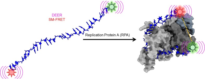
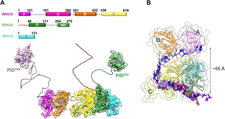
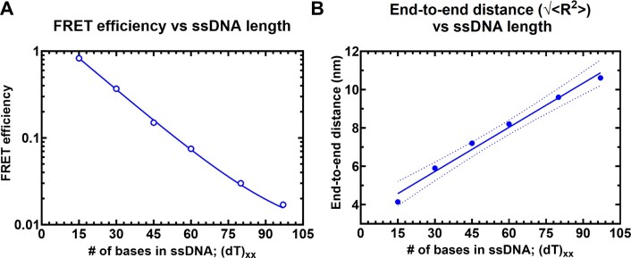
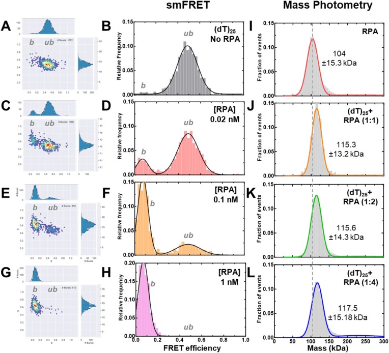
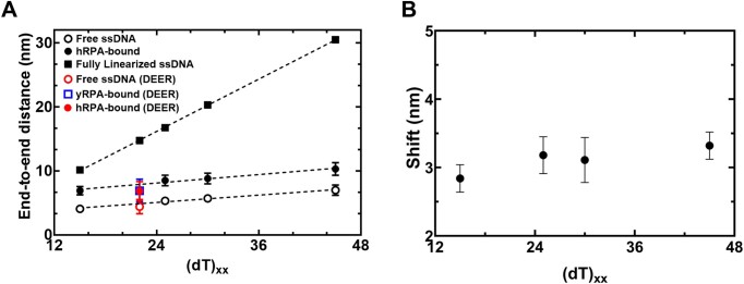
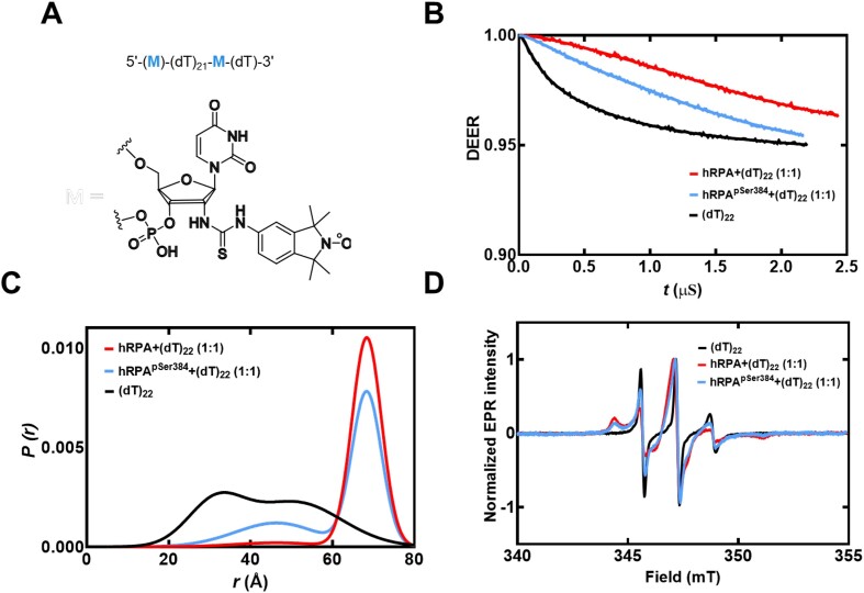
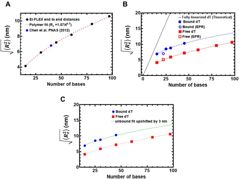
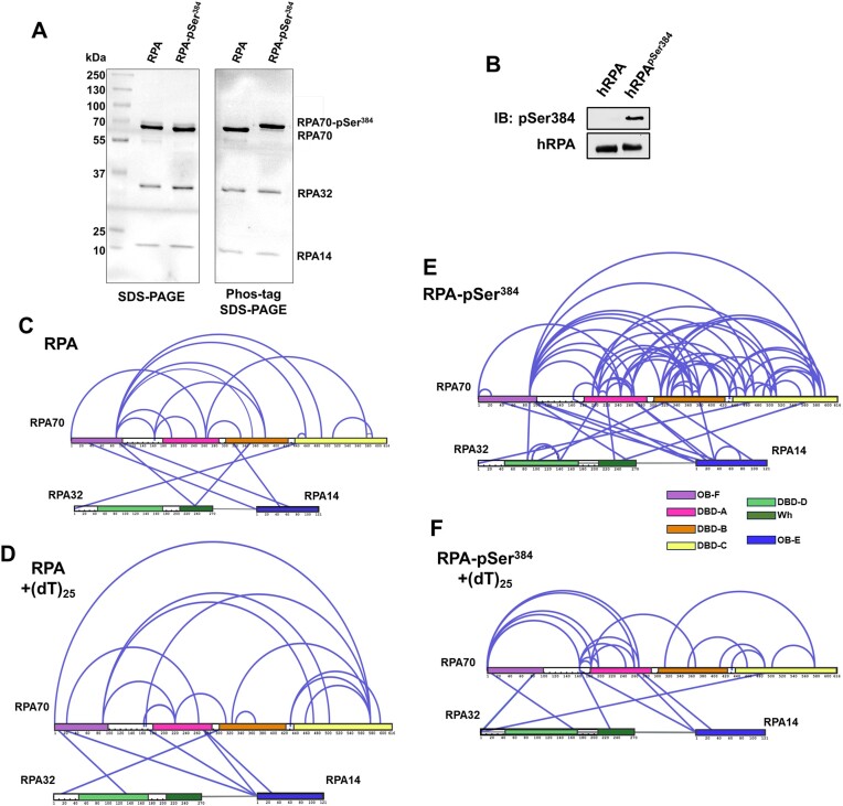
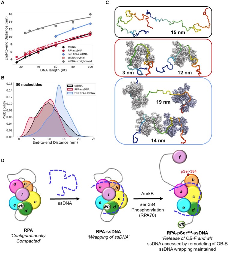
Single-stranded DNA (ssDNA) intermediates which emerge during DNA metabolic processes are shielded by replication protein A (RPA). RPA binds to ssDNA and acts as a gatekeeper to direct the ssDNA towards downstream DNA metabolic pathways with exceptional specificity. Understanding the mechanistic basis for such RPA-dependent functional specificity requires knowledge of the structural conformation of ssDNA when RPA-bound. Previous studies suggested a stretching of ssDNA by RPA. However, structural investigations uncovered a partial wrapping of ssDNA around RPA. Therefore, to reconcile the models, in this study, we measured the end-to-end distances of free ssDNA and RPA-ssDNA complexes using single-molecule FRET and double electron-electron resonance (DEER) spectroscopy and found only a small systematic increase in the end-to-end distance of ssDNA upon RPA binding. This change does not align with a linear stretching model but rather supports partial wrapping of ssDNA around the contour of DNA binding domains of RPA. Furthermore, we reveal how phosphorylation at the key Ser-384 site in the RPA70 subunit provides access to the wrapped ssDNA by remodeling the DNA-binding domains. These findings establish a precise structural model for RPA-bound ssDNA, providing valuable insights into how RPA facilitates the remodeling of ssDNA for subsequent downstream processes.


Figures

Graphical Abstract
Graphical Abstract
Figure 1.
Figure 1.
Architecture of the replication protein A (RPA) – ssDNA complex. (A) The three subunits of RPA—RPA70, RPA32 and RPA14 house several oligonucleotide/oligosaccharide binding (OB) domains that are classified as either DNA binding domains (DBDs: DBD-A, DBD-B, DBD-C and DBD-D) or protein-interaction domains (OB-F/PID70N and winged helix (wh)/PID32C). A structural model of RPA is shown and was generated using information from known structures of the OB-domains and AlphaFold2 models of the linkers. (B) Crystal structure of Ustilago maydis RPA bound to ssDNA is shown (PDB 4GNX). This structure lacks OB-F, the F-A linker, wh and the D-wh linker. ssDNA from CryoEM structures of Saccharomyces cerevisiae RPA (PDB 6I52) and Pyrococcus abyssi RPA (8OEL) are superimposed and reveal an end-to-end DNA distance of ∼55 Å. Structural data supports partial wrapping of ssDNA around the OB domains.
Figure 2.
Figure 2.
Solution confocal-based FRET measurements accurately report on ssDNA end-to-end distances. (A) FRET efficiencies were calculated based on single molecule measurements of Cy3 (donor) and Cy5 (acceptor) fluorescence. Data were collected on a series of ssDNA substrates (30 pM) of increasing lengths. Energy transfer events were detected as bursts of photons as the molecules transited the confocal volume. The mean of the distribution is plotted in panel A as a function of length of poly-thymidine (dTxx). (B) The end-to-end distances were calculated based on a R0 value of 5.4 nm for the Cy3/Cy5 pair and Eq. (3) described in the Methods. The dotted lines represent a 95% confidence interval for the linear fit.
Figure 3.
Figure 3.
Concentration dependence of RPA–ssDNA complexes. A-H) FRET analysis of (dT)25 ssDNA bound to increasing concentrations of RPA show a shift from the unbound to bound complex. As RPA concentrations are increased, a complete shift to the bound population is observed. Ratios are defined as one molecule of ssDNA: number of RPA trimers (molar ratio). I-L) Mass photometry analysis of RPA and RPA-(dT)25 complexes show formation of predominantly single RPA bound (dT)25 complexes. The dotted line serves as a reference point for the mass of free RPA in solution as seen in panel I. The measured mass for RPA and the RPA-DNA complexes (1:1 stoichiometry) are noted. In all conditions tested here, one RPA molecule binds to one molecule of ssDNA.
Figure 4.
Figure 4.
RPA binding produces a modest 3 nm increase in end-to-end distance. (A) The estimated end-to-end distance between the 3' and 5' ends of ssDNA is plotted as a function of nucleotides which make up the chain. Closed squares represent theoretically calculated end-to-end distances assuming the ssDNA was completely linearized. Open and closed circles represent experimental end-to end distance measurements from smFRET analysis ssDNA -/+ RPA, respectively. The figure also shows that end-to-end distances of yRPA-bound, hRPA-bound, and free (dT)22 ssDNA, measured using DEER spectroscopy. The DEER measurements fall on the respective trend lines suggesting good agreement between experimental measurements performed using two independent biophysical approaches. (B) A ∼3.1 ± 0.2 nm shift is observed in the end-to-end distances between the free ssDNA and RPA-bound ssDNA. This ssDNA-independent uniform expansion suggests that the average dimensions of the DNA ensemble are increasing due to interactions with RPA.
Figure 5.
Figure 5.
DEER spectroscopy of RPA and RPA-pSer384 bound to ssDNA. (A) Structure of the isoindoline nitroxide spin label and the position on the (dT) oligonucleotide. (B) Raw DEER decays and fits are presented for the experimentally determined distance distributions P(r) (C). (D) CW EPR spectra of labeled ssDNA in the absence and presence of RPA and RPA-pSer384.
Figure 6.
Figure 6.
Computational predictions of ssDNA wrapping agree well with experimental measurements. (A) Black circles are data, and the red dashed line is a fit to those data using a polymer scaling model. The blue circle is data reporting on the end-to-end distance for (dT)40 made by Chen et. al. 2011. (B) Blue circles are from RPA-bound (dT) oligonucleotides, red squares are from RPA-free (dT) oligonucleotides. Data include distances measured by both EPR and smFRET experiments. Blue and red dashed lines are polymer model fits to the data, respectively. The black line is the theoretical expected end-to-end distance if the DNA were fully linearized and stretched to its contour length. (C) The root mean squared end-to-end distance dependence on the number of bases is essentially identical for the bound and unbound states, as highlighted by the fact that the exact polymer fit upshifted by 3.1 ± 0.2 nm fully describes the bound dT dependency.
Figure 7.
Figure 7.
Phosphorylation at a single position in RPA70 alters access to the ssDNA with minimal alternations of end-to-end distance. (A) SDS-PAGE and Phos-tag SDS-PAGE analysis of RPA and RPA-pSer384 proteins show selective shift of the RPA70 band only in the RPA-pSer384 sample confirming 100% incorporation of pSer. (B) Western blotting of RPA and RPA-pSer384 with an antibody specific to pSer384 confirms the site-specific phosphorylation. (C, D) Cross-linking mass spectrometry (XL-MS) analysis of RPA in the absence or presence of ssDNA (dT)25. A direct comparison of the XLs between RPA and the RPA-(dT)25 experiments are presented with XLs unique to each condition denoted in blue. (F) A similar comparative XL-MS analysis RPA-pSer384 and the RPA-pSer384-(dT)25 complex is shown and the XLs unique to each condition is shown in blue. The XL patterns show differences in ssDNA driven changes upon phosphorylation.
Figure 8.
Figure 8.
Conformational analysis of ssDNA-RPA complexes from coarse-grained molecular dynamics simulation and a model for ssDNA wrapping by RPA. (A) Mean end-to-end distances for ssDNA of varying lengths in different conditions: free ssDNA (black), ssDNA bound to a single RPA (red), and ssDNA bound to two RPAs (blue). Additionally, ssDNA was studied in isolation (i.e. in the absence of explicit RPA) but with a restraint applied on the end-to-end distance of the middle 23 nucleotides, which was set to be 55 Å (mimicking curved binding to RPA) or 138 Å (mimicking stretched conformations of the ssDNA). The coarse-grained simulations were based on Ustilago maydis RPA (PDB: 4GNX), but the five OB-domains were considered as non-dynamic for simplicity. (B) Distribution of end-to-end distances of ssDNA (dT)80 nucleotides in length, modeled as free ssDNA (black), ssDNA bound to a single RPA (red), and ssDNA bound to two RPAs (blue). The distributions are based on multiple long simulations for each system. The distribution of the end-to-end distances for ssDNA interacting with two RPA molecules combines simulations in which the excess ssDNA is placed either as a linker between the two RPAs or as flanking ssDNA at the ends. (C) Snapshots from coarse-grained simulations showing varying end-to-end distances for 80-nucleotide ssDNA in different states: free ssDNA, bound to a single RPA, and bound to two RPAs. Please also refer to videos provided in the Supplemental Information. (D) The OB-domains of RPA are depicted along with the connecting disordered linkers. The XL-MS data suggest that the domains are compacted together, and ssDNA is wrapped around this architecture. The domains are remodeled upon phosphorylation by Aurora kinase B at position Ser-384 in the RPA70 subunit. This modification releases the OB-F and wh domains and promotes RPA interactions with other proteins. While the wrapping of ssDNA is not altered, remodeling allows access to the ssDNA.

Update of

References

    1. Caldwell C.C., Spies M.. Dynamic elements of replication protein A at the crossroads of DNA replication, recombination, and repair. Crit. Rev. Biochem. Mol. Biol. 2020; 55:482–507. - PMC - PubMed
    1. Wold M.S., Kelly T.. Purification and characterization of replication protein A, a cellular protein required for in vitro replication of simian virus 40 DNA. Proc. Natl. Acad. Sci. U.S.A. 1988; 85:2523–2527. - PMC - PubMed
    1. Wold M.S. Replication protein A: A heterotrimeric, single-stranded DNA-binding protein required for eukaryotic DNA metabolism. Annu. Rev. Biochem. 1997; 66:61–92. - PubMed
    1. Iftode C., Daniely Y., Borowiec J.A.. Replication protein A (RPA): the eukaryotic SSB. Crit. Rev. Biochem. Mol. Biol. 1999; 34:141–180. - PubMed
    1. Kim C., Paulus B.F., Wold M.S.. Interactions of human replication protein A with oligonucleotides. Biochemistry. 1994; 33:14197–14206. - PubMed

MeSH terms

LinkOut - more resources