. 2024 Oct;13(27):e2400522.
doi: 10.1002/adhm.202400522. Epub 2024 Jul 11.

Amniotic Membrane-Derived Multichannel Hydrogels for Neural Tissue Repair

Affiliations

Amniotic Membrane-Derived Multichannel Hydrogels for Neural Tissue Repair

Joana P M Sousa et al. Adv Healthc Mater. 2024 Oct.

Abstract

In the pursuit of advancing neural tissue regeneration, biomaterial scaffolds have emerged as promising candidates, offering potential solutions for nerve disruptions. Among these scaffolds, multichannel hydrogels, characterized by meticulously designed micrometer-scale channels, stand out as instrumental tools for guiding axonal growth and facilitating cellular interactions. This study explores the innovative application of human amniotic membranes modified with methacryloyl domains (AMMA) in neural stem cell (NSC) culture. AMMA hydrogels, possessing a tailored softness resembling the physiological environment, are prepared in the format of multichannel scaffolds to simulate native-like microarchitecture of nerve tracts. Preliminary experiments on AMMA hydrogel films showcase their potential for neural applications, demonstrating robust adhesion, proliferation, and differentiation of NSCs without the need for additional coatings. Transitioning into the 3D realm, the multichannel architecture fosters intricate neuronal networks guiding neurite extension longitudinally. Furthermore, the presence of synaptic vesicles within the cellular arrays suggests the establishment of functional synaptic connections, underscoring the physiological relevance of the developed neuronal networks. This work contributes to the ongoing efforts to find ethical, clinically translatable, and functionally relevant approaches for regenerative neuroscience.

Keywords: amniotic membrane; hydrogel; multichannel; nervous system; neural stem cells; scaffolds; tissue regeneration.


Conflict of interest statement

The authors declare no conflict of interest.

Figures

Figure 1
Figure 1
Proteomic analysis of dAM matrisome. A) Matrisome‐associated, core matrisome, and non‐matrisome protein abundance percentages relative to total protein number. B) Protein abundance percentages of matrisome‐associated protein subtypes (ECM regulators, ECM‐affiliated proteins, and secreted factors) and core matrisome protein subtypes (collagens, ECM glycoproteins, and proteoglycans) as annotated using the Matrisome database[ 32 , 33 ] (https://matrisome.org). C) Identification of dAM matrisome proteins containing integrin‐binding domains as accessed through Gene Ontology Ribbon analysis[ 34 , 35 ] (https://geneontology.org/ribbon.html). Gene Ontology data from the 2024‐01‐17 release (https://doi.org/10.5281/zenodo.10536401) is made available under the terms of the CC BY 4.0 license.
Figure 2
Figure 2
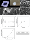
Fabrication process and structural details of AMMA multichannel hydrogels. A) 3D reconstruction of micro‐CT scans of the mold fabricated by 3D printing. B) Macroscopic hydrogel architecture. SEM images reveal C) hydrogel‐embedded OCT surface, D) a top view of a channel clear from OCT, E) a longitudinal section displaying a vertical channel, and F) the internal microstructure of the hydrogel. G) Swelling properties of AMMA 0.75% w/v hydrogel. H) Degradation of AMMA hydrogels in vitro. I) Representative compressive stress‐strain curve for AMMA hydrogels. J) Young's modulus, ultimate strain, and ultimate stress for AMMA hydrogels.
Figure 3
Figure 3
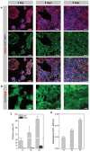
Viability and proliferative capacity of NSC cultured on AMMA hydrogel films. A) Representative confocal images of NSC stained with anti‐Nestin, anti‐Ki67, and DAPI at 1, 3, and 5 days of culture. Scale bars = 50 µm. B) Fluorescence images of Live/Dead staining of NSC cultured on hydrogel films for 1, 3, and 5 days. Scale bars = 50 µm. C) Quantification of the area covered by live and dead cells over time; and D) metabolic activity analysis through Alamar Blue assay.
Figure 4
Figure 4
Differentiation studies of NSC cultured on AMMA hydrogel films and PLL‐coated coverslips. Neurons are stained for TUJ‐1, in green, astrocytes for GFAP, in red, and the nuclei with DAPI, in blue. A) In the hydrogels, neurons were predominantly organized into clusters from which neurites extended establishing connections among distant cells. The presence of GFAP+ cells is residual. B) Zoom‐in micrograph of the marked region in (A). C) In the PLL‐coated coverslip, smaller neuronal clusters formed on top of an astrocyte layer. D) Zoom‐in micrograph of the marked region in (C). Scale bars = 50 µm.
Figure 5
Figure 5
Synapse formation on AMMA hydrogel films and on PLL‐coated coverslips. Representative confocal images showing staining for TUJ‐1 (green), synaptophysin (red) and DAPI (blue) are shown. Scale bars = 50 µm.
Figure 6
Figure 6
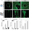
Viability and proliferation of NSC cultured on AMMA multichannel hydrogels. (A) Confocal images of Live/Dead staining of NSC cultured for 1, 4 and 7 days. The dotted circumferences and dotted lines represent the channels on top view and cross section images, respectively. Scale bars = 100 µm. Quantification of the area covered by live and dead cells from (B) top view and (C) cross section Live/Dead images. (D) Metabolic activity results from Alamar Blue assay.
Figure 7
Figure 7
Phalloidin (red) and DAPI (blue) staining for NSC seeded on multichannel hydrogels for 7 days. A) Top view of the hydrogel shows three channels encircled by cells. B) Zoomed in picture a channel. C) Cross section view displaying a channel filled with cells and cellular infiltration into the hydrogel microstructure. D) 3D rendering of a channel. Scale bars = 100 µm.
Figure 8
Figure 8
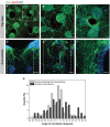
Differentiation studies of NSC cultured on AMMA multichannel hydrogels. Neurons are labelled for TUJ‐1 (green), astrocytes for GFAP (red), and cell nuclei with DAPI (blue). A) A neuronal network formed on the top a hydrogel spanning around four distinct channels. B) Zoom‐in micrograph of the marked region in (A) showing a neurosphere positioned at the boundary between the surface and a channel. C) The presence of neurospheres and neurites surrounding the channel led to a reduction in its overall diameter. D) Neurospheres positioned atop the hydrogel extending neurites into the channels and forming connections with other neurospheres adhered to the channel walls. E) Neurosphere situated at the center of a channel, extending neurites longitudinally. F) A channel densely populated with both cell bodies and neurites. The dashed lines try to define the channel. Scale bars = 50 µm. G) Histogram comparing angle of neurite orientation extending from neurospheres and in cell columns measured from cross‐section images.
Figure 9
Figure 9
Examination of synaptophysin protein expression. Neurons are stained for TUJ‐1, in green, synapses for synaptophysin, in red, and the nuclei with DAPI, in blue. A) A neuronal network formed on the top a hydrogel showing a neurosphere entering a channel. B) Neurospheres inside a channel projecting neurites. C) A channel densely populated with both cell bodies and neurites. Scale bars = 50 µm.

References

    1. Cohen‐Adad J., NeuroImage 2018, 182, 169. - PubMed
    1. Uckermann O., Hirsch J., Galli R., Bendig J., Later R., Koch E., Schackert G., Steiner G., Tanaka E., Kirsch M., Sci. Rep. 2019, 9, 12641. - PMC - PubMed
    1. Barros C. S., Franco S. J., Müller U., Cold Spring Harb Perspect Biol 2011, 3, a005108. - PMC - PubMed
    1. Hu X., Xu W., Ren Y., Wang Z., He X., Huang R., Ma B., Zhao J., Zhu R., Cheng L., Signal Transduct Target Ther 2023, 8, 245. - PMC - PubMed
    1. Bradbury E. J., Burnside E. R., Nat. Commun. 2019, 10, 3879. - PMC - PubMed

MeSH terms

LinkOut - more resources