. 2024 Jul 3;41(7):msae141.
doi: 10.1093/molbev/msae141.

Following the Evolutionary Paths of Dscam1 Proteins toward Highly Specific Homophilic Interactions

Affiliations

Following the Evolutionary Paths of Dscam1 Proteins toward Highly Specific Homophilic Interactions

Gil Wiseglass et al. Mol Biol Evol. .

Abstract

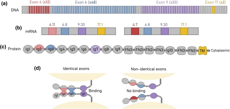
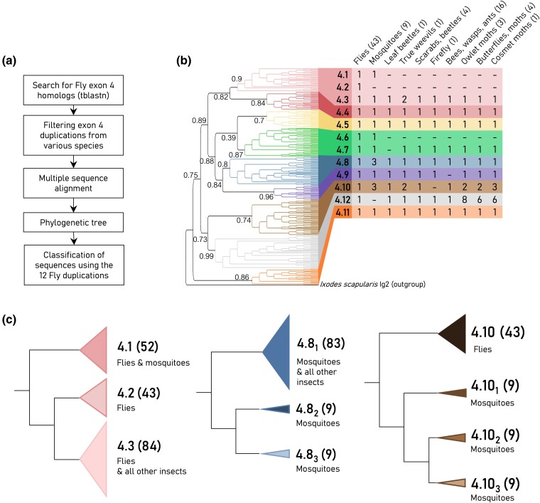
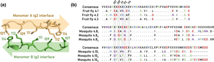
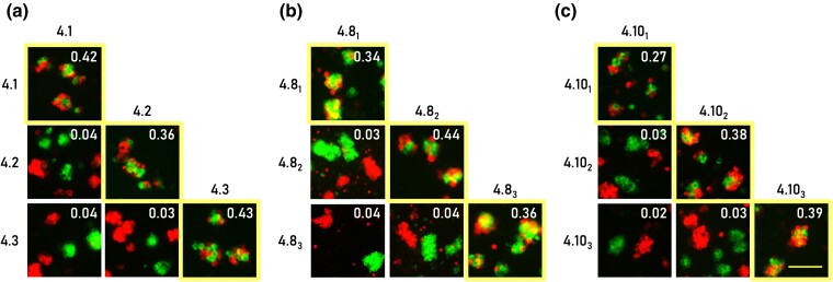
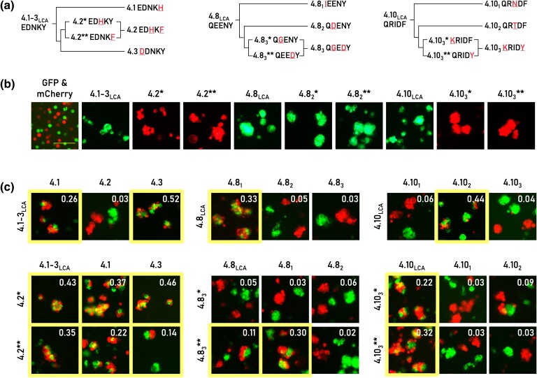
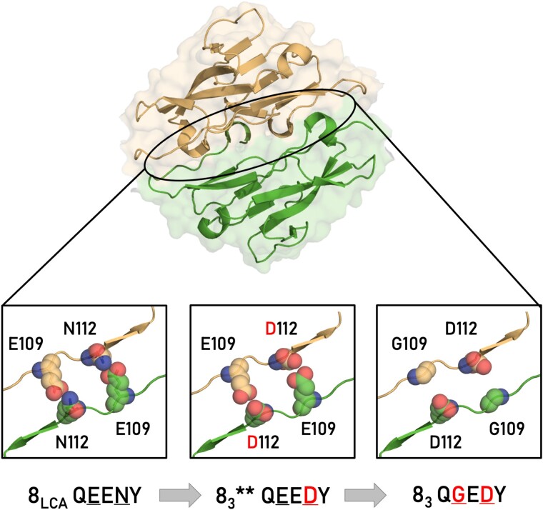
Many adhesion proteins, evolutionarily related through gene duplication, exhibit distinct and precise interaction preferences and affinities crucial for cell patterning. Yet, the evolutionary paths by which these proteins acquire new specificities and prevent cross-interactions within their family members remain unknown. To bridge this gap, this study focuses on Drosophila Down syndrome cell adhesion molecule-1 (Dscam1) proteins, which are cell adhesion proteins that have undergone extensive gene duplication. Dscam1 evolved under strong selective pressure to achieve strict homophilic recognition, essential for neuronal self-avoidance and patterning. Through a combination of phylogenetic analyses, ancestral sequence reconstruction, and cell aggregation assays, we studied the evolutionary trajectory of Dscam1 exon 4 across various insect lineages. We demonstrated that recent Dscam1 duplications in the mosquito lineage bind with strict homophilic specificities without any cross-interactions. We found that ancestral and intermediate Dscam1 isoforms maintained their homophilic binding capabilities, with some intermediate isoforms also engaging in promiscuous interactions with other paralogs. Our results highlight the robust selective pressure for homophilic specificity integral to the Dscam1 function within the process of neuronal self-avoidance. Importantly, our study suggests that the path to achieving such selective specificity does not introduce disruptive mutations that prevent self-binding but includes evolutionary intermediates that demonstrate promiscuous heterophilic interactions. Overall, these results offer insights into evolutionary strategies that underlie adhesion protein interaction specificities.

Keywords: adhesion proteins; cell-cell recognition; down syndrome cell adhesion molecule, DSCAM; gene duplication; protein evolution; protein–protein interactions.


Figures

Fig. 1.
Fig. 1.
Dscam1 gene has the capacity to transcribe tens of thousands of strictly homophilic isoforms. a) In insects, exons 4, 6, and 9 of the Dscam1 gene have undergone extensive tandem duplications, contributing to the vast diversity of isoforms produced by the gene. b) Through stochastic alternative splicing, a single exon from each cluster is retained in the mature transcript. In Drosophila, 38,016 unique isoforms can be expressed, with 19,008 unique extracellular domains. Each neuron expresses a different set of Dscam1 isoforms. c) Exons 4, 6, and 9 encode partial (exons 4 and 6) or an entire (exon 9) Ig domain. These domains determine the binding specificity of the Dscam1 protein. d) For a Dscam1 dimer to form between two cell membranes, all domains must fully match.
Fig. 2.
Fig. 2.
Phylogenetic analysis of insect exon 4. a) Illustration of the workflow for phylogenetic analysis. b) The phylogenetic tree is constructed from 266 exon 4 sequences from 83 representative species (left). The tree topology preserves the organization of the major ortholog clusters, which are highlighted by different colors and are notated according to the Drosophila exon 4 nomenclature. The tree was generated using FastTree with FastTree local support values shown. The table summarizes the number of duplications (paralogs) per species group for each cluster (right). The number of species per group is denoted within brackets. c) Simplified phylogenetic subtrees of three recent duplications (4.1 to 4.3, 4.81-3, and 4.101-3, colors corresponding to the main tree) for both flies and mosquitoes. The number of sequences per cluster is denoted within the brackets. This number includes the redundant sequences that were not used in reconstructing the main tree.
Fig. 3.
Fig. 3.
Three recent duplications for both mosquitos and flies. a) Structure of the Ig2 homodimer interface (PDB 3DMK) encoded by Drosophila exon 4.1. The interface spans amino acid positions 107 to 114 and aligns in an antiparallel fashion. b) Multiple sequence alignments of the fruit fly D. melanogaster exons 4.1 to 4.3 and the yellow fever mosquito A. aegypti exons 4.81 to 3 and 4.101 to 3. A period (i.e. “.”) in the multiple sequence alignments indicates invariant positions, and only residues that deviate from the consensus sequence are shown. The Ig2:Ig2 interface residues are highlighted in gray, and the sequence position is indicated at the top of the alignment.
Fig. 4.
Fig. 4.
Recent duplications in mosquito Dscam1 engage in highly specific homophilic interactions. Pairwise combinations within each exon paralog cluster were assessed for their interaction specificity. HEK293F cells expressing identical isoforms formed mixed red and green aggregates (as marked by both a yellow boundary and an aggregation score exceeding 0.1), while cells expressing different isoforms formed separate red and green aggregates. a) Binding assay for the fruit fly D. melanogaster exons 4.1 to 4.3. b and c) Binding assays for the yellow fever mosquito A. aegypti exons 4.81 to 3 and 4.101 to 3, respectively. The aggregate mixing score is presented in the right corner of each image. Scale 100 µm.
Fig. 5.
Fig. 5.
Tracing evolutionary paths of binary specificities of Dscam1. a) Predicted mutational pathways from ancestor to contemporary extant isoforms. The Ig2:Ig2 interface residues corresponding to sequence positions 107, 109, 111, 112, and 114 are shown for each isoform. Exon duplications are indicated with diverging lines. Mutations are highlighted in red and is also underscored. b) Resurrected ancestral (“LCA”) and intermediate (“*”/”**”) proteins mediate cell aggregation, while the control cells expressing GFP and mCherry do not mediate aggregation. c) Binding preferences of resurrected and contemporary isoforms demonstrate heterophilic interactions in many cases (mixing score >0.1, highlighted by the yellow boundary). The aggregate mixing score is presented in the right corner of each image. Scale 100 µm.
Fig. 6.
Fig. 6.
Dscam1 exon 83 mutational path. The top image presents the structural model of the Ig2:Ig2 dimer complex with Ig2 encoded by exon 4.8 ancestor and highlighted by a black oval. At the bottom, three close-up views of the homophilic Ig2:Ig2 interface for 4.8 LCA (left), 4.8 intermediate isoform (middle), and 4.83 current isoform (right). The structural models' backbones are shown as cartoon, and mutated residues are shown with Van der Waals spheres and colored based on chain origin and atom type. Ig2:Ig2 interface residues are noted at the bottom.

Similar articles

References

    1. Aakre CD, Herrou J, Phung TN, Perchuk BS, Crosson S, Laub MT. Evolving new protein-protein interaction specificity through promiscuous intermediates. Cell. 2015:163(3):594–606. 10.1016/j.cell.2015.09.055. - DOI - PMC - PubMed
    1. Aharoni A, Gaidukov L, Khersonsky O, Gould SM, Roodveldt C, Tawfik DS. The ‘evolvability’ of promiscuous protein functions. Nat Genet. 2005:37(1):73–76. 10.1038/ng1482. - DOI - PubMed
    1. Armitage SAO, Freiburg RY, Kurtz J, Bravo IG. The evolution of Dscam genes across the arthropods. BMC Evol Biol. 2012:12(1):53. 10.1186/1471-2148-12-53. - DOI - PMC - PubMed
    1. Bisogni AJ, Ghazanfar S, Williams EO, Marsh HM, Yang JY, Lin DM. Tuning of delta-protocadherin adhesion through combinatorial diversity. Elife. 2018:7:e41050. 10.7554/eLife.41050. - DOI - PMC - PubMed
    1. Boucard AA, Maxeiner S, Südhof TC. Latrophilins function as heterophilic cell-adhesion molecules by binding to teneurins: regulation by alternative splicing. J Biol Chem. 2014:289(1):387–402. 10.1074/jbc.M113.504779. - DOI - PMC - PubMed

Substances

LinkOut - more resources