. 2024 Aug 7;68(8):e0002924.
doi: 10.1128/aac.00029-24. Epub 2024 Jul 11.

Zafirlukast induces DNA condensation and has bactericidal effect on replicating Mycobacterium abscessus

Affiliations

Zafirlukast induces DNA condensation and has bactericidal effect on replicating Mycobacterium abscessus

Sanne van der Niet et al. Antimicrob Agents Chemother. .

Abstract

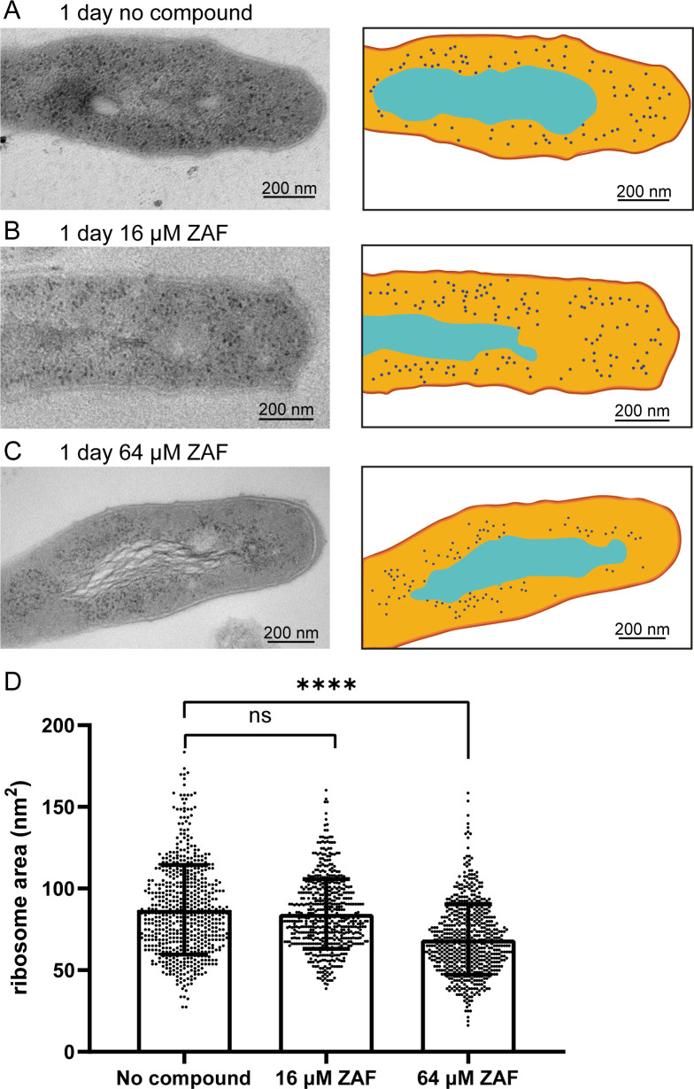
Mycobacterium abscessus infections are emerging in cystic fibrosis patients, and treatment success rate in these patients is only 33% due to extreme antibiotic resistance. Thus, new treatment options are essential. An interesting target could be Lsr2, a nucleoid-associated protein involved in mycobacterial virulence. Zafirlukast is a Food and Drug Administration (FDA)-approved drug against asthma that was shown to bind Lsr2. In this study, zafirlukast treatment is shown to reduce M. abscessus growth, with a minimal inhibitory concentration of 16 µM and a bactericidal concentration of 64 µM in replicating bacteria only. As an initial response, DNA condensation, a known stress response of mycobacteria, occurs after 1 h of treatment with zafirlukast. During continued zafirlukast treatment, the morphology of the bacteria alters and the structural integrity of the bacteria is lost. After 4 days of treatment, reduced viability is measured in different culture media, and growth of M. abscessus is reduced in a dose-dependent manner. Using transmission electron microscopy, we demonstrated that the hydrophobic multilayered cell wall and periplasm are disorganized and ribosomes are reduced in size and relocalized. In summary, our data demonstrate that zafirlukast alters the morphology of M. abscessus and is bactericidal at 64 µM. The bactericidal concentration of zafirlukast is relatively high, and it is only effective on replicating bacteria but as zafirlukast is an FDA-approved drug, and currently used as an anti-asthma treatment, it could be an interesting drug to further study in in vivo experiments to determine whether it could be used as an antibiotic for M. abscessus infections.

Keywords: Mycobacterium abscessus; antibiotics; electron microscopy; zafirlukast.


Conflict of interest statement

The authors declare no conflict of interest.

Figures

Fig 1
Fig 1
Growth curves of ZAF-treated M. abscessus in different culture media. M. abscessus was treated with 2 to 128 µM ZAF, and absorption was measured every 24 h for a period of 96 h. Bacteria were cultured in (A) MH2 medium, (B) 7H9 medium, (C) MH2 medium supplemented with DC, (D) 7H9 medium supplemented with DC, (E) MH2 medium supplemented with ADC, and (F) 7H9 medium supplemented with DC. Graphs represent the mean of two individual experiments, and the error bars represent the standard error of the mean. Abs(600 nm) is 600-nm absorbance value.
Fig 2
Fig 2
CFU of M. absessus treated with ZAF. (A) M. abscessus was grown in MH2 medium and treated with 0 (gray)-, 16 (light blue)-, and 64 (turquoise) µM ZAF. CFU were determined at 24, 48, 72, 96, and 168 h. (B) M. abscessus was grown in PBS to achieve a non-replicating state and was treated with 0 (gray)-, 16 (light blue)-, and 64 (turquoise) µM ZAF. CFU were determined at 24, 48, 72, 96, and 168 h. Error bars represent the variation of two independent cultures.
Fig 3
Fig 3
ZAF induces rapid DNA condensation. (A) Representative deconvoluted widefield fluorescent images of M. abscessus treated with no compound, 16 or 64 µM ZAF for 1 h, 1 day, and 4 days. DNA is stained with Hoechst (cyan) and lipids with Nile red (red). Scale bars represent 2 µm. (B) Quantification of DNA pattern of M. abscessus in n = 3 experiments and n = 2 experiments for 64 µM day 1. DNA was defined as condensed when it was present in one compact nucleoid. Per condition, 40–479 M. abscessus were analyzed. *P < 0.05, and error bars represent the standard error of the mean.
Fig 4
Fig 4
Organization of condensed DNA in M. abscessus. Representative TEM images of M. abscessus treated with no compound, 16 and 64 µM ZAF. (A) One-hour untreated M. abscessus have DNA dispersed throughout the majorority of the bacterium. The DNA is present in in the cytosol of the bacterium as electron-dense fiber-like structures. DNA is indicated by a white dotted line. Scale bar indicates 200 nm. (B) Schematic representation of A, capsular layer is indicated in dark orange, myco-membrane in light orange, DNA in cyan, and ribosomes in dark blue. (C) M. abscessus treated with 16 µM ZAF for 1 h. The DNA is present as a small clump of electron-dense fibers close together in the center of the bacterium. DNA is indicated by a white dotted line. (E) M. abscessus treated with 64 µM ZAF for 1 h. The DNA is present in a small part in the center of the bacterium. DNA is indicated by a white dotted line. scale bar indicates 200 nm. (F) Schematic representation of E with the same color code as in B. (G) Left panel: high-magnification TEM image of uncondensed DNA in an untreated bacterium. The DNA is present as thin electron-dense fibers with space in between the fibers. The individual DNA fibers have a random orientation. Right panel: DNA fibers are schematically represented with the same color code as B. (H) Left panel: high-magnification TEM image of condensed DNA of M. abscessus treated with 16 µM ZAF for 1 h. The DNA fibers are densely clumped together, the individual fibers are aligned, and some DNA fibers stick out of the clump. Right panel: organization of the DNA is schematically represented with the same color code as B. (I–L) Tomography reconstruction of M. abscessus treated with 64 µM ZAF for 1 h. (I) Top view of a slice of the tomogram with (J) a 3D reconstruction of the DNA on top. DNA is indicated in cyan. (K) Side view of 3D-reconstructed DNA from the tomogram displayed in I. (L) 3D reconstruction of DNA only, from condensed DNA displayed in I–K.
Fig 5
Fig 5
ZAF induces structural abnormalities in M. abscessus. (A) Representative TEM images of resin embedded M. abscessus at 1 h, day 1, and day 4, treated with 16 and 64 µM ZAF. Black arrows indicate membrane damage at 1 day of ZAF treatment, and cyan arrows indicate condensed DNA. All scale bars represent 500 nm. Representative TEM image and schematic representation of (B) intact M. abscessus, (C) damaged M. abscessus, and (D) empty/deformed M. abscessus. Scale bars represent 200 nm. Capsular layer is indicated in dark orange, myco-membrane in light orange, DNA in cyan, and ribosomes in dark blue. (E) Quantification of M. abscessus morphology at day 4. Untreated M. abscessus are indicated in dark grey and 16 µM ZAF in light grey. Bars represent the mean and standard deviation of two experiments.
Fig 6
Fig 6
ZAF affects ribosome size after 1 day. Representative TEM image and schematic representation of M. abscessus (A) 1 day untreated, (B) 1-day 16 µM ZAF and (C) 1-day 64 µM ZAF. Capsular layer is indicated in dark orange, myco-membrane in orange, cytosol light orange, DNA in cyan, and ribosomes in dark blue. Scale bars represent 200 nm. (D) Quantification of the area of the ribosomes from two independent experiments. Per condition, n = 174–435 ribosomes were analyzed. A Mann–Whitney test was performed on the data, ****P < 0.0001 and ns = not significant. The graph represents all data points of the two experiments and mean with standard deviation.

Similar articles

References

    1. Martiniano SL, Nick JA, Daley CL. 2019. Nontuberculous mycobacterial infections in cystic fibrosis. Thorac Surg Clin 29:95–108. doi:10.1016/j.thorsurg.2018.09.008 - DOI - PubMed
    1. Kwak N, Dalcolmo MP, Daley CL, Eather G, Gayoso R, Hasegawa N, Jhun BW, Koh W-J, Namkoong H, Park J, Thomson R, van Ingen J, Zweijpfenning SMH, Yim J-J. 2019. Mycobacterium abscessus pulmonary disease: individual patient data meta-analysis. Eur Respir J 54:1801991. doi:10.1183/13993003.01991-2018 - DOI - PubMed
    1. Thomson R, Tolson C, Sidjabat H, Huygens F, Hargreaves M. 2013. Mycobacterium abscessus isolated from municipal water - a potential source of human infection. BMC Infect Dis 13:1. doi:10.1186/1471-2334-13-241 - DOI - PMC - PubMed
    1. Bryant JM, Grogono DM, Rodriguez-Rincon D, Everall I, Brown KP, Moreno P, Verma D, Hill E, Drijkoningen J, Gilligan P, et al. . 2016. Population-level genomics identifies the emergence and global spread of a human transmissible multidrug-resistant nontuberculous mycobacterium. Science 354:751–757. doi:10.1126/science.aaf8156 - DOI - PMC - PubMed
    1. Nessar R, Cambau E, Reyrat JM, Murray A, Gicquel B. 2012. Mycobacterium abscessus: a new antibiotic nightmare. J Antimicrob Chemother 67:810–818. doi:10.1093/jac/dkr578 - DOI - PubMed

Publication types

MeSH terms

LinkOut - more resources