Comparative Study
. 2024 Aug;6(4):e230068.
doi: 10.1148/ryct.230068.

Hypersensitivity Pneumonitis on Thin-Section Chest CT Scans: Diagnostic Performance of the ATS/JRS/ALAT versus ACCP Imaging Guidelines

Affiliations
Comparative Study

Hypersensitivity Pneumonitis on Thin-Section Chest CT Scans: Diagnostic Performance of the ATS/JRS/ALAT versus ACCP Imaging Guidelines

Lydia Chelala et al. Radiol Cardiothorac Imaging. 2024 Aug.

Abstract

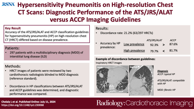
Purpose To compare the diagnostic performance of the American Thoracic Society, Japanese Respiratory Society, and Asociación Latinoamericana del Tórax (ATS/JRS/ALAT) versus the American College of Chest Physicians (ACCP) imaging classifications for hypersensitivity pneumonitis (HP). Materials and Methods Patients in the institutional review board-approved Interstitial Lung Disease (ILD) registry referred for multidisciplinary discussion (MDD) at the authors' institution (January 1, 2006-April 1, 2021) were included in this retrospective study when ILD was diagnosed at MDD. MDD diagnoses included HP, connective tissue disease-ILD, and idiopathic pulmonary fibrosis. Retrospective review of thin-section CT images was performed in consensus by two cardiothoracic radiologists blinded to the diagnosis. Diagnostic patterns were determined for thin-section CT images using both classifications. Discordance rates were determined. Sensitivity, specificity, positive predictive value, negative predictive value, and accuracy were assessed using MDD diagnosis as the reference standard. Results A total of 297 patients were included in the study: 200 (67%) with HP, 49 (16%) with connective tissue disease-ILD, and 48 (16%) with idiopathic pulmonary fibrosis at MDD. The discordance rate between the two classifications was 21%. Assuming low HP prevalence (10%), ATS/JRS/ALAT classification outperformed ACCP classification, with greater accuracy (92.3% vs 87.6%) and greater positive predictive value (60.7% vs 42.9%). Assuming high prevalence (50%), accuracy and negative predictive value were superior using ACCP classification (81.7% vs 79.7% and 77.7% vs 72.6%, respectively), and positive predictive value was superior using ATS/JRS/ALAT classification (93.3% vs 87.1%). Conclusion Accuracy of the ATS/JRS/ALAT and ACCP HP classifications was greater in settings with low and high HP prevalence, respectively. Diagnostic performance of both classifications was discordant in a minority of cases. Keywords: CT, Thorax, Hypersensitivity Pneumonitis, Interstitial Lung Disease Supplemental material is available for this article. © RSNA, 2024.

Keywords: CT; Hypersensitivity Pneumonitis; Interstitial Lung Disease; Thorax.


Conflict of interest statement

Disclosures of conflicts of interest: L.C. No relevant relationships. A.A. NIH grant K23HL146942-01A1; grants/contracts from Pulmonary Fibrosis Foundation, the American College of Chest Physicians, and the National Institutes of Health; consulting fees from Roche, Boehringer Ingelheim, Inogen, PatientMpower, AbbVie, and Medscape; payment or honoraria from Boehringer Ingelheim; participation on data safety monitoring board or advisory board for Roche, Boehringer Ingelheim, Inogen, and PatientMpower; leadership role with American Thoracic Society. M.S. Grants/contracts to institution from Boehringer Ingelheim Pharmaceuticals, NIH, and Pulmonary Fibrosis Foundation; honoraria for Self-Education and Evaluation of Knowledge (SEEK) Pulmonary Board Review textbook (CHEST), medical writing support provided by Fleishman Hillard; honoraria for CME lectures (CHEST), medical writing support provided by Fleishman Hillard; payment/ honoraria from Boehringer Ingelheim Pharmaceuticals; payment to author from participation on Fibrinogen Adjudication committee and BMS Data Monitoring committee; leadership role for American Thoracic Society on Clinical Problems Committee and Research Innovation Summit Planning Committee; leadership role for Pulmonary Fibrosis Foundation on Scientific Review Committee. C.T.L. Grant from Pulmonary Fibrosis Foundation. R.J. Participation on data safety monitoring board/advisory board for AbbVie on adjudication committee for ILD events for chemotherapeutic drug (unrelated to study). A.N.H. No relevant relationships. I.U. No relevant relationships. J.H.C. Senior consulting editor for Radiology: Cardiothoracic Imaging.

Figures

None
Graphical abstract
Inspiratory (A) axial and (C) sagittal CT images in the lung window in
a 76-year-old woman show lower lung and peripheral predominant reticulation
(circle, A and C) and traction bronchiectasis (arrow, A). (B) Expiratory
axial CT image shows air trapping (dashed circle, B). Diagnostic patterns
were typical hypersensitivity pneumonitis per ACCP and compatible
hypersensitivity pneumonitis per ATS/JRS/ALAT guidelines. A higher level of
confidence could not be reached with ATS/JRS/ALAT given lower lobe
distribution. A diagnosis of fibrotic hypersensitivity pneumonitis was made
during multidisciplinary discussion. ACCP = American College of Chest
Physicians, ATS/JRS/ALAT = American Thoracic Society/Japanese Respiratory
Society/Asociación Latinoamericana del Tórax.
Figure 1:
Inspiratory (A) axial and (C) sagittal CT images in the lung window in a 76-year-old woman show lower lung and peripheral predominant reticulation (circle, A and C) and traction bronchiectasis (arrow, A). (B) Expiratory axial CT image shows air trapping (dashed circle, B). Diagnostic patterns were typical hypersensitivity pneumonitis per ACCP and compatible hypersensitivity pneumonitis per ATS/JRS/ALAT guidelines. A higher level of confidence could not be reached with ATS/JRS/ALAT given lower lobe distribution. A diagnosis of fibrotic hypersensitivity pneumonitis was made during multidisciplinary discussion. ACCP = American College of Chest Physicians, ATS/JRS/ALAT = American Thoracic Society/Japanese Respiratory Society/Asociación Latinoamericana del Tórax.
(A) Inspiratory axial, (B) expiratory axial, and (C) sagittal CT
images in a lung window in a 61-year-old woman show lower lobe predominant
ground-glass opacity with reticulation (dashed circles, A and C), traction
bronchiectasis (long arrows, A and C), and subpleural reticulation with
honeycombing (arrowheads, A) (short arrow, C). Inspiratory mosaicism (white
circle, A) is associated with air trapping on expiratory axial CT image
(white circles, B). The combination of ground-glass opacity indicative of
infiltration and fibrosis, mosaicism indicative of air trapping, and
intervening normal parenchymal attenuation constitute the three-attenuation
pattern. Diagnostic patterns were typical hypersensitivity pneumonitis per
ACCP and compatible hypersensitivity pneumonitis per ATS/JRS/ALAT
guidelines. A higher level of confidence could not be reached with
ATS/JRS/ALAT given lower lobe predominance. A diagnosis of fibrotic
hypersensitivity pneumonitis was made during multidisciplinary discussion.
ACCP = American College of Chest Physicians, ATS/JRS/ALAT = American
Thoracic Society/Japanese Respiratory Society/Asociación
Latinoamericana del Tórax.
Figure 2:
(A) Inspiratory axial, (B) expiratory axial, and (C) sagittal CT images in a lung window in a 61-year-old woman show lower lobe predominant ground-glass opacity with reticulation (dashed circles, A and C), traction bronchiectasis (long arrows, A and C), and subpleural reticulation with honeycombing (arrowheads, A) (short arrow, C). Inspiratory mosaicism (white circle, A) is associated with air trapping on expiratory axial CT image (white circles, B). The combination of ground-glass opacity indicative of infiltration and fibrosis, mosaicism indicative of air trapping, and intervening normal parenchymal attenuation constitute the three-attenuation pattern. Diagnostic patterns were typical hypersensitivity pneumonitis per ACCP and compatible hypersensitivity pneumonitis per ATS/JRS/ALAT guidelines. A higher level of confidence could not be reached with ATS/JRS/ALAT given lower lobe predominance. A diagnosis of fibrotic hypersensitivity pneumonitis was made during multidisciplinary discussion. ACCP = American College of Chest Physicians, ATS/JRS/ALAT = American Thoracic Society/Japanese Respiratory Society/Asociación Latinoamericana del Tórax.
Inspiratory (A) axial and (C) sagittal CT images in the lung window in
a 28-year-old man show basilar and peribronchovascular predominant
ground-glass opacity (black circles, A and C) and subtle traction
bronchiectasis (arrow, A) with nonprofuse ground-glass nodularity (white
circles, A and C). (B) There is no air trapping on the expiratory axial CT
image. Diagnostic patterns were compatible hypersensitivity pneumonitis per
ACCP and indeterminate hypersensitivity pneumonitis per ATS/JRS/ALAT
guidelines. A higher level of confidence could not be reached with
ATS/JRS/ALAT given lower lobe distribution of nonprofuse ground-glass
opacity in a nonusual interstitial pneumonia pattern. A diagnosis of
connective tissue disease–interstitial lung disease was made during
multidisciplinary discussion. ACCP = American College of Chest Physicians,
ATS/JRS/ALAT = American Thoracic Society/Japanese Respiratory Society/
Asociación Latinoamericana del Tórax.
Figure 3:
Inspiratory (A) axial and (C) sagittal CT images in the lung window in a 28-year-old man show basilar and peribronchovascular predominant ground-glass opacity (black circles, A and C) and subtle traction bronchiectasis (arrow, A) with nonprofuse ground-glass nodularity (white circles, A and C). (B) There is no air trapping on the expiratory axial CT image. Diagnostic patterns were compatible hypersensitivity pneumonitis per ACCP and indeterminate hypersensitivity pneumonitis per ATS/JRS/ALAT guidelines. A higher level of confidence could not be reached with ATS/JRS/ALAT given lower lobe distribution of nonprofuse ground-glass opacity in a nonusual interstitial pneumonia pattern. A diagnosis of connective tissue disease–interstitial lung disease was made during multidisciplinary discussion. ACCP = American College of Chest Physicians, ATS/JRS/ALAT = American Thoracic Society/Japanese Respiratory Society/ Asociación Latinoamericana del Tórax.

Similar articles

Cited by

  • Radiology: Cardiothoracic Imaging Highlights 2024.
    Catania R, Mukherjee A, Chamberlin JH, Calle F, Philomina P, Mastrodicasa D, Allen BD, Suchá D, Abbara S, Hanneman K. Catania R, et al. Radiol Cardiothorac Imaging. 2025 Jun;7(3):e250064. doi: 10.1148/ryct.250064. Radiol Cardiothorac Imaging. 2025. PMID: 40471074 Review.

References

    1. Bourke SJ , Dalphin JC , Boyd G , McSharry C , Baldwin CI , Calvert JE . Hypersensitivity pneumonitis: current concepts . Eur Respir J Suppl 2001. ; 32 : 81s – 92s . - PubMed
    1. Vasakova M , Selman M , Morell F , Sterclova M , Molina-Molina M , Raghu G . Hypersensitivity Pneumonitis: Current Concepts of Pathogenesis and Potential Targets for Treatment . Am J Respir Crit Care Med 2019. ; 200 ( 3 ): 301 – 308 . - PubMed
    1. Fernández Pérez ER , Swigris JJ , Forssén AV , et al. . Identifying an inciting antigen is associated with improved survival in patients with chronic hypersensitivity pneumonitis . Chest 2013. ; 144 ( 5 ): 1644 – 1651 . - PMC - PubMed
    1. Adegunsoye A , Oldham JM , Bellam SK , et al. . Computed Tomography Honeycombing Identifies a Progressive Fibrotic Phenotype with Increased Mortality across Diverse Interstitial Lung Diseases . Ann Am Thorac Soc 2019. ; 16 ( 5 ): 580 – 588 . - PMC - PubMed
    1. Adegunsoye A , Freiheit E , White EN , et al. . Evaluation of Pulmonary Fibrosis Outcomes by Race and Ethnicity in US Adults . JAMA Netw Open 2023. ; 6 ( 3 ): e232427 . - PMC - PubMed

Publication types