. 2024 Jul 23;43(7):114486.
doi: 10.1016/j.celrep.2024.114486. Epub 2024 Jul 10.

Glucose transporter 1 is essential for the resolution of methicillin-resistant S. aureus skin and soft tissue infections

Affiliations

Glucose transporter 1 is essential for the resolution of methicillin-resistant S. aureus skin and soft tissue infections

Srijon K Banerjee et al. Cell Rep. .

Abstract

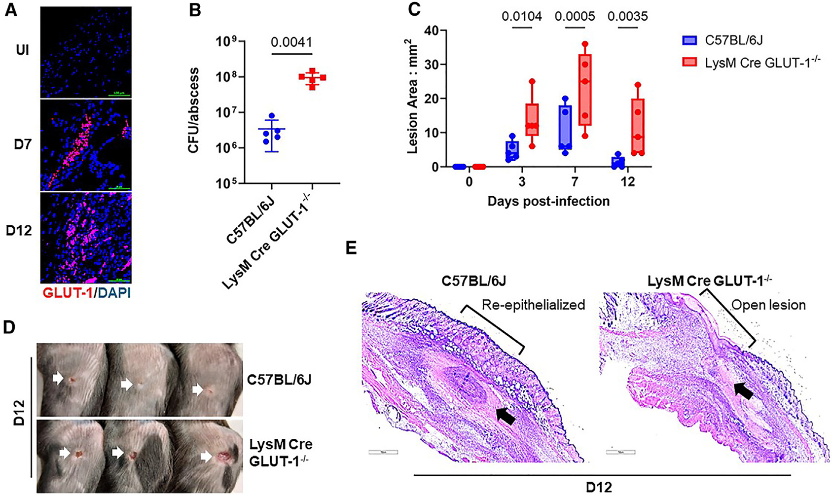
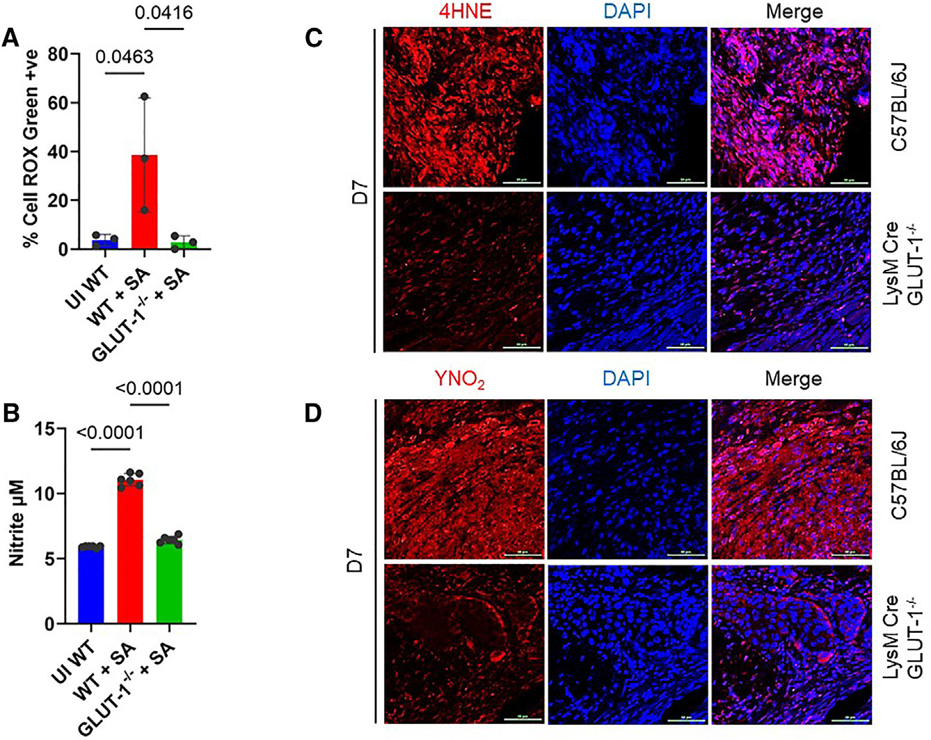
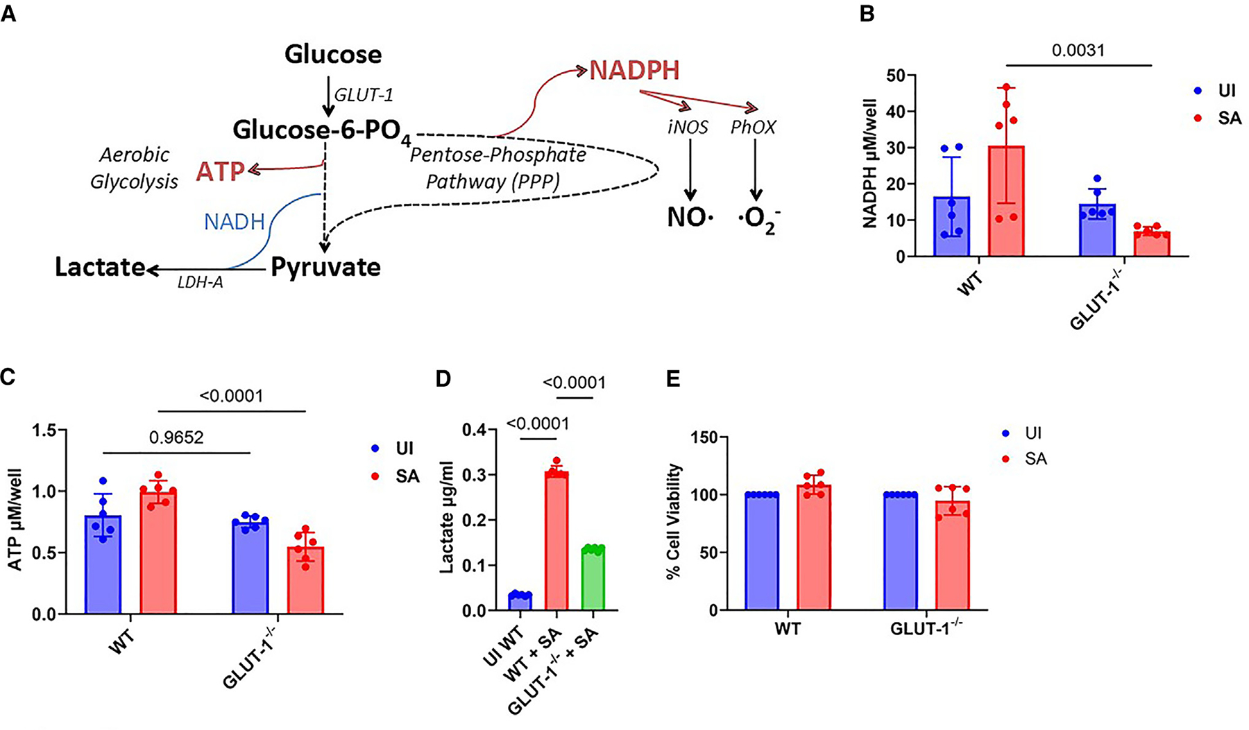
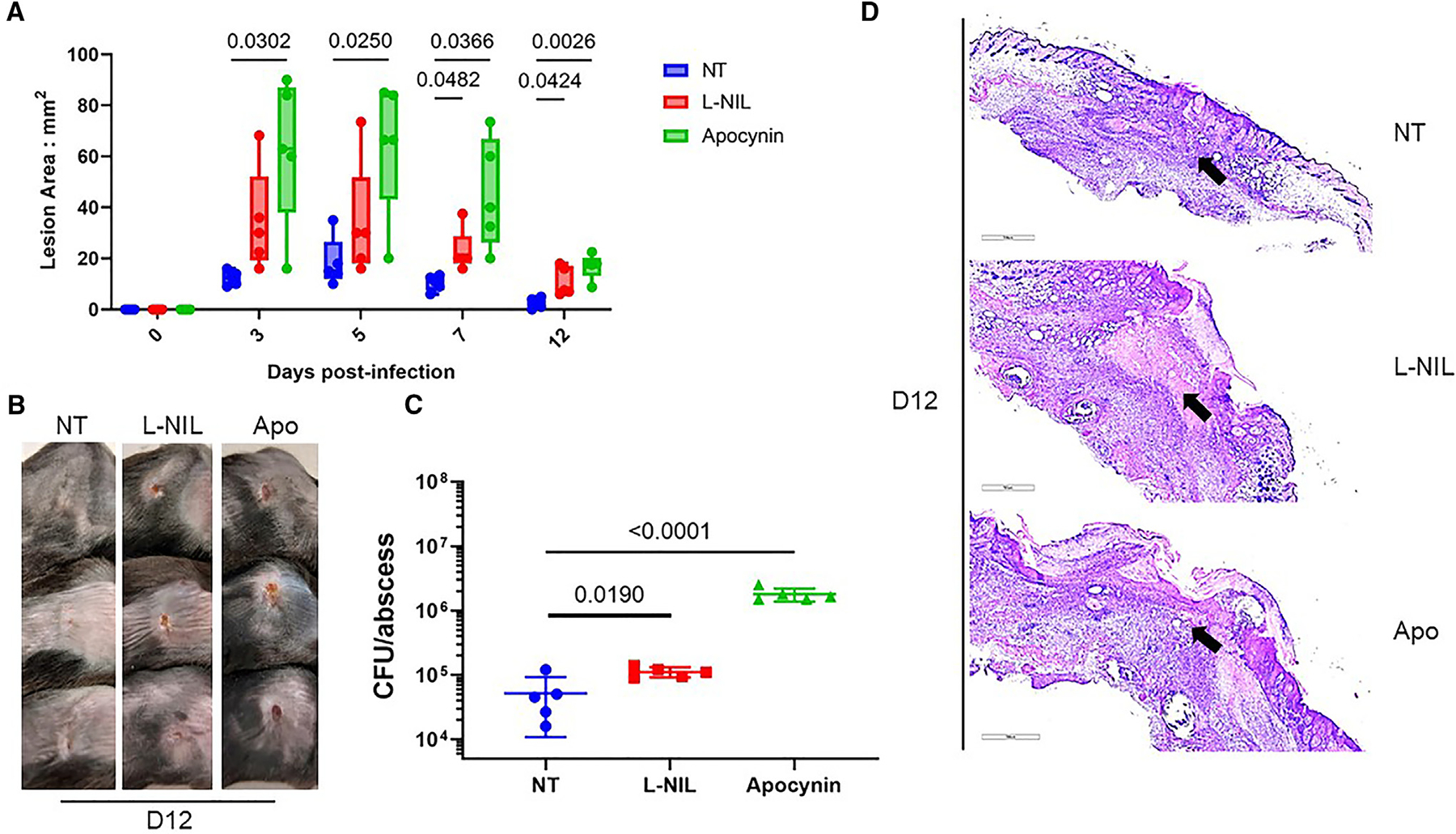
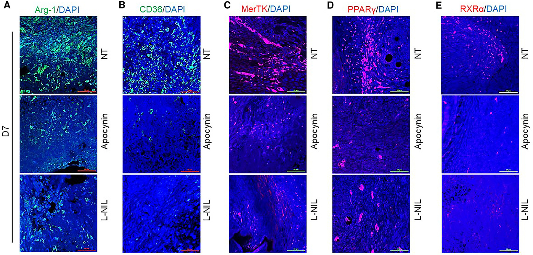
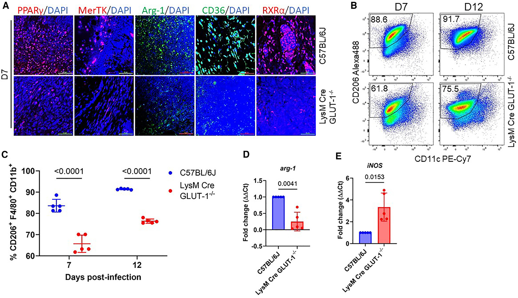
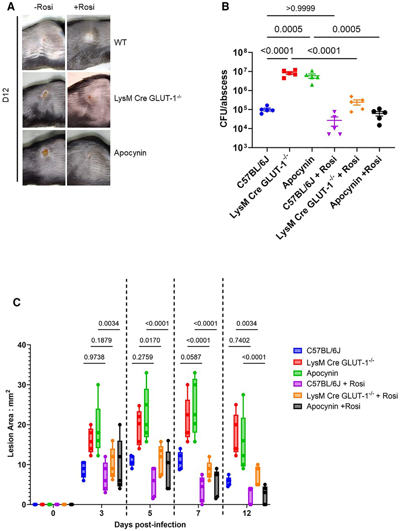
Skin/soft tissue infections (SSTIs) caused by methicillin-resistant Staphylococcus aureus (MRSA) pose a major healthcare burden. Distinct inflammatory and resolution phases comprise the host immune response to SSTIs. Resolution is a myeloid PPARγ-dependent anti-inflammatory phase that is essential for the clearance of MRSA. However, the signals activating PPARγ to induce resolution remain unknown. Here, we demonstrate that myeloid glucose transporter 1 (GLUT-1) is essential for the onset of resolution. MRSA-challenged macrophages are unsuccessful in generating an oxidative burst or immune radicals in the absence of GLUT-1 due to a reduction in the cellular NADPH pool. This translates in vivo as a significant reduction in lipid peroxidation products required for the activation of PPARγ in MRSA-infected mice lacking myeloid GLUT-1. Chemical induction of PPARγ during infection circumvents this GLUT-1 requirement and improves resolution. Thus, GLUT-1-dependent oxidative burst is essential for the activation of PPARγ and subsequent resolution of SSTIs.

Keywords: CP: Metabolism; CP: Microbiology; aureus; immunometabolism.


Conflict of interest statement

Declaration of interests The authors declare no competing interests.

Figures

Figure 1.
Figure 1.. GLUT-1 is essential for the resolution of MRSA SSTIs
(A) Immunofluorescent staining of skin abscess tissue slices from uninfected (UI) and CA-MRSA strain LAC infected (SA) C57BL/6J mice showing GLUT-1 (red) expression on days (D) 7 and 12. Counterstained with DAPI (blue). Images are 1 of n = 3 subjects. Scale bar, 100 μm. (B) Total MRSA CFU per abscess from LysM Cre GLUT-1−/− mice is ~2log higher than in WT C57BL/6J mice on day 12. n = 5 per group, Student’s t test with Welch’s correction for p value. (C) Lesion area in mm2 of abscesses from C57BL/6J and LysM Cre GLUT-1−/− mice over a period of 12 days shows larger lesions in the absence of GLUT-1. n = 5 per group, 2-way ANOVA for p value. (D) Gross pathology of lesions areas on C57BL/6J and LysM Cre GLUT-1−/− mice on day 12 shows larger unresolved lesions in the GLUT-1−/− mice. (E) Histopathology of lesions from C57BL/6J and LysM Cre GLUT-1−/− mice. Tissue slices were stained with H&E. Arrows and brackets show regions of open unresolved lesions in the absence of GLUT-1, whereas WT mice have re-epithelialized skin and closed abscess. Scale bar, 700 μm. All error bars are mean ± SD.
Figure 2.
Figure 2.. Attenuated oxidative burst in the absence of GLUT-1 in MRSA infection
(A) Fluorescent detection of ROS produced by MRSA-infected WT and GLUT-1−/− BMMs 4 h post-infection. Enumeration of % cell ROX green positive cells shows reduced ROS production in GLUT-1−/− macrophages. Representative images can be found in Figure S2D n = 3 per group, Student’s t test with Welch’s correction for p value. (B) Determination of extracellular nitrite produced by MRSA-infected WT and GLUT-1−/− BMMs 4 h post-infection. GLUT-1−/− macrophages produce less nitrite than the WT macrophages. n = 6 per group, Student’s t test with Welch’s correction for p value. (C and D) Immunofluorescent staining of day 7 lesion slices from C57BL/6J and LysM Cre GLUT-1−/− mice with antibodies against 4HNE (C) and YNO2 (D) seen in red. Slices were counterstained with DAPI (blue). Both 4HNE and YNO2 accumulation was reduced in LysM Cre GLUT-1−/− lesions. Images are 1 of n = 3 subjects. Scale bar, 50 μm. All error bars are mean ± SD.
Figure 3.
Figure 3.. GLUT-1-mediated oxidative burst is via the PPP
(A) Schematic representation of glucose metabolism via the glycolytic pathway and PPP in macrophages upon MRSA infection. The PPP generates NADPH, which is required for the function of NOX and iNOS to generate ROS and NO. (B) Comparison of intracellular NADPH concentrations (μM/well) in uninfected (UI) and MRSA-infected (SA) WT and GLUT-1−/− BMMs at 6 h post-infection shows an ~3-fold decrease in NADPH in GLUT-1−/− BMMs. n = 6 per group, 1-way ANOVA used for p value. (C) Comparison of intracellular ATP concentrations (μM/well) in uninfected (UI) and LAC-infected (SA) WT and GLUT-1−/− BMMs at 6 h post-infection shows an ~2-fold reduction in ATP in GLUT-1−/− BMMs. n = 6 per group, 1-way ANOVA used for p value. (D) Measurement of lactate released from uninfected (UI) and MRSA-infected (SA) WT and GLUT-1−/− BMMs at 6 h post-infection. Lactate release is downregulated ~3-fold in GLUT-1−/− BMMs. n = 6, Student’s t test used for p value. (E) Calcein-AM uptake-based survival assay shows no difference in % cell viability of WT and GLUT-1−/− BMMs at 6 h post-infection with LAC. All error bars are mean ± SD.
Figure 4.
Figure 4.. SSTI resolution is attenuated in the absence of oxidative burst
Untreated (NT), apocynin (Apo)-, and L-NIL hydrochloride (L-NIL)-treated C57BL/6J mice were infected subcutaneously with 108 CFU of MRSA and monitored over time. (A) Lesion areas (mm2) were measured over a period of 12 days and show larger lesions that remain unresolved in apocynin- and L-NIL-treated groups. n = 5 per group, 2-way ANOVA used for p value. (B) Gross pathology of the infected areas supports the lesion area measurement data, showing large open lesions on day 12 in apocynin- and L-NIL-treated mice, while NT mice have healed lesions. (C) Day 12 CFU of MRSA from the infected abscesses shows higher CFUs in apocynin- and L-NIL-treated mice than in NT mice. n = 5, 1-way ANOVA used for p value. (D) H&E-stained slices of day 12 lesions from the infected mice show open, unresolved lesions in apocynin- and L-NIL-treated groups, while the NT group has a walled-off abscess with re-epithelialized skin (arrows). Scale bar, 700 μm. All error bars are mean ± SD.
Figure 5.
Figure 5.. Absence of oxidative burst abrogates the emergence of anti-inflammatory and pro-efferocytotic markers in MRSA SSTIs
Immunofluorescent staining of (A) Arg-1 (green), (B) CD36 (green), (C) MerTK (red), (D) PPARγ (red), and (E) RXRα (red) in day 7 abscess tissue slices from MRSA-infected untreated (NT) and apocynin- and L-NIL-treated C57BL/6J mice shows lower expression of all markers in the apocynin- and L-NIL-treated groups compared to the NT group. Slices were counterstained with DAPI (blue). Panels show merged images only. Single-channel images can be found in Figure S4. Images are 1 of n = 3 subjects. Scale bar, 50 μm.
Figure 6.
Figure 6.. GLUT-1 is essential for the emergence of anti-inflammatory and pro-efferocytotic markers in MRSA SSTIs
(A) Immunofluorescent detection of PPARγ (red), MerTK (red), Arg-1 (green), CD36 (green), and RXRα (red) in day 7 lesions from WT C57BL/6J and LysM Cre GLUT-1−/− mice showing reduced expression of these markers in the absence of myeloid GLUT-1. Slices were counterstained with DAPI (blue). Panels show merged images only. Single-channel images can be found in Figure S6. Images are 1 of n = 3 subjects. Scale bar, 50 μm. (B) Flow cytometric plots showing a larger population of CD206+ macrophages in day 7 and day 12 abscesses of WT mice compared to LysM Cre GLUT-1−/− mice. Gates show CD45+F4/80+CD11b+CD11cCD206+ cells. (C) Quantification plots of flow cytometric data showing a significant reduction in CD206+F4/80+CD11b+ cells in LysM Cre GLUT-1−/− abscesses on both days 7 and 12. n = 5, 2-way ANOVA used for p values. (D and E) CD45+F4/80+CD11b+ populations were sorted from day 7 abscesses of WT C57BL/6J and LysM Cre GLUT-1−/− mice, and (D) arg1 and (E) iNOS transcripts were quantified. LysM Cre GLUT-1−/− abscesses had lower arg1 and higher iNOS transcripts than their WT counterparts. n = 5, Student’s t test used for p values. All error bars are mean ± SD.
Figure 7.
Figure 7.. PPARγ agonist restores resolution of MRSA SSTIs in the absence of myeloid GLUT-1
C57BL/6J, LysM Cre GLUT-1−/−, and apocynin-treated mice were either treated with rosiglitazone (+Rosi) or left untreated (−Rosi), followed by subcutaneous infection with 108 CFU MRSA and monitoring over time. (A) Gross pathology on day 12 shows rosiglitazone-treated LysM Cre GLUT-1−/− and apocynin groups have resolved lesions similar to the WT-rosiglitazone group. (B) Enumeration of MRSA CFUs from abscesses shows significant control of MRSA growth in rosiglitazone-treated LysM Cre GLUT-1−/− and apocynin groups compared to the untreated groups. n = 5, 1-way ANOVA used for p values. (C) Measurement of lesion areas (mm2) over a period of 12 days shows smaller lesions upon treatment with rosiglitazone in both LysM Cre GLUT-1−/− and apocynin groups than what is observed when rosiglitazone is not administered. n = 5, 2-way ANOVA used for p values. All error bars are mean ± SD.

Similar articles

References

    1. Talan DA, Krishnadasan A, Gorwitz RJ, Fosheim GE, Limbago B, Albrecht V, and Moran GJ; for The EIDNSG (2011). Comparison of Staphylococcus aureus From Skin and Soft-Tissue Infections in US Emergency Department Patients, 2004 and 2008. Clin. Infect. Dis. 53, 144–149. - PubMed
    1. Lee BY, Singh A, David MZ, Bartsch SM, Slayton RB, Huang SS, Zimmer SM, Potter MA, Macal CM, Lauderdale DS, et al. (2013). The economic burden of community-associated methicillin-resistant Staphylococcus aureus (CA-MRSA). Clin. Microbiol. Infect. 19, 528–536. - PMC - PubMed
    1. Pillai SK, Sakoulas G, Wennersten C, Eliopoulos GM, Moellering RC Jr., Ferraro MJ, and Gold HS (2002). Linezolid Resistance in Staphylococcus aureus: Characterization and Stability of Resistant Phenotype. J. Infect. Dis. 186, 1603–1607. - PubMed
    1. Turner NA, Sharma-Kuinkel BK, Maskarinec SA, Eichenberger EM, Shah PP, Carugati M, Holland TL, and Fowler VG (2019). Methicillin-resistant Staphylococcus aureus: an overview of basic and clinical research. Nat. Rev. Microbiol. 17, 203–218. - PMC - PubMed
    1. Larsen J, Raisen CL, Ba X, Sadgrove NJ, Padilla-González GF, Simmonds MSJ, Loncaric I, Kerschner H, Apfalter P, Hartl R, et al. (2022). Emergence of methicillin resistance predates the clinical use of antibiotics. Nature 602, 135–141. - PMC - PubMed

Publication types

MeSH terms

LinkOut - more resources