. 2024 Jul 11;19(7):e0306862.
doi: 10.1371/journal.pone.0306862. eCollection 2024.

The antimicrobial activity of an antiseptic soap against Candida Albicans and Streptococcus Mutans single and dual-species biofilms on denture base and reline acrylic resins

Affiliations

The antimicrobial activity of an antiseptic soap against Candida Albicans and Streptococcus Mutans single and dual-species biofilms on denture base and reline acrylic resins

Camilla Olga Tasso et al. PLoS One. .

Erratum in

Abstract

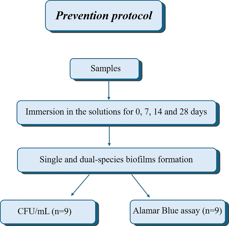
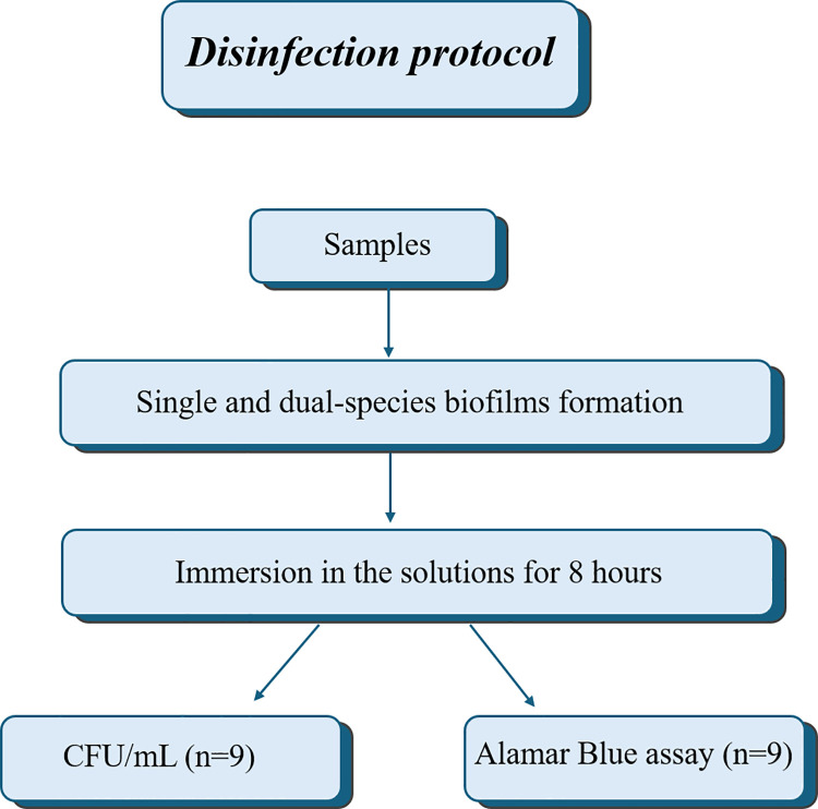
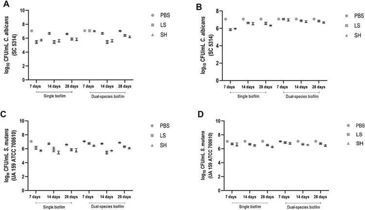
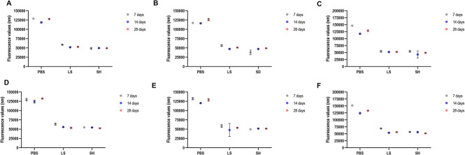
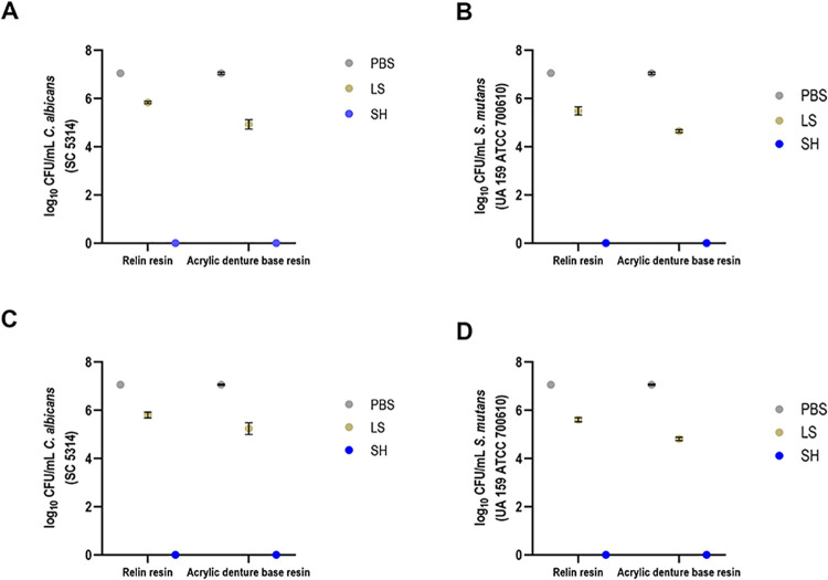
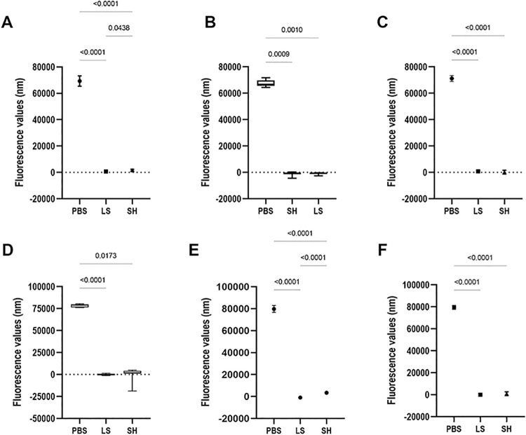
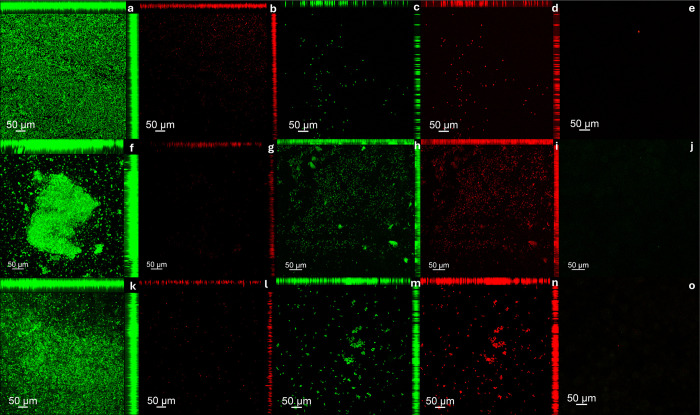
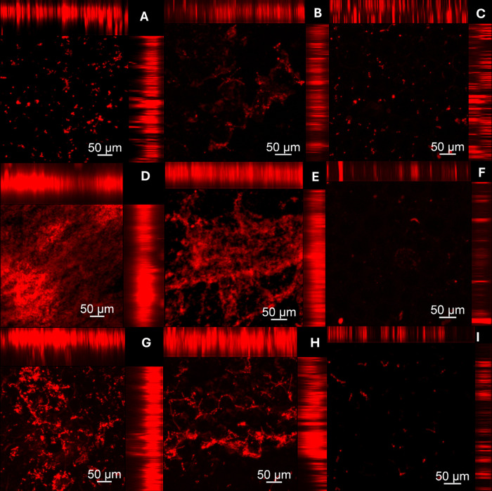
To evaluate the effect of antiseptic soap on single and dual-species biofilms of Candida albicans and Streptococcus mutans on denture base and reline resins. Samples of the resins were distributed into groups (n = 9) according to the prevention or disinfection protocols. In the prevention protocol, samples were immersed in the solutions (Lifebuoy, 0.5% sodium hypochlorite solution and PBS) for 7, 14 and 28 days before the single and dual-species biofilms formation. Overnight denture disinfection was simulated. In the disinfection protocol, samples were immersed in the same solutions during 8 hours after the single and dual-species biofilms formation. Antimicrobial activity was analyzed by counting colony-forming units (CFU/mL) and evaluating cell metabolism. Cell viability and protein components of the biofilm matrix were evaluated using confocal laser scanning microscopy (CLSM). Data were submitted to ANOVA, followed by Tukey's post-test (α = 0.05) or Dunnett's T3 multiple comparisons test. In the prevention protocol, Lifebuoy solution effectively reduced the number of CFU/mL of both species. In addition, the solution decreased the cell metabolism of the microorganisms. Regarding disinfection protocol, the Lifebuoy solution was able of reduce approximately of 2-3 logs for all the biofilms on the denture base and reline resin. Cellular metabolism was also reduced. The images obtained with CLSM corroborate these results. Lifebuoy solution was effective in reducing single and dual-species biofilms on denture base and reline resins.


Conflict of interest statement

The authors have declared that no competing interests exist.

Figures

Fig 1
Fig 1. Sequence for the prevention protocol.
Fig 2
Fig 2. Sequence for the disinfection protocol.
Fig 3
Fig 3
CFU/mL values of C. albicans and S mutans cells (single and dual-species biofilms) referring to biofilm formation on denture base (A and C) and reline acrylic resin (B and D) samples after immersion in PBS, Lifebuoy, and 0.5% sodium hypochlorite solutions.
Fig 4
Fig 4
Results of the cellular metabolism of the C. albicans and S. mutans cells (single and dual-species biofilms) referring to biofilm formation on denture base (A-C) and reline acrylic resin (D-F) samples after immersion in PBS, Lifebuoy, and 0.5% sodium hypochlorite solutions.
Fig 5
Fig 5
CFU/mL values of single (A and B) and dual-species (C and D) biofilms formed on the denture base and reline acrylic resin samples, submitted to the disinfection procedure.
Fig 6
Fig 6
Results of the cellular metabolism of the C. albicans and S. mutans cells (single and dual-species biofilms) formed on denture base (A-C) and reline acrylic resins (D-F) samples.
Fig 7
Fig 7. Cell viability of C. albicans and S. mutans single and dual-specie biofilms analyzed by CLSM on denture base acrylic resin samples.
Samples labeled with Syto-9 (cells marked in green) and propidium iodide (cells marked in red). (A, B) C. albicans biofilm–PBS (Negative control); (C, D) C. albicans biofilm–Lifebuoy solution; (E) C. albicans biofilm–Sodium Hypochlorite 0,5% (Positive control); (F, G) S. mutans biofilm—PBS (Negative control); (H, I) S. mutans biofilm—Lifebuoy solution; (J) S. mutans biofilm—Sodium Hypochlorite 0,5% (Positive control); (K, L) Dual-species biofilm—PBS (Negative control); (M, N) Dual-species biofilm—Lifebuoy solution; (O) Dual-species biofilm–Sodium Hypochlorite 0,5% (Positive control).
Fig 8
Fig 8. Cell viability of C. albicans and S. mutans single and dual-specie biofilms analyzed by CLSM on hard reline acrylic resin samples.
Samples labeled with Syto-9 (cells marked in green) and propidium iodide (cells marked in red). (a, b) C. albicans biofilm–PBS (Negative control); (c, d) C. albicans biofilm–Lifebuoy solution; (e) C. albicans biofilm–Sodium Hypochlorite 0,5% (Positive control); (f, g) S. mutans biofilm—PBS (Negative control); (h, i) S. mutans biofilm—Lifebuoy solution; (j) S. mutans biofilm—Sodium Hypochlorite 0,5% (Positive control); (k, l) Dual-species biofilm—PBS (Negative control); (m, n) Dual-species biofilm—Lifebuoy solution; (o) Dual-species biofilm–Sodium Hypochlorite 0,5% (Positive control).
Fig 9
Fig 9. Assessment of damage to the protein components of the biofilm extracellular matrix after treatment protocols on denture base acrylic resin samples.
Components visualized in red were marked with Sypro Ruby Biofilm Matrix Stain. (A) C. albicans biofilm–PBS (Negative control); (B) C. albicans biofilm–Lifebuoy solution; (C) C. albicans biofilm–Sodium Hypochlorite 0,5% (Positive control); (D) S. mutans biofilm—PBS (Negative control); (E) S. mutans biofilm—Lifebuoy solution; (F) S. mutans biofilm—Sodium Hypochlorite 0,5% (Positive control); (G) Dual-species biofilm—PBS (Negative control); (H) Dual-species biofilm—Lifebuoy solution; (I) Dual-species biofilm–Sodium Hypochlorite 0,5% (Positive control).
Fig 10
Fig 10. Assessment of damage to the protein components of the biofilm extracellular matrix after treatment protocols on hard reline acrylic resin samples.
Components visualized in red were marked with Sypro Ruby Biofilm Matrix Stain. (a) C. albicans biofilm–PBS (Negative control); (b) C. albicans biofilm–Lifebuoy solution; (c) C. albicans biofilm–Sodium Hypochlorite 0,5% (Positive control); (d) S. mutans biofilm—PBS (Negative control); (e) S. mutans biofilm—Lifebuoy solution; (f) S. mutans biofilm—Sodium Hypochlorite 0,5% (Positive control); (g) Dual-species biofilm—PBS (Negative control); (h) Dual-species biofilm—Lifebuoy solution; (i) Dual-species biofilm–Sodium Hypochlorite 0,5% (Positive control).

Similar articles

Cited by

References

    1. Gendreau L, Loewy ZG. Epidemiology and etiology of denture stomatitis. J Prosthodont. 2011;20(4):251–260. doi: 10.1111/j.1532-849X.2011.00698.x - DOI - PubMed
    1. Budtz-Jörgensen E. Clinical aspects of Candida infection in denture wearers. J Am Dent Assoc. 1978; 96(3):474–479. doi: 10.14219/jada.archive.1978.0088 - DOI - PubMed
    1. Arendorf TM, Walker D. M. Denture stomatitis: a review. J Oral Rehabil. 1987;14(3):217–227. doi: 10.1111/j.1365-2842.1987.tb00713.x - DOI - PubMed
    1. Jeganathan S, Lin CC. Denture stomatitis—a review of the aetiology, diagnosis and management. Aust Dent J. 1992;37(2):107–114. doi: 10.1111/j.1834-7819.1992.tb03046.x - DOI - PubMed
    1. Reichart PA. Oral mucosal lesions in a representative cross-sectional study of aging Germans. Community Dent Oral Epidemiol. 2000;28(5):390–398. doi: 10.1034/j.1600-0528.2000.028005390.x - DOI - PubMed

MeSH terms

Substances