. 2024 Jul 16;121(29):e2317977121.
doi: 10.1073/pnas.2317977121. Epub 2024 Jul 11.

Point mutations at specific sites of the nsp12-nsp8 interface dramatically affect the RNA polymerization activity of SARS-CoV-2

Affiliations

Point mutations at specific sites of the nsp12-nsp8 interface dramatically affect the RNA polymerization activity of SARS-CoV-2

Cristina Ferrer-Orta et al. Proc Natl Acad Sci U S A. .

Abstract

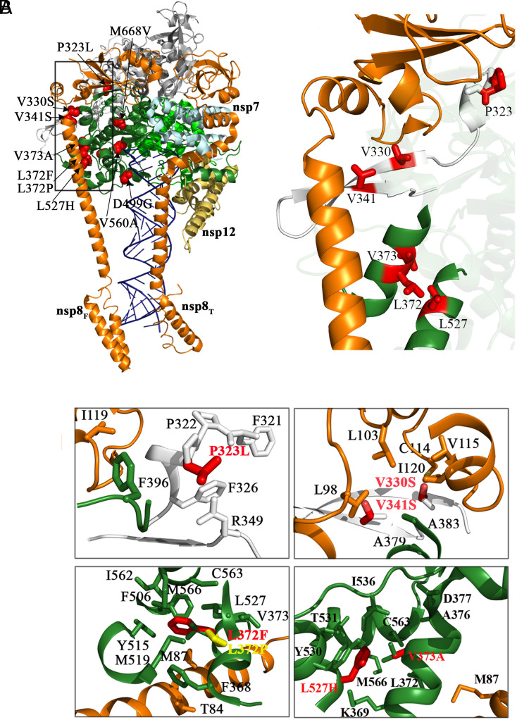
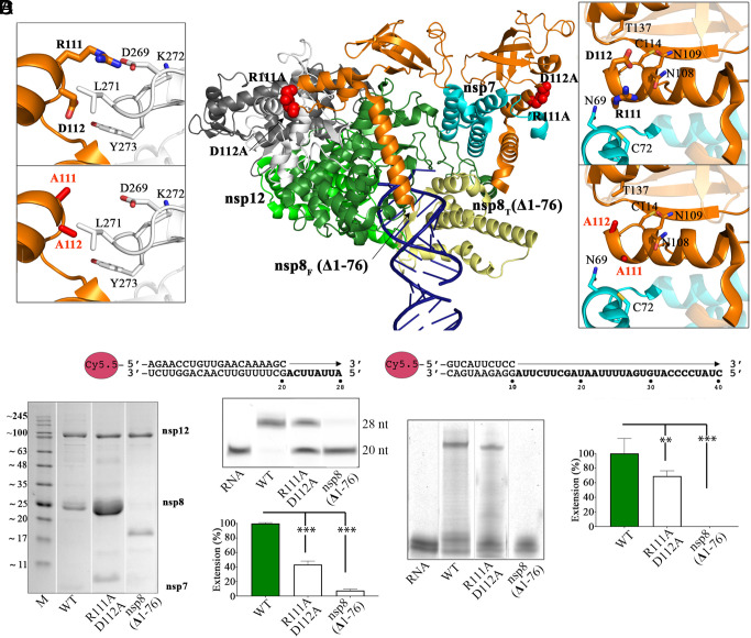
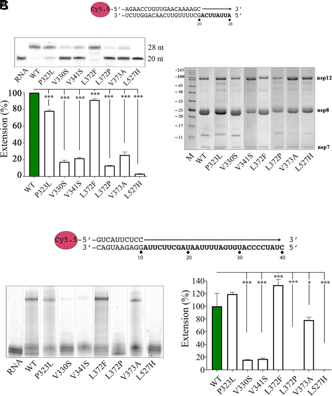
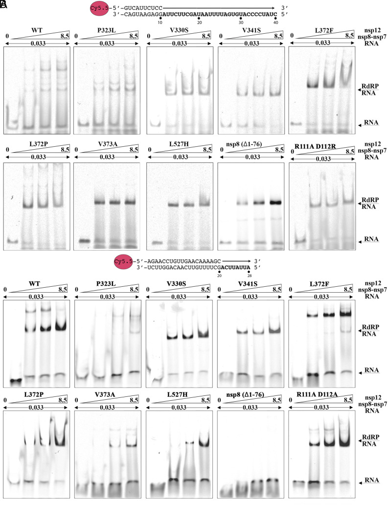
In a recent characterization of the Severe Acute Respiratory Syndrome Coronavirus 2 (SARS-CoV-2) variability present in 30 diagnostic samples from patients of the first COVID-19 pandemic wave, 41 amino acid substitutions were documented in the RNA-dependent RNA polymerase (RdRp) nsp12. Eight substitutions were selected in this work to determine whether they had an impact on the RdRp activity of the SARS-CoV-2 nsp12-nsp8-nsp7 replication complex. Three of these substitutions were found around the polymerase central cavity, in the template entry channel (D499G and M668V), and within the motif B (V560A), and they showed polymerization rates similar to the wild type RdRp. The remaining five mutations (P323L, L372F, L372P, V373A, and L527H) were placed near the nsp12-nsp8F contact surface; residues L372, V373, and L527 participated in a large hydrophobic cluster involving contacts between two helices in the nsp12 fingers and the long α-helix of nsp8F. The presence of any of these five amino acid substitutions resulted in important alterations in the RNA polymerization activity. Comparative primer elongation assays showed different behavior depending on the hydrophobicity of their side chains. The substitution of L by the bulkier F side chain at position 372 slightly promoted RdRp activity. However, this activity was dramatically reduced with the L372P, and L527H mutations, and to a lesser extent with V373A, all of which weaken the hydrophobic interactions within the cluster. Additional mutations, specifically designed to disrupt the nsp12-nsp8F interactions (nsp12-V330S, nsp12-V341S, and nsp8-R111A/D112A), also resulted in an impaired RdRp activity, further illustrating the importance of this contact interface in the regulation of RNA synthesis.

Keywords: RNA synthesis; RNA-dependent RNA polymerase; nsp12–nsp8–nsp7 complex; primer extension; protein–protein interactions.


Conflict of interest statement

Competing interests statement:The authors declare no competing interest.

Figures

Fig. 1.
Fig. 1.
Location and functional implication of the selected amino acid substitutions in the nsp12 protein of SARS-CoV-2. (A) The Left panel shows the location of the amino acid substitutions studied here (P323L, V330S, V341S, L372F, L372P, V373A, D499G, L527H, V560A, and M668V) in the three-dimensional structure of nsp12. The structure used as reference is that of the nsp12–nsp8–nsp7–RNA complex (PDB: 6YYT), depicted as a cartoon representation with nsp12 colored by domains (NiRAN in gray, interface in white, and the RdRp domain in dark green, green, and dark yellow for the fingers, palm, and thumb subdomains, respectively). The cofactors nsp8 and nsp7 are colored in orange and cyan, respectively, and the RNA is depicted in dark blue. Substituted amino acids are shown as red spheres and explicitly labeled. The Right panel shows a close-up view, highlighting the positions of the amino acid substitutions in the nsp12–nsp8F contact interface (side chains in red sticks). (B) Detailed views of the interactions around the mutated positions. Side chains of substituted amino acids and neighboring residues are shown as sticks in different colors (white for residues in the nsp12 interface domain, green for residues in the nsp12 fingers, and orange for nsp8), and explicitly labeled. Both wild type and mutated side chains are seen in the different panels. The Upper Left panel shows the P323L substitution placed in the nsp12 interface (depicted in white for proline and in red for the mutated leucine). The Upper Right panel shows substitutions V330S and V341S, within the nsp12 interface (white for the valine side chains and red for the mutated serine residues). The Bottom Left panel shows L372F and L372P side chain substitutions, within de nsp12 fingers (side chain depicted in green for leucine, red for phenylalanine, and yellow for proline). The Bottom Right panel shows V373A and L527H substitutions, also within the nsp12 fingers (green for valine and leucine side chains and red for alanine and histidine).
Fig. 2.
Fig. 2.
Location and functional implication of the SARS-CoV-2 nsp12–nsp8–nsp7 complex, harboring amino acid substitutions in nsp8, specially designed to weaken the nsp12–nsp8 interaction interface. (A) Mapping of the nsp8-R111A/D112A double mutant. The central image shows a cartoon representation of the nsp12–nsp7–nsp8–RNA complex colored as in Fig. 1. In this representation the first 76 amino acids of nsp8 have been removed to show a model of the nsp8(Δ1-76) construct. The position of the double mutation R111A/D112A is shown as red spheres in the two nsp8 molecules of the complex. The two Insets at the Left and Right sides of the central image show close-up views of the environment and interactions of the replaced residues in nsp8F (Left side Insets) and nsp8T (Right side Insets). Side chains of the original (Upper Insets), mutated residues (Lower Insets), and interacting amino acids (within a 5 Å radius) are shown as sticks in different colors, and explicitly labeled. (B–D) Comparative in vitro RNA synthesis activities of the SARS-CoV-2 nsp12–nsp7–nsp8 WT and mutants nsp8(R111A/D112A) and nsp8(Δ1-76). Reactions were performed at 33 °C, and activities were measured in gel-based nonradioactive primer extension assays. (B) P/T duplexes used in the assays, with the arrow indicating the location and direction of primer extension. (C) SDS–PAGE analysis of the purified SARS-CoV-2 RdRp WT and mutant complexes, employed in the primer-extension assays. (D) Primer-extension activities of the different RdRp complexes on a representative PAGE, 18% polyacrylamide, 7M urea/Tris–Borate–EDTA (TBE). The Bottom panels show the relative efficiencies of full-length RNA synthesis by the different RdRp mutants compared to the WT. SDs from three independent experiments are shown. Statistical significance of the differences was calculated by one-way ANOVA followed by Dunnett’s multiple comparison test (SI Appendix, Table S1 in https://saco.csic.es/index.php/s/Xp33GCyPMp8Lif7).
Fig. 3.
Fig. 3.
Comparative in vitro RNA synthesis activities of the SARS-CoV-2 nsp12–nsp7–nsp8 complex, WT and nsp12 mutants harboring amino acid substitutions in the nsp12–nsp8F interface. Activities were measured in gel-based nonradioactive primer extension assays. (A) The Top panel shows the P/T 20/28 nt RNA used in the assays. The Middle panel shows the primer-extension activities of the different RdRp complexes on a representative PAGE, 18% polyacrylamide, and 7M urea/TBE. Reactions were performed at 33 °C, and samples were collected at 10 min intervals. The Bottom panel shows the relative efficiencies of full-length RNA synthesis by the different RdRp mutants compared to the WT. SDs from three independent experiments are shown. Statistical significance of the differences was calculated by one-way ANOVA followed by Dunnett’s multiple comparison test (see also SI Appendix, Table S1 in https://saco.csic.es/index.php/s/Xp33GCyPMp8Lif7). (B) SDS–PAGE analysis of the purified SARS-CoV-2 RdRp WT and mutant complexes, employed in the primer-extension assays. (C) The Top panel shows the P/T 10/40 RNA used in the assays, with the arrow indicating the location and direction of primer extension. The Right panel shows the primer-extension activities of the different RdRp complexes on a representative PAGE, performed as in A. The Left panel shows the relative efficiencies of full-length RNA synthesis by the different RdRp mutants compared to the WT. SDs from three independent experiments are shown. Statistical significances are calculated as in A (SI Appendix, Table S1 in https://saco.csic.es/index.php/s/Xp33GCyPMp8Lif7).
Fig. 4.
Fig. 4.
RNA-binding capacity of the nsp12–nsp7–nsp8 complexes, WT and mutants. EMSA were performed to measure the capacity of the different replication complexes to bind the RNAs P/T 10/40 (A) and P/T 20/28 (B). WT and mutant nsp12–nsp7–nsp8 complexes at increasing concentrations (from 0 to 8.5 pmol) were incubated with constant amounts of the template-primer RNA used in the polymerizations assays (RNA concentration 0.033 pmol, in 50 mM NaCl, 20 mM HEPES pH 8, 5 mM MgCl2, and 20 mM DTT buffer). This would correspond to RNA–protein ratios of 1:0, 1:64, 1:128, and 1:256, in the different lanes. The protein–RNA complexes were resolved by electrophoresis in a nondenaturing 8% polyacrylamide gel, run at 4 °C in 0.5× TBE.

Similar articles

Cited by

References

    1. Karamitros T., et al. , SARS-CoV-2 exhibits intra-host genomic plasticity and low-frequency polymorphic quasispecies. J. Clin. Virol. 131, 104585 (2020). - PMC - PubMed
    1. Jary A., et al. , Evolution of viral quasispecies during SARS-CoV-2 infection. Clin. Microbiol. Infect. 26, 1560.e1–1560.e4 (2020). - PMC - PubMed
    1. Rueca M., et al. , Compartmentalized replication of SARS-Cov-2 in upper vs. lower respiratory tract assessed by whole genome quasispecies analysis. Microorganisms. 8, 1302 (2020). - PMC - PubMed
    1. Andino R., Domingo E., Viral quasispecies. Virology 479–480, 46–51 (2015), 10.1016/j.virol.2015.03.022. - DOI - PMC - PubMed
    1. Domingo E., Sabo D., Taniguchi T., Weissmann C., Nucleotide sequence heterogeneity of an RNA phage population. Cell 13, 735–744 (1978), 10.1016/0092-8674(78)90223-4. - DOI - PubMed

MeSH terms

Substances

LinkOut - more resources