. 2024 Jul 16;121(29):e2404551121.
doi: 10.1073/pnas.2404551121. Epub 2024 Jul 11.

Mechanical stress during confined migration causes aberrant mitoses and c-MYC amplification

Affiliations

Mechanical stress during confined migration causes aberrant mitoses and c-MYC amplification

Giulia Bastianello et al. Proc Natl Acad Sci U S A. .

Abstract

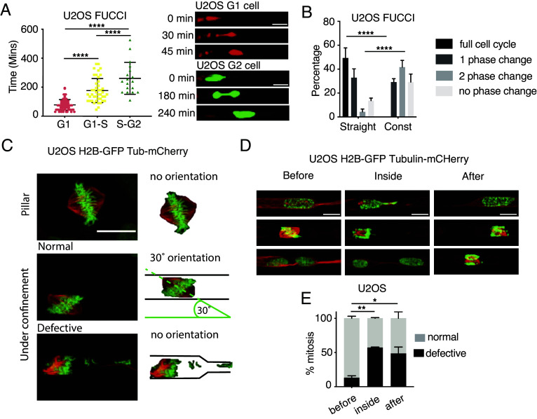
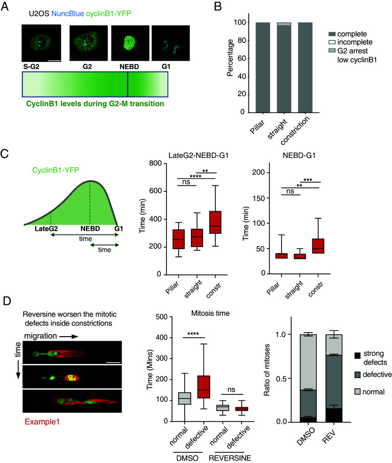
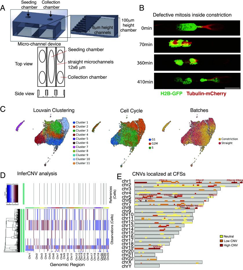
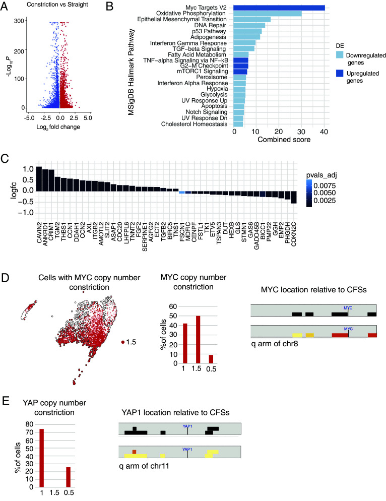
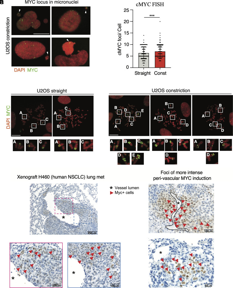
Confined cell migration hampers genome integrity and activates the ATR and ATM mechano-transduction pathways. We investigated whether the mechanical stress generated by metastatic interstitial migration contributes to the enhanced chromosomal instability observed in metastatic tumor cells. We employed live cell imaging, micro-fluidic approaches, and scRNA-seq to follow the fate of tumor cells experiencing confined migration. We found that, despite functional ATR, ATM, and spindle assembly checkpoint (SAC) pathways, tumor cells dividing across constriction frequently exhibited altered spindle pole organization, chromosome mis-segregations, micronuclei formation, chromosome fragility, high gene copy number variation, and transcriptional de-regulation and up-regulation of c-MYC oncogenic transcriptional signature via c-MYC locus amplifications. In vivo tumor settings showed that malignant cells populating metastatic foci or infiltrating the interstitial stroma gave rise to cells expressing high levels of c-MYC. Altogether, our data suggest that mechanical stress during metastatic migration contributes to override the checkpoint controls and boosts genotoxic and oncogenic events. Our findings may explain why cancer aneuploidy often does not correlate with mutations in SAC genes and why c-MYC amplification is strongly linked to metastatic tumors.

Keywords: aneuploidy; chromosome segregation; mechanical stress; mitosis; mitotic spindle.


Conflict of interest statement

Competing interests statement:M.P. is of the board of Directors and stakeholder of CheckmAb s.r.l. and is a recipient of grants under a research agreement with Bristol-Myers Squibb and Macomics.

Figures

Fig. 1.
Fig. 1.
Interstitial migration delays cell cycle progression, induces mitotic defects, and chromosome missegregation. (A) Cell cycle phase determines the speed of migration across constrictions. Analysis of the time to cross the constriction in U2OS cells labeled with FUCCI cell cycle marker. G1-phase cells (Red) pass-through the fastest while G2 cells (Green) take the longest. ****P value < 0.0001 one-way ANOVA. Bars show mean ± SD. (Scale bars: 20 μm.) (B) Mechanical constraints delay cell cycle progression. Cell cycle analysis in FUCCI-U2OS cells under various mechanically constrained environments (straight channels vs. channels with constrictions). ****P value < 0.0001 two-way ANOVA. Error bars are SEM. (C) Mitotic spindle organization in U2OS H2B-GFP Tubulin-mCherry migrating inside microchannels. The mitotic spindle orients following a 30° angle in cells undergoing mitosis inside straight channels; the presence of constrictions causes defective mitoses with altered spindle orientation and chromosome missegregations. (Scale bar: 20 μm.) (D and E) U2OS cells undergo defective mitoses while dividing across constrictions. (D) Examples of U2OS H2B-GFP Tubulin-mCherry cells dividing before, inside or after the constriction in channels. (Scale bars: 10 μm.) (E) Quantification of defective mitoses w.r.t their position in the channel. *P value < 0.05, **P value < 0.01 two-way ANOVA. Error bars are SEM.
Fig. 2.
Fig. 2.
Confined migration causes defective mitosis and aneuploidy in spite of a functional SAC. (A) Schematic representation of cyclinB1 expression during cell cycle along with example images from U2OS Cyclin B1-YFP cells where DNA was stained using the NuncBlue dye. Cyclin B1-YFP fluorescence signal increases at the end of S-phase and continues to show higher intensity though G2-phase until mitosis. Upon NEBD cyclin B1 enters the nucleus and is decayed as the sister chromatids separate into daughter cells. (Scale bar: 20 μm.) (B) Analysis of mitotic events in U2OS cells expressing Cyclin B1-YFP and migrating outside or inside microchannels. (C) Analysis of cyclin B1-YFP lifespan during mitosis in U2OS cells migrating inside channels. Cyclin B1-lifespan is increased in U2OS cells dividing inside constrictions (LateG2-G1) suggesting a mitotic stress. Mitosis inside constrictions also causes a mild delay in cyclin B1 degradation following NEBD in U2OS cells (NEBD-G1). ****P value < 0.0001, **P value < 0.01 ***P value < 0.001 one-way ANOVA. (D) Time-lapse images showing increased mitotic defects in U2OS H2B-GFP Tubulin-mCherry cells dividing inside microchannels (Left). Reversine abolishes the mitotic delay in U2OS H2B-GFP Tubulin-mCherry cells undergoing defective mitoses during interstitial migration suggesting that SAC is activated by chromosome missegregations caused by mechanical stress. Mitotic time is measured as the time-window between chromosome condensation in prophase and the generation of G1 daughter cells (Central graph). Reversine 0.5 μM treatment significantly worsens the mitotic defects in U2OS H2B-GFP Tubulin-mCherry cells dividing inside microchannels (Right graph). ****P value< 0.0001 one-way ANOVA. Error bars in the bar graph are SDs.
Fig. 3.
Fig. 3.
Interstitial migration across one single constriction results in mitotic defects and copy number variation that affect also chromosome fragile sites (CFSs). (A) Schematic representation of the custom-made microchannel device system designed to collect cells migrated across straight channels or channels with 3,5 μm constrictions. (B) Example of U2OS H2B-GFP Tubulin-mCherry undergoing defective mitosis during migration across constriction inside the custom-made microchannel device. (Scale bar: 20 μm.) (C) Low dimensional UMAP embeddings of single-cell RNA-seq data showing automatic clustering based on transcriptomic profile of each cell (Left), cell cycle phase clustering (Middle) and identification of cells belonging to each batch, straight or constriction (Right). (D) InferCNV analysis of single-cell RNA-seq from U2OS clone migrated in the device with straight (reference) or constriction channels. Cells migrated across one single constriction show increase CNV at several genomic regions. (E) CNVs also localize at CFSs in U2OS cells migrated across constriction. Schematic representation showing all the individual chromosomes (gray bars) and the relative fragile sites (yellow to red bars). Color code refer to the frequency of CNV identified on each single fragile site (red: higher amount of CNV). The highlighted red names indicate fragile sites with the highest frequency of CNV across all CFSs.
Fig. 4.
Fig. 4.
Interstitial migration across one single constriction results transcriptional upregulation of pathways involving c-MYC, YAP, inflammation, and cell metabolism. (A) Volcano plot showing differential expression analysis comparing straight and constriction samples showing that cells migrated across constrictions have several genes deregulated at the transcriptional level. (B) Enrichr analysis of the transcriptionally up-regulated (blue) and down-regulated (light-blue) pathways in U2OS cells migrated across constrictions. c-MYC targets appear as the top-ranked transcriptionally up-regulated genes. (C) Graphs showing transcriptional upregulation of the YAP signature (52) in U2OS cells migrated across one constriction. (D) Identification of cells with gain of MYC copy number in the BBrowser map and bar-graph showing MYC CNV in cells migrated across constrictions (Left). Graphical representation highlighting the c-MYC locus on the q arm of chr8 located near two CFSs (FRA8E and FRA8C) that are affected by CNV following interstitial migration (Right). (E) Graphs showing absence of YAP copy number gain in U2OS cells migrated across one constriction (Left) and graphical representation highlighting the YAP1 locus on the q arm of chr11 located far from CFSs and in a region poorly affected by CNV following interstitial migration (Right).
Fig. 5.
Fig. 5.
Interstitial migration causes c-MYC locus amplifications. (A) FISH of c-MYC locus localized in micronuclei of U2OS cells migrated across constrictions. (Scale bar: 20 μm.) (B) c-MYC FISH showing increased number of c-MYC foci in U2OS cells migrated across channels with constrictions. ***P value < 0.001 unpaired t test, error bars are SDs. (C) Representative FISH images showing c-MYC amplifications found in metaphase spreads of U2OS cells migrated across constriction. The regular c-MYC locus configuration in U2OS cells migrated across straight channels is shown on the Left. (Scale bars: 20 μm.) (D) Immunohistochemistry images of lung metastasis derived from a xenograft model of human NSCLC stained for c-MYC. H460 cells metastasized in the lungs show increased heterogeneity for c-MYC protein expression.

Similar articles

Cited by

References

    1. Turner K. M., et al. , Extrachromosomal oncogene amplification drives tumour evolution and genetic heterogeneity. Nature 543, 122–125 (2017). - PMC - PubMed
    1. Bach D. H., Zhang W., Sood A. K., Chromosomal instability in tumor initiation and development. Cancer Res. 79, 3995–4002 (2019). - PMC - PubMed
    1. Mitelman F., Johansson B., Mertens F., The impact of translocations and gene fusions on cancer causation. Nat. Rev. Cancer 7, 233–245 (2007). - PubMed
    1. Garribba L., et al. , Short-term molecular consequences of chromosome mis-segregation for genome stability. Nat. Commun. 14, 1353 (2023). - PMC - PubMed
    1. Lara-Gonzalez P., Westhorpe F. G., Taylor S. S., The spindle assembly checkpoint. Curr. Biol. 22, R966–R980 (2012). - PubMed

Substances