

Epileptic activity on foramen ovale electrodes is associated with sleep and tau pathology in Alzheimer's disease
- PMID: 38990981
- PMCID: PMC11788210
- DOI: 10.1093/brain/awae231
Epileptic activity on foramen ovale electrodes is associated with sleep and tau pathology in Alzheimer's disease
Abstract
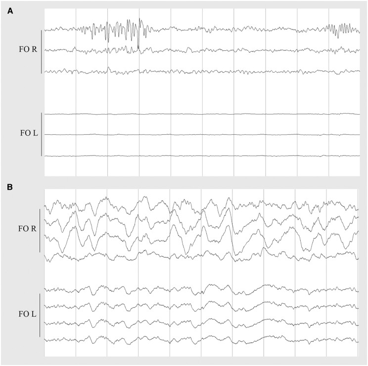
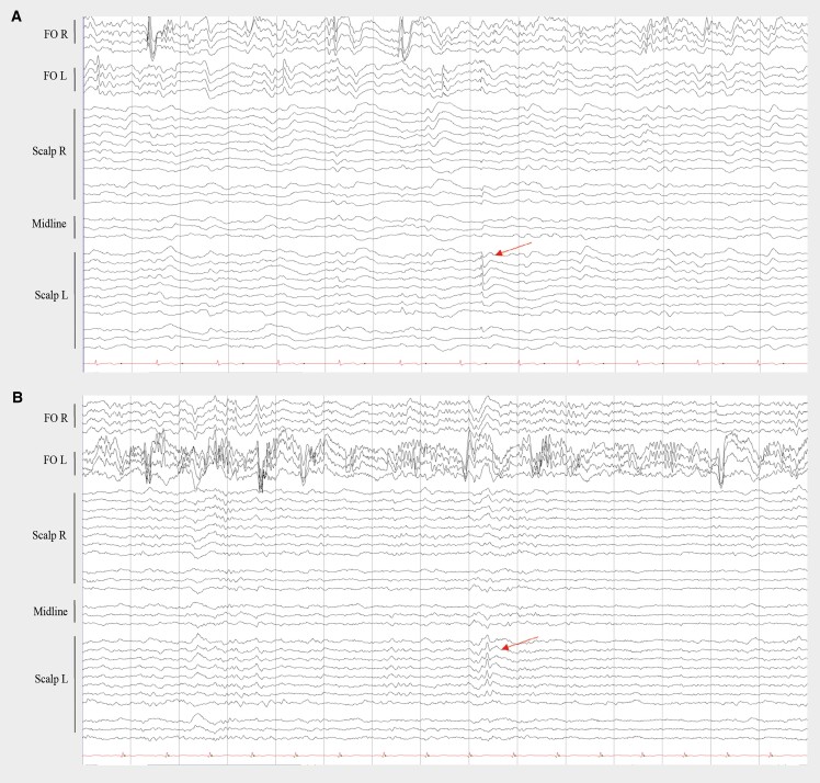
Both sleep alterations and epileptiform activity are associated with the accumulation of amyloid-β and tau pathology and are currently investigated for potential therapeutic interventions in Alzheimer's disease. However, a bidirectional intertwining relationship between sleep and neuronal hyperexcitability might modulate the effects of Alzheimer's disease pathology on the corresponding associations. To investigate this, we performed multiple day simultaneous foramen ovale (FO) plus scalp EEG and polysomnography recordings and acquired 18F-MK6240 tau PET-MR in three patients in the prodromal stage of Alzheimer's disease and in two patients with mild and moderate dementia due to Alzheimer's disease, respectively. As an eligibility criterion for the present study, subjects either had a history of a recent seizure (n = 2) or subclinical epileptiform activity (SEA) on a previous scalp EEG taken in a research context (n = 3). The 18F-MK6240 standard uptake value ratio (SUVR) and asymmetry index (AI) were calculated in a priori-defined volumes of interest. Linear mixed-effects models were used to study associations between interictal epileptiform discharges (IEDs), polysomnography parameters and 18F-MK6240 SUVR. Epileptiform activity was bilateral but asymmetrically present on FO electrodes in all patients and ≥95% of IEDs were not visible on scalp EEG. In one patient, two focal seizures were detected on FO electrodes, both without visual scalp EEG correlate. We observed lateralized periodic discharges, brief potentially ictal rhythmic discharges and lateralized rhythmic delta activity on FO electrodes in four patients. Unlike scalp EEG, intracranial electrodes showed a lateralization of epileptiform activity. Although the amount of IEDs on intracranial electrodes was not associated to the 18F-MK6240 SUVR binding in different volumes of interest, there was a congruent asymmetry of the 18F-MK6240 binding towards the most epileptic hemisphere for the mesial (P = 0.007) and lateral temporal cortex (P = 0.006). IEDs on intracranial electrodes were most abundant during slow wave sleep (SWS) (92/h) and non-REM sleep 2 (N2, 81/h), followed by non-REM sleep 1 (N1, 33/h) and least frequent during wakefulness (17/h) and REM sleep (9/h). The extent of IEDs during sleep was not reflected in the relative time in each sleep stage spent [REM% (P = 0.415), N1% (P = 0.668), N2% (P = 0.442), SWS% (P = 0.988)], and not associated with the arousal index (P = 0.317), apnoea-hypopnoea index (P = 0.846) or oxygen desaturation index (P = 0.746). Together, our observations suggest a multi-directional interaction between sleep, epileptiform activity and tau pathology in Alzheimer's disease.
Keywords: 18F-MK6240 tau-PET; epileptiform activity; mesial temporal lobe; neuronal hyperexcitability in Alzheimer’s disease; polysomnography; scalp-intracranial EEG.
© The Author(s) 2024. Published by Oxford University Press on behalf of the Guarantors of Brain.
Conflict of interest statement
K.V.L. is advisory board member for Cerveau/Lantheus and has performed contract research through Leuven Research and Development with Cerveau/Lantheus, Janssen Pharmaceuticals, BMS, Cerevel, Merck, Biogen and GE Healthcare. R.V. was PI of the phase 1 study with 18F-MK6240 and of the phase 1 and 2 studies with 18F-flutemetamol. R.V.'s institution has clinical trial agreements (R.V. as PI) with Alector, Biogen, Denali J&J, Prevail, and UCB. R.V.'s institution has consultancy agreements (R.V. as DSMB member) with AC Immune and Novartis. The other authors report no competing interests.
Figures


References
MeSH terms
Substances
Grants and funding
LinkOut - more resources
Full Text Sources
Medical
Research Materials
