. 2024 Oct 10;64(4):2301441.
doi: 10.1183/13993003.01441-2023. Print 2024 Oct.

A range of 30-62% of functioning multiciliated airway cells is sufficient to maintain ciliary airway clearance

Affiliations

A range of 30-62% of functioning multiciliated airway cells is sufficient to maintain ciliary airway clearance

Niki T Loges et al. Eur Respir J. .

Abstract

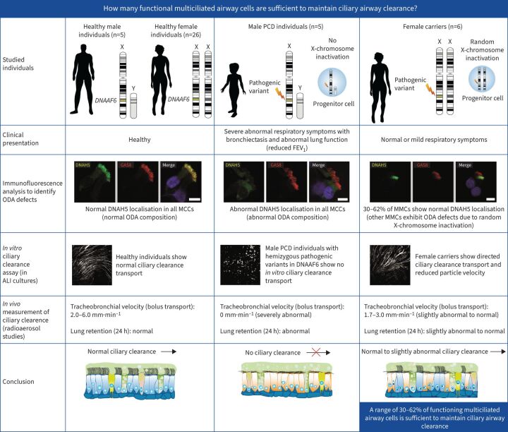
Background: Primary ciliary dyskinesia is a genetic disorder caused by aberrant motile cilia function that results in defective ciliary airway clearance and subsequently leads to recurrent airway infections and bronchiectasis. We aimed to determine: how many functional multiciliated airway cells are sufficient to maintain ciliary airway clearance?

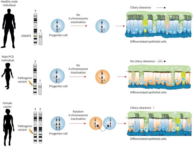
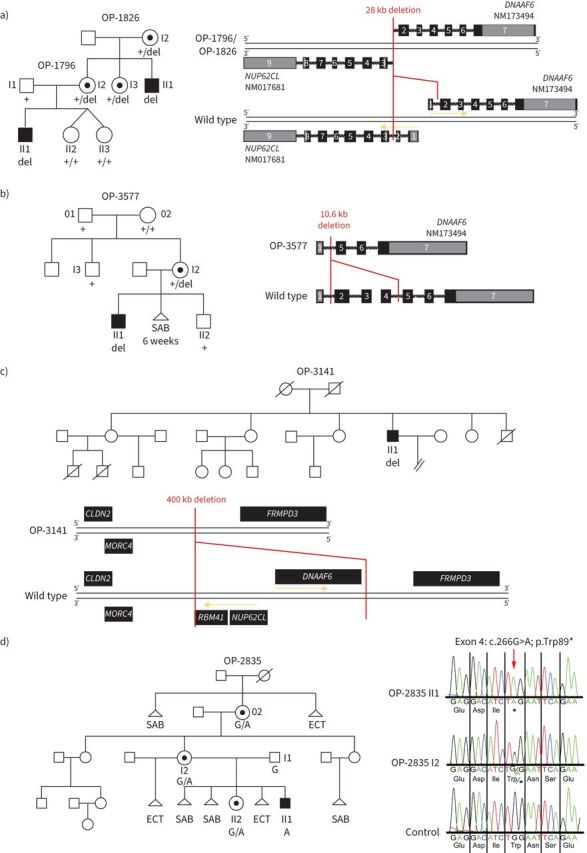
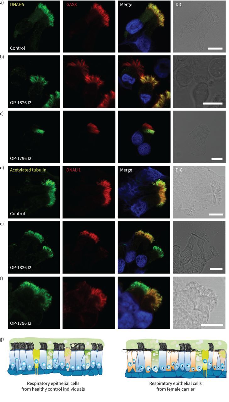
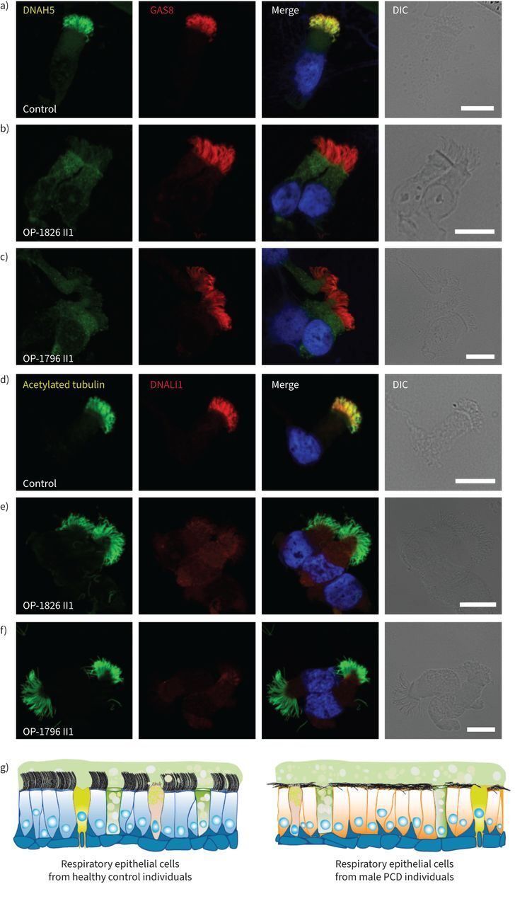
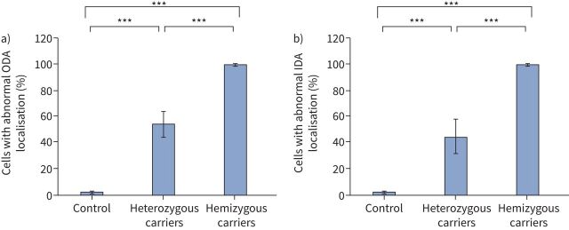
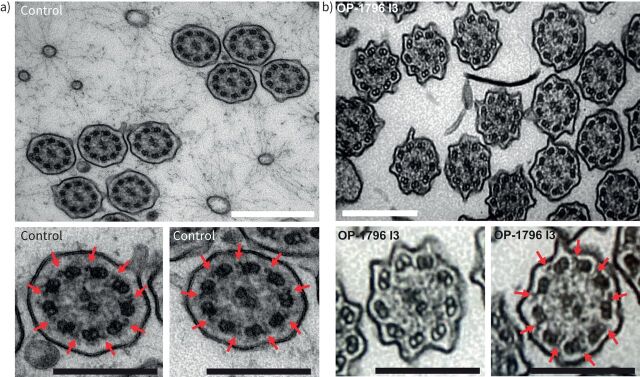
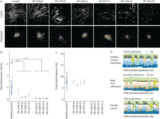
Methods: To answer this question we exploited the molecular defects of the X-linked recessive primary ciliary dyskinesia variant caused by pathogenic variants in DNAAF6 (PIH1D3), characterised by immotile cilia in affected males. We carefully analysed the clinical phenotype and molecular defect (using immunofluorescence and transmission electron microscopy) and performed in vitro studies (particle tracking in air-liquid interface cultures) and in vivo studies (radiolabelled tracer studies) to assess ciliary clearance of respiratory cells from female individuals with heterozygous and male individuals with hemizygous pathogenic DNAAF6 variants.

Results: Primary ciliary dyskinesia male individuals with hemizygous pathogenic DNAAF6 variants displayed exclusively immotile cilia, absence of ciliary clearance and severe primary ciliary dyskinesia symptoms. Owing to random or skewed X-chromosome inactivation in six female carriers with heterozygous pathogenic DNAAF6 variants, 54.3±10% (range 38-70%) of multiciliated cells were defective. Nevertheless, in vitro and in vivo assessment of the ciliary airway clearance was normal or slightly abnormal. Consistently, heterozygous female individuals showed no or only mild respiratory symptoms.

Conclusions: Our findings indicate that having 30-62% of multiciliated respiratory cells functioning can generate either normal or slightly reduced ciliary clearance. Because heterozygous female carriers displayed either no or subtle respiratory symptoms, complete correction of 30% of cells by precision medicine could improve ciliary airway clearance in individuals with primary ciliary dyskinesia, as well as clinical symptoms.


Conflict of interest statement

Conflict of interest: The authors have no potential conflicts of interest to declare.

Figures

None
Overview of the study. PCD: primary ciliary dyskinesia; FEV1: forced expiratory volume in 1 s; ODA: outer dynein arm; MCC: multiciliated cell.
FIGURE 1
FIGURE 1
Random X-chromosome inactivation (XCI) in female carriers of heterozygous pathogenic DNAAF6 variants results in two populations of respiratory ciliated cells. The differentiated respiratory epithelium consists of different type of cells: non-ciliated cells (e.g. ionocytes, goblet and club cells) and ciliated cells. DNAAF6 is located on the X chromosome. In contrast to female individuals, in male individuals there is no XCI. Male individuals with a hemizygous pathogenic DNAAF6 variant display exclusively immotile cilia in the airways, resulting in primary ciliary dyskinesia. In contrast, due to XCI, female individuals have a mosaic of two populations of cells that differ in the X-chromosome that is active. Thus, in female individuals with a heterozygous pathogenic DNAAF6 variant, it is expected that half of all ciliated respiratory cells are lined with immotile cilia. In this study, we performed detailed in vitro and in vivo studies to assess the impact on ciliary clearance.
FIGURE 2
FIGURE 2
Pathogenic variants in DNAAF6 located on the X chromosome were identified in four families. a) In family OP-1826/OP-1796 we found a 28 kb deletion (del) including the first exon of DNAAF6 and the first two exons of NUP62CL. b) In family OP-3577, a 10.6 kb deletion comprising DNAAF6 exons 2–4 was identified in OP-3577 I2. Segregation analyses revealed that neither her brother (OP-3577 I3) nor grandmother (OP-3577 02) carry the deletion, indicating that this is a de novo variant. OP-3577 I2 reported difficulties in getting pregnant and had one spontaneous abortion in the sixth week of pregnancy. c) In OP-3141 we identified a 400 kb deletion comprising DNAAF6, NUP62CL and RBM41. OP-3141 II1 is the fourth of six children. He has three older and one younger sister. The only brother died 10 days after birth owing to cardiorespiratory failure of unknown cause. His oldest sister gave birth to two male individuals: the first born died 3 months after birth, the second born died 4 months after birth. Both died of unknown causes with cardiorespiratory failure. With another partner, she gave birth to a healthy boy. The second oldest sister has three healthy children, two girls and a boy. The third oldest sister gave birth to a healthy boy. The younger sister has no children. d) In family OP-2835, a transition from G to A (c.266G>A) was identified in DNAAF6 exon 4, predicting premature stop of translation (p.Trp89*). Segregation analyses revealed that the mother (OP-2835 I2) and the grandmother (OP-2835 02) carry this variant in a heterozygous status whereas the father (OP-2835 I1) is unaffected. The great-grandmother of OP-2835 II1 had a spontaneous abortion (SAB) and gave birth to OP-2835 02, who inherited the DNAAF6 variant. The great-grandmother died owing to complications of an ectopic (ECT) pregnancy. OP-2835 02 gave birth to three daughters. The oldest one has two healthy children, a girl, who already has another healthy daughter, and a boy. The second oldest one, OP-2835 I2, is a carrier of the DNAAF6 variant and has two children. She had one ectopic pregnancy with a previous partner and with her current partner two spontaneous abortions and an ectopic pregnancy. OP-2835 II2 was her fourth pregnancy and is also a carrier of the DNAAF6 variant. In addition, she gave birth to the index primary ciliary dyskinesia (PCD) male individual OP-2835 II1, who was her sixth pregnancy and who carries the hemizygous DNAAF6 variant. The younger sister had one abortion and no children. PCD-affected males are shaded black and unaffected siblings are shaded white. Heterozygous female carriers are indicated by a central dot. Genotypes are indicated in the pedigrees.
FIGURE 3
FIGURE 3
Heterozygous DNAAF6 pathogenic variants in female individuals result in a mosaic of cells with and without axonemal outer and inner dynein arm defects, respectively. a–c) Respiratory epithelial cells from female control individuals and female carriers with a heterozygous pathogenic DNAAF6 variant were double-labelled with antibodies directed against the outer dynein arm protein DNAH5 (green) and GAS8 (red). Both proteins colocalise (yellow) along the ciliary axonemes in all cells from the unaffected control (a). In female carriers with a heterozygous pathogenic DNAAF6 variant, DNAH5 showed a normal localisation in some cells or was completely absent or severely reduced in the ciliary axonemes of other (b, c). d–f) Respiratory epithelial cells from a female control and female carriers with a heterozygous pathogenic DNAAF6 variant were double-labelled with antibodies directed against acetylated tubulin (green) and the inner dynein arm protein DNALI1 (red). Both proteins colocalise (yellow) along the ciliary axonemes in all cells from the unaffected control (d). In female carriers with heterozygous pathogenic DNAAF6 variant, DNALI1 showed a normal localisation in some cells or was completely absent or severely reduced in the ciliary axonemes of other cells (e, f). Nuclei were stained with Hoechst 33342 (blue). g) Schematic summarising the immunofluorescence microscopy results. In female control cells, cytoplasmic preassembly of dynein arms was not altered and all cells showed normal axonemal composition of inner (DNALI1) and outer (DNAH5) dynein arms. In contrast, female carriers with a heterozygous pathogenic DNAAF6 variant show a mosaic of cells with normal axonemal composition of dynein arms (blue) and abnormal composition dynein arms (yellow), respectively. Scale bars: 10 μm. DIC: differential interference contrast.
FIGURE 4
FIGURE 4
Hemizygous pathogenic variants in DNAAF6 result in outer and inner dynein arm defects in primary ciliary dyskinesia (PCD)-affected males. a–c) Respiratory epithelial cells from control individuals and male PCD individuals OP-1826 II1 and OP-1796 II1 with a hemizygous pathogenic DNAAF6 variant were double-labelled with antibodies directed against the outer dynein arm protein DNAH5 (green) and GAS8 (red). Both proteins colocalise (yellow) along the ciliary axonemes in cells from the unaffected control (a). In cells from PCD individuals OP-1826 II1 (b) and OP-1796 II1 (c), DNAH5 was completely absent or severely reduced from the ciliary axonemes of all cells analysed. d–f) Respiratory epithelial cells from a control and male PCD individuals OP-1826 II1 and OP-1796 II1 with hemizygous pathogenic DNAAF6 variants were double-labelled with antibodies directed against acetylated tubulin (green) and the inner dynein arm protein DNALI1 (red). Both proteins colocalise (yellow) along the ciliary axonemes in all cells from the unaffected control (d). In cells from the male PCD individuals OP-1826 II1 (e) and OP-1796 II1 (f), DNALI1 was completely absent or severely reduced in the ciliary axonemes of all cells analysed. Nuclei were stained with Hoechst 33342 (blue). g) Schematic summarising the immunofluorescence microscopy results. In all control cells (blue), cytoplasmic preassembly of dynein arms was not altered and all cells showed normal axonemal composition of inner (DNALI1) and outer (DNAH5) dynein arms. In contrast, in male PCD individuals with hemizygous DNAAF6 pathogenic variants, all cells show abnormal composition of dynein arms (yellow). Scale bars: 10 μm. DIC: differential interference contrast.
FIGURE 5
FIGURE 5
Pooled statistical analysis of immunofluorescence (IF) microscopy findings using antibodies directed against outer dynein arm (ODA) component DNAH5 (a) and the inner dynein arm (IDA) protein DNALI1 (b) in control respiratory cells, cells from female carriers with a heterozygous pathogenic DNAAF6 variant (OP-1796 I2, OP-1796 I3, OP-1826 I2, OP-3577 I2 and OP-2835 I2) and male primary ciliary dyskinesia (PCD) individuals with a hemizygous pathogenic DNAAF6 variant (OP-1796 II1, OP-1826 II1, OP-3577 II1, OP-2835 II1 and OP-2835 II2). a) Analyses of ciliated respiratory cells of females with a heterozygous pathogenic DNAAF6 variant revealed that 54.3±10% (range 38–70%, table 1) show abnormal axonemal localisation of DNAH5 when compared to control respiratory cells (1.8±0.67%). In male PCD individuals with a hemizygous pathogenic DNAAF6 variant, almost all respiratory cells showed absence of DNAH5 from the ciliary axonemes (99.8±0.37%). b) Analyses of ciliated respiratory cells of female carriers with a heterozygous DNAAF6 pathogenic variant revealed that 45.2±13% (range 32–66%, table 1) show abnormal localisation of DNALI1 when compared to control respiratory cells (2.4±1.6%). In male PCD individuals with hemizygous pathogenic DNAAF6 variants, almost all respiratory cells showed axonemal absence of DNALI1 (99.6±0.4%). For male (n=1) and female controls (n=2) and for OP-1796 I2, OP-1796 I3, OP-1796 II1, OP-1826 I2, OP-1826 II1, OP-3577 I2 and OP-3577 II1, a total of 500 cells per IF staining were counted. For OP-2835 II1, 200 cells were counted. For OP-2835 I2 and OP-2835 II2, 75 cells were counted. Error bars denote standard deviation. p-values were generated by the ANOVA two-tailed tests. ***: p<0.001.
FIGURE 6
FIGURE 6
Heterozygous pathogenic DNAAF6 variants in female carriers result in a mosaic of cells with and without axonemal outer dynein arm (ODA) defects. Transmission electron microscopy analysis of ciliary cross-sections from a) a control and b) female individual OP-1796 I3 carrying a heterozygous DNAAF6 variant show the absence of ODAs in more than half of the ciliated cells (n=391 of 581 sections, 67%) from the female carrier. Red arrows mark ODAs. A total of 581 ciliary cross-sections from OP-1796 I3 were analysed. White scale bars: 500 nm; black scale bars: 250 nm.
FIGURE 7
FIGURE 7
Air–liquid interface cultures of respiratory epithelial cells from female carriers with heterozygous pathogenic DNAAF6 variants are able to generate a directed fluid flow while respiratory epithelial cells from male individuals with hemizygous pathogenic DNAAF6 variants are unable to produce a fluid flow. a) After complete cell differentiation and ciliation (30 days after airlift), fluorescent particles were added to the apical compartments of the respiratory epithelial cells to assess ciliary clearance capacity. Tracking videos are represented as z-stack projections, while the transport direction of each particle is summarised in polargraphs. b) Respiratory cells from a control group (n=19) and female carriers with heterozygous pathogenic DNAAF6 variants (OP-1796 I2, OP-1796 I3, OP-1826 I2 and OP-3577 I2) transported fluorescent particles in a linear direction along the cell layer. The velocity of this flow was significantly slower than that of the healthy controls (p=0.009, t-test). In contrast, the particle transport was non-oriented and significantly slower in male primary ciliary dyskinesia (PCD) individuals carrying hemizygous pathogenic DNAAF6 variants (OP-1796 II1, OP-1826 II1, OP-2835 II1 and OP-3141 II1; p=0.0003, t-test). Circles indicate values measured with 2 µm beads while triangles indicate values measured with 0.5 µm beads. Because two different bead sizes were used for the analyses, values were normalised against the mean value of the healthy control group. Exact values are shown in supplementary figure S15. The significantly reduced particle transport measured in the male PCD individuals (dashed red line) is a thermal-driven background flow (Brownian movement). c) Measurement of the ciliary beat frequency of healthy controls (8.5 Hz), female carriers with heterozygous variants in DNAAF6 (OP-1796 I2=7.6 Hz, OP-1796 I3=6.3 Hz, OP-1826 I2=7.7 Hz, OP-3577 I2=7.5 Hz) and male PCD individuals with hemizygous pathogenic DNAAF6 variants (0 Hz). There was a subtle reduction in the ciliary beat frequency in female carriers with heterozygous DNAAF6 variants compared to healthy controls (p=0.04, t-test). In total, 30 videos per individuals were analysed for statistical evaluation and 253 particles were tracked per video on average from three different cell culture inserts from each individual. d) Schematic summarising the particle tracking results. Ciliated cells from female control individuals (blue) were able to produce a directed flow. In contrast, in male PCD individuals with pathogenic DNAAF6 variants (orange), all cells display cilia immotility due to lack of functional outer and inner dynein arms and the absence of directed particle transport.

Comment in

Similar articles

Cited by

References

    1. Fliegauf M, Benzing T, Omran H. When cilia go bad: cilia defects and ciliopathies. Nat Rev Mol Cell Biol 2007; 8: 880–893. doi:10.1038/nrm2278 - DOI - PubMed
    1. Wallmeier J, Nielsen KG, Kuehni CE,et al. . Motile ciliopathies. Nat Rev Dis Primers 2020; 6: 77. doi:10.1038/s41572-020-0209-6 - DOI - PubMed
    1. Nielsen KG, Holgersen MG, Crowley S, et al. . Chronic airway disease in primary ciliary dyskinesia-spiced with geno-phenotype associations. Am J Med Genet C Semin Med Genet 2022; 190: 20–35. doi:10.1002/ajmg.c.31967 - DOI - PMC - PubMed
    1. Desai PB, Dean AB, Mitchell DR. Cytoplasmic preassembly and trafficking of axonemal dyneins. In: King SM, ed. Dyneins: Structure, Biology and Disease (Second Edition). London, Academic Press, 2018; pp. 140–161.
    1. Braschi B, Omran H, Witman GB, et al. . Consensus nomenclature for dyneins and associated assembly factors. J Cell Biol 2022; 221: e202109014. doi:10.1083/jcb.202109014 - DOI - PMC - PubMed

LinkOut - more resources