Review
. 2024 Aug 16;106(3):129-144.
doi: 10.1124/molpharm.124.000949.

Get Ready to Sharpen Your Tools: A Short Guide to Heterotrimeric G Protein Activity Biosensors

Affiliations
Review

Get Ready to Sharpen Your Tools: A Short Guide to Heterotrimeric G Protein Activity Biosensors

Remi Janicot et al. Mol Pharmacol. .

Abstract

G protein-coupled receptors (GPCRs) are the largest class of transmembrane receptors encoded in the human genome, and they initiate cellular responses triggered by a plethora of extracellular stimuli ranging from neurotransmitters and hormones to photons. Upon stimulation, GPCRs activate heterotrimeric G proteins (Gαβγ) in the cytoplasm, which then convey signals to their effectors to elicit cellular responses. Given the broad biological and biomedical relevance of GPCRs and G proteins in physiology and disease, there is great interest in developing and optimizing approaches to measure their signaling activity with high accuracy and across experimental systems pertinent to their functions in cellular communication. This review provides a historical perspective on approaches to measure GPCR-G protein signaling, from quantification of second messengers and other indirect readouts of activity to biosensors that directly detect the activity of G proteins. The latter is the focus of a more detailed overview of the evolution of design principles for various optical biosensors of G protein activity with different experimental capabilities. We will highlight advantages and limitations of biosensors that detect different G protein activation hallmarks, like dissociation of Gα and Gβγ or nucleotide exchange on Gα, as well as their suitability to detect signaling mediated by endogenous versus exogenous signaling components or in physiologically relevant systems like primary cells. Overall, this review intends to provide an assessment of the state-of-the-art for biosensors that directly measure G protein activity to allow readers to make informed decisions on the selection and implementation of currently available tools. SIGNIFICANCE STATEMENT: G protein activity biosensors have become essential and widespread tools to assess GPCR signaling and pharmacology. Yet, investigators face the challenge of choosing from a growing list of G protein activity biosensors. This review provides an overview of the features and capabilities of different optical biosensor designs for the direct detection of G protein activity in cells, with the aim of facilitating the rational selection of systems that align with the specific scientific questions and needs of investigators.


Figures

Fig. 1.
Fig. 1.
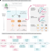
G protein activation cycle and canonical signaling pathways. (A) GPCR-mediated activation of G proteins. Upon stimulation, GPCRs promote the exchange of GDP for GTP on the Gα subunit of inactive Gαβγ heterotrimer, triggering the dissociation of Gβγ from Gα-GTP. Both Gα-GTP and free Gβγ modulate their respective effectors to propagate signals to downstream cascades. Signaling is turned off once Gα hydrolyzes GTP back to GDP, leading to reassociation with Gβγ into an inactive heterotrimeric complex. (B) G protein families and their canonical signaling pathways. G proteins are subcategorized into four families based on the identity of their Gα subunits, which in turn have specific modulatory effects on different types of effectors.
Fig. 2.
Fig. 2.
Different approaches to assess GPCR-G protein signaling. (A) Left, schematic representation of GPCR-G protein signaling mechanisms and classification of different potential readouts as “DIRECT” (blue) or “INDIRECT” (red). Right, representation of potential distortions associated with indirect readouts, including changes in response kinetics, amplification events, or pathway crosstalk. (B) Timeline for the development of assays and sensor designs to monitor GPCR-G protein signal transduction.
Fig. 3.
Fig. 3.
Design of RET-based sensors to directly monitor G protein activity in cells. (A) Gα-Gβγ dissociation sensors. In the inactive state, a Gα fused to a RET donor (blue) interacts with Gβγ fused to a RET acceptor (yellow), leading to high RET. After G protein activation, the Gα and Gβγ subunits dissociate (or rearrange), leading to a decrease in RET. (B) Free Gβγ detection sensors. In the inactive state, Gβγ fused to a RET acceptor (yellow) is associated with Gα, preventing its binding to GRK3ct fused to a RET donor (blue) and attached to the plasma membrane. After G protein activation, Gβγ is released, allowing it to interact with GRK3ct and leading to an increase in RET. (C) Gα-GTP detection sensors. Upon G protein activation, a Gα fused to a BRET acceptor (yellow) binds to detector modules fused to a BRET donor (blue) and attached to the plasma membrane that specifically bind to Gα-GTP species, leading to high BRET. (D) BERKY sensors. As in (C), this biosensor design relies on binding of active G proteins to specific detector modules, but BRET increases arise from intramolecular interactions between a BRET donor (blue) and a BRET acceptor (yellow) within the same biosensor construct, which is attached to the plasma membrane. (E) EMTA sensors. Upon G protein activation, an effector molecule fused to a BRET donor (blue) is recruited form the cytosol to membranes via binding to Gα subunits, which leads to increased BRET with a bystander membrane-anchored BRET acceptor (yellow). (F) ONE-GO sensors. Biosensor designs analogous to those in (C) are delivered to cells as a single genetic payload, enabling robust activity measurements across diverse cell types.
Fig. 4.
Fig. 4.
Comparisons of the features of direct G protein activity biosensors. The table in this figure summarizes the characteristics of the RET biosensors highlighted in this review. # indicates demonstrated use of targetability feature, whereas others indicated as “Yes” are only theoretically possible.

References

    1. Adjobo-Hermans MJW, Goedhart J, van Weeren L, Nijmeijer S, Manders EMM, Offermanns S, Gadella TWJ Jr (2011) Real-time visualization of heterotrimeric G protein Gq activation in living cells. BMC Biol 9:32. - PMC - PubMed
    1. Aittaleb M, Boguth CA, Tesmer JJG (2010) Structure and function of heterotrimeric G protein-regulated Rho guanine nucleotide exchange factors. Mol Pharmacol 77:111–125. - PMC - PubMed
    1. Akgoz M, Kalyanaraman V, Gautam N (2004) Receptor-mediated reversible translocation of the G protein betagamma complex from the plasma membrane to the Golgi complex. J Biol Chem 279:51541–51544. - PubMed
    1. Alvaro CG, Thorner J (2016) Heterotrimeric G protein-coupled receptor signaling in yeast mating pheromone response. J Biol Chem 291:7788–7795. - PMC - PubMed
    1. Audet N, Galés C, Archer-Lahlou E, Vallières M, Schiller PW, Bouvier M, Pineyro G (2008) Bioluminescence resonance energy transfer assays reveal ligand-specific conformational changes within preformed signaling complexes containing delta-opioid receptors and heterotrimeric G proteins. J Biol Chem 283:15078–15088. - PMC - PubMed

MeSH terms

Substances

LinkOut - more resources