. 2024 Jul 12;15(1):5849.
doi: 10.1038/s41467-024-50064-y.

Harnessing virus flexibility to selectively capture and profile rare circulating target cells for precise cancer subtyping

Affiliations

Harnessing virus flexibility to selectively capture and profile rare circulating target cells for precise cancer subtyping

Hui-Da Li et al. Nat Commun. .

Abstract

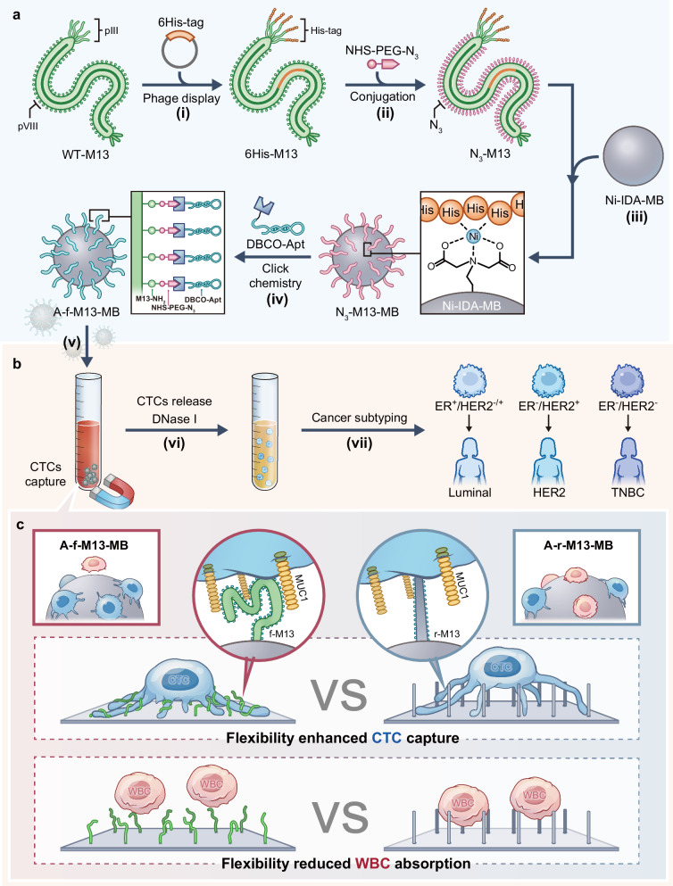
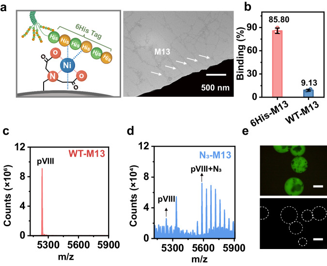
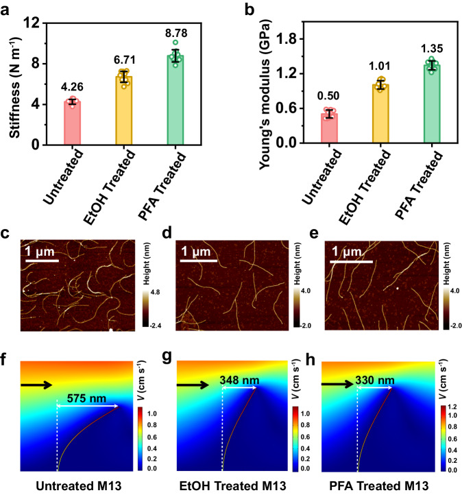
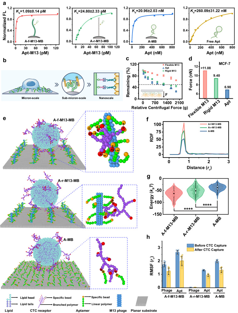
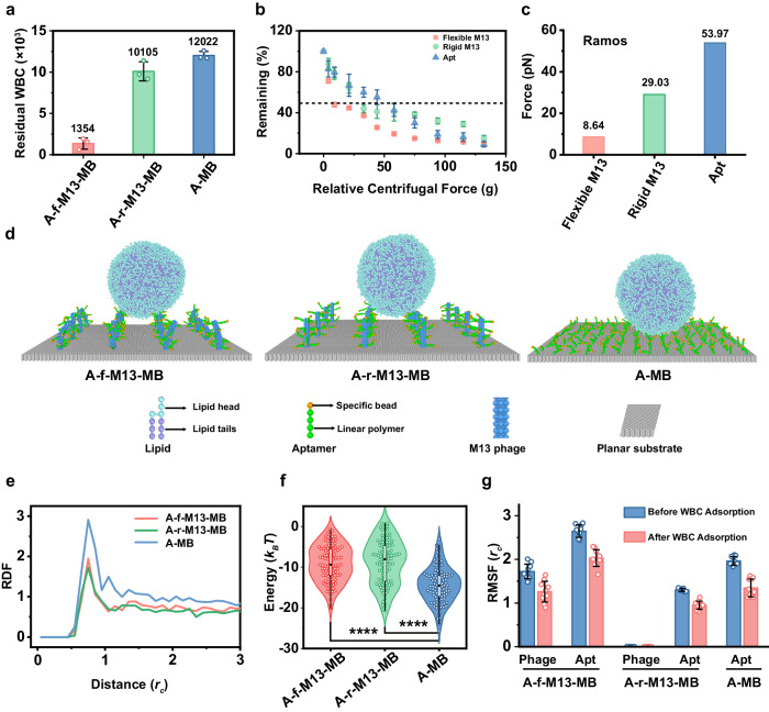
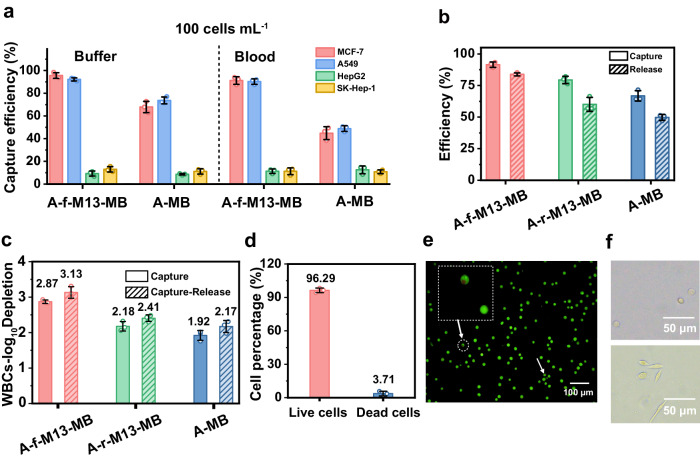
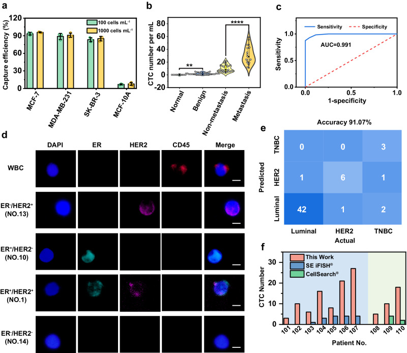
The effective isolation of rare target cells, such as circulating tumor cells, from whole blood is still challenging due to the lack of a capturing surface with strong target-binding affinity and non-target-cell resistance. Here we present a solution leveraging the flexibility of bacterial virus (phage) nanofibers with their sidewalls displaying target circulating tumor cell-specific aptamers and their ends tethered to magnetic beads. Such flexible phages, with low stiffness and Young's modulus, can twist and adapt to recognize the cell receptors, energetically enhancing target cell capturing and entropically discouraging non-target cells (white blood cells) adsorption. The magnetic beads with flexible phages can isolate and count target cells with significant increase in cell affinity and reduction in non-target cell absorption compared to magnetic beads having rigid phages. This differentiates breast cancer patients and healthy donors, with impressive area under the curve (0.991) at the optimal detection threshold (>4 target cells mL-1). Immunostaining of captured circulating tumor cells precisely determines breast cancer subtypes with a diagnostic accuracy of 91.07%. Our study reveals the power of viral mechanical attributes in designing surfaces with superior target binding and non-target anti-fouling.


Conflict of interest statement

The authors declare no competing interests.

Figures

Fig. 1
Fig. 1. Schematic illustration of the fabrication of the virus nanofiber-based deformable surface for highly efficient circulating tumor cell (CTC) isolation from whole blood.
a Engineering of the M13 virus nanofibers. Wild type M13 phage (WT-M13) was first genetically engineered to introduce a 6His tag at the N-terminus of the pIII minor capsid protein, generating 6His-M13 (Step (i)). The azide moiety was then introduced to the sidewall of the nanofibers by reacting NHS-PEG-N3 with the N-terminal NH2 of pVIII protein, producing N3-M13 (Step (ii)). Thereafter, the N3-M13 nanofiber was immobilized on the surface of Ni-IDA -grafted magnetic beads (Ni-IDA MB) in an end- on manner through the affinitive interaction between Ni and 6His, producing N3-M13-MB (Step (iii)). Finally, the DBCO-labeled aptamer (DBCO-Apt) was conjugated with N3-M13 via click reaction on N3-M13-MB, forming Apt-M13-MB (Step (iv)), which was termed aptamer-flexible-M13-MB (A-f-M13-MB) to emphasize the flexibility of M13 phage. b The whole process of breast cancer CTC capture and profiling. Breast cancer CTCs in patients’ whole blood were selectively captured by A-f-M13-MB and isolated via magnetic separation (Step (v)). The captured CTCs were released by DNase I and immuno-stained for the profiling of surface proteins including estrogen receptor protein (ER) and human epidermal growth factor receptor 2 (HER2) (Step) (vi)-(vii). CTCs stained as ER+/HER2+or-, ER-/HER2+, and ER-/HER2- were recognized as luminal subtype cell, HER2 positive subtype, and basal-like subtype, respectively. c Flexibility enhanced CTC capture and reduced white blood cell (WBC) absorption. With low stiffness and Young’s modulus, M13 phage exhibits a deformable feature with much configurational freedom in flowing solutions. This feature facilitated M13 on the A-f-M13-MB to adapt its configuration to promote the multivalent CTC-binding interactions to energetically enhance the CTC capture ability, whereas the rigid M13, formed by treatment of flexible M13 with paraformaldehyde (PFA), on the aptamer-rigid-M13-MB (A-r-M13-MB) could only offer limited sites for binding CTCs. Meanwhile, the flexibility feature of A-f-M13-MB led to the increase of the binding free energy for the non-specific adsorption of WBCs due to the entropy loss, thus guaranteeing a low WBC background.
Fig. 2
Fig. 2. Characterization of aptamer-displaying, flexible M13 nanofibers on magnetic beads (A-f-M13-MB).
a The cartoon illustration indicated the specific interaction between Ni-IDA on MBs and 6His tag on pIII of phage. Cartoon shown in Fig. 2a created with BioRender.com released under a Creative Commons Attribution-NonCommercial-NoDerivs 4.0 International license (https://creativecommons.org/licenses/by-nc-nd/4.0/deed.en). TEM images revealed that M13 nanofibers were anchored on MBs in an end-on manner. M13 nanofibers are indicated by arrows. b The much higher binding efficiency of 6His-M13 on Ni-IDA MBs compared to wild type (WT) M13 indicated the successful construction of 6His-M13 (n = 3 samples, mean ± s.d.). The Q-TOF LC/MS spectra of WT-M13 phage (c) and N3-M13 phage (d). The peak at 5238 m/z confirmed the presence of pVIII in WT-M13, while the peak difference between 5238 m/z and 5585 m/z fitted the molecular weight of N3-PEG, indicating presence of N3-PEG. e Fluorescence microscopic images of FAM-A-f-M13-MB (upper) and control (lower). Aptamer was labeled with FAM, thus those MBs anchored with A-f-M13 emitted green fluorescence whereas the control MBs that anchored with WT-M13 didn’t. Scale bar: 20 μm. Source data are provided as a Source Data file. WT-M13: wild type M13 phage; 6His-M13: M13 with 6His tag displayed on pIII; N3-M13: 6His-M13 with N3-PEG decorated on pVIII; Apt-M13: N3-M13 with aptamer “clicked” onto pVIII.
Fig. 3
Fig. 3. Mechanical properties of M13 phage.
Stiffness (a) and Young’s modulus (b) of untreated M13, EtOH-treated M13 and PFA-treated M13 measured by AFM under Hertzian mode (n = 10 samples, mean ± s.d.). M13 nanofibers were hardened to restrict their motion in solution by being treated with 100% ethanol (EtOH) or 4% paraformaldehyde (PFA) to produce EtOH-treated M13 and PFA-treated M13 for the comparison of mechanical properties. The stiffness of EtOH-treated M13 and PFA-treated M13 was 1.58-fold and 2.06-fold higher than that of untreated M13, whereas the Young’s modulus of EtOH-treated M13 and PFA-treated M13 was 2.02-fold and 2.70-fold higher than that of untreated M13, respectively. AFM images of untreated M13 (c), EtOH-treated M13 (d) and PFA-treated M13 (e). AFM images revealed that these treated rigid M13 presented a less twisty morphology than untreated M13. The loading amount of three types of M13 phages was identical (109 pfu). Numerical simulations results for untreated M13 (f), EtOH-treated M13 (g) and PFA-treated M13 (h) under a flow field, wherein the arrow indicates the flow direction. Subjected flow speed: 2 cm/s. When M13 was subjected to the same force from fluid shear stress, the deformation of untreated M13 was 1.74 and 1.65 folds larger than that of EtOH-treated M13 and PFA-treated M13, respectively. Source data are provided as a Source Data file. EtOH-treated M13: M13 phage treated with 100% ethanol (EtOH); PFA-treated M13: M13 phage treated with 4% paraformaldehyde (PFA).
Fig. 4
Fig. 4. Flexible M13 enhanced CTCs capture performance.
a The dissociation constants (Kd) of A-f-M13-MB, A-r-M13-MB, A-MB and free aptamer. The Kd value reflects the affinity between CTCs and affinity ligands, with a lower Kd corresponding to a higher CTC binding affinity. b Illustration of the molecular matching between VNTR repeats in MUC1 and aptamers on Apt-M13, sub-micron-scale matching between MUC1 (200–500 nm in size) and M13 nanofibers (880 nm long), and micron-scale matching between CTCs and M13 nanofiber-anchored topological interfaces. c Percentages of cells remaining on the slide under different relative centrifugal forces (n = 3 samples, mean ± s.d.). d The detachment forces required for separating MCF-7 cells from different slides’ surface calculated from centrifugation-based cell adhesion assay indicated that the CTC binding force (MCF-7 as CTC model) was decreasing in the order of flexible M13 > rigid M13 > aptamer. e Representative final snapshots of different MB surfaces interacting with CTCs in the simulations. The arrows indicate the aptamer-receptor binding sites. f Radial distribution function (RDF) of aptamer-specific-bead/receptor-specific-bead pairs in the three cases. RDF indicates the relative density of aptamers around receptors, which was decreased in the order of A-f-M13-MB > A-r-M13-MB > A-MB. g Total energy of receptor-aptamer interaction was increased in the order of A-f-M13-MB < A-r-M13-MB < A-MB, as more contacts between the specific beads constituting the aptamers and the receptors resulted in lower total energy of the receptor-aptamer interaction (n = 101 tests). Unpaired two-sided Student’s t test. The central dot is the median; box bounds are 25th and 75th percentiles, upper and lower limits of whiskers are 1.5 × interquartile ranges. Values outside of the upper and lower limits are defined as outliers. *p  <  0.05, **p  <  0.01, ***p  <  0.001, ****p  <  0.0001. h The root mean square fluctuation (RMSF) of the aptamer and/or the M13 of the three MBs before and after the CTC adsorption (n = 10 tests, mean ±s .d.). Source data are provided as a Source Data file. Cartoons shown in (a) and (c) were created with BioRender.com released under a Creative Commons Attribution-NonCommercial-NoDerivs 4.0 International license (https://creativecommons.org/licenses/by-nc-nd/4.0/deed.en). A-MB: aptamer-modified-magnetic beads; A-f-M13-MB: Ni-IDA-MBs anchored with aptamer-modified-flexible M13; A-r-M13-MB: Ni-IDA-MBs anchored with aptamer-modified-rigid M13 (PFA-treated M13 as a rigid M13 model); Apt aptamer.
Fig. 5
Fig. 5. Flexible M13 reduced non-specific absorption of WBCs.
a Number of WBCs adsorbed on the A-f-M13-MB, A-r-M13-MB and A-MB after 30-min incubation (n = 3 samples, mean ± s.d.). b Percentages of residual WBCs on the slide under different relative centrifugal forces (n = 3 samples, mean ± s.d.). Either flexible or rigid N3-M13 were anchored on Ni-IDA glass slides and loaded with aptamers. WBCs were first incubated with M13 slides for 30 min, and a reversed centrifugation force was then applied on the WBC-attached slide for 5 min. c The detachment forces required for separating WBCs from the surface of different slides calculated from the centrifugation-based cell adhesion assay indicated that the WBC binding force (Ramos cell as a model) was increased in the order of flexible M13<rigid M13 <aptamer. d Representative final snapshots of different MB surfaces interacting with WBCs in the simulations. The results indicated that WBCs could be adsorbed onto all three MB surfaces. e Radial distribution function (RDF) of aptamer-specific-bead/receptor-specific-bead pairs in the three cases. RDF indicates the relative density of aptamers around receptors. Higher RDF corresponds to a lower total energy of the receptor-aptamer interaction. f Total energy of receptor-aptamer interaction in three cases (n = 101 tests). Unpaired two-sided Student’s t test. The central dot is the median; box bounds are 25th and 75th percentiles, upper and lower limits of whiskers are 1.5 × interquartile ranges. Values outside of the upper and lower limits are defined as outliers. *p  <  0.05, **p  <  0.01, ***p  <  0.001, ****p  <  0.0001. Results from (e) and (f) revealed the adsorption of WBCs on A-MB was the most energy-favorable, compared to the reduced adsorption of WBCs on A-f-M13-MB and A-r-M13-MB. (n = 101 tests). g The root mean square fluctuation (RMSF) of the aptamer and/or the M13 of the three MBs before and after the WBC adsorption, indicating that the adsorption of WBCs onto the A-f-M13-MB surface is the least entropy-favorable (n = 10 tests, mean ± s.d.). Source data are provided as a Source Data file. A-MB: aptamer-modified-magnetic beads; A-f-M13-MB: Ni-IDA-MBs anchored with aptamer-modified-flexible M13; A-r-M13-MB: Ni-IDA-MBs anchored with aptamer-modified-rigid M13 (PFA-treated M13 as a rigid M13 model); Apt aptamer.
Fig. 6
Fig. 6. Capture and release performance of A-f-M13-MB.
a Capture efficiency of MUC1-positive cancer cells (MCF-7 and A549) and MUC1-negative cancer cells (HepG2 and SK-Hep-1) using A-f-M13-MB and A-MB in buffer and blood, respectively (n = 3 samples, mean ± s.d.). The results indicated that A-f-M13-MB can effectively capture the MUC1 positive cancer cells (MCF-7, A549), but barely adsorbed negative cells (HepG2, SK-Hep-1) both in buffer and whole blood. b Capture and release performance of A-f-M13-MB, A-r-M13-MB and A-MB towards MCF-7 cells (n = 3 samples, mean ± s.d.). c Depletion index value of WBCs with A-f-M13-MB, A-r-M13-MB and A-MB (n = 3 samples, mean ± s.d.). The depletion index value reflects the purity of CTCs after capture and release, with a higher log10-depletion index value indicating a higher CTC purity. The results revealed a higher CTC purity after A-f-M13-MB capture compared to other two MBs, as well as an improved CTC purity after CTC release. d Viability of the isolated MCF-7 cells calculated from PI/AO staining results (n = 3 samples, mean ± s.d.). e Representative fluorescence microscope images of the isolated live cells and dead cells. The released CTCs were stained by PI/AO, and the live cells emitted green fluorescence and dead cells (indicated by arrows) emitted red fluorescence. Scale bar: 100 μm. Insert is the enlarged dead cells. f Bright-filed images of the released cells (upper) and the cells after being further cultured for 36 h (lower). The images revealed the released cells were well alive and could be adhered on the well surface 36 h after in vitro cultivation. Source data are provided as a Source Data file. A-MB: aptamer-modified-magnetic beads; A-f-M13-MB: Ni-IDA-MBs anchored with aptamer-modified-flexible M13; A-r-M13-MB: Ni-IDA-MBs anchored with aptamer-modified-rigid M13 (PFA-treated M13 as a rigid M13 model).
Fig. 7
Fig. 7. CTC isolation and diagnosis performance in clinical applications.
a The capture efficiency of different breast cell lines indicated that the CTC affinity interfaces could selectively capture several subtypes of breast cancer cells but not normal breast mammary epithelial cells (n = 3 samples, mean ± s.d.). b Distinct difference in the enumeration of CTCs in 1 mL blood samples from healthy volunteers (n = 10), benign patients (n = 34), non-metastatic breast cancer (BC) patients (n = 32), and metastatic BC patients (n = 24). Unpaired two-sided Student’s t test. The central dot is the median; box bounds are 25th and 75th percentiles, upper and lower limits of whiskers are 1.5 × interquartile ranges. Values outside of the upper and lower limits are defined as outliers. *p  <  0.05, **p  <  0.01, ***p  <  0.001, ****p  <  0.0001. c ROC analysis of CTC numbers between the cancer patient groups and healthy/benign groups. The receiver operating characteristic (ROC) curve showed good diagnostic performance (AUC = 0.991) in differentiating cancer patient groups and healthy/benign groups. d Immunofluorescence staining (DAPI/ER/HER2/CD45) of CTCs isolated from patients. Scale bar: 10 μm. CD45 was the biomarker for WBC and CK was the biomarker for CTC, with DAPI+/CK+/CD45- cells recognized as CTCs and DAPI+/CK-/CD45+ cells identified as WBCs. ER and HER2 were used for the profiling of three subtypes of BC CTCs, including luminal (HER2+ or -/ER+), HER2-positive (HER2+/ER-) and triple negative breast cancer (TNBC, belongs to basal-like subtype) (HER2-/ER-). 51 out of 56 molecular profiling results were in accordance with clinical diagnostic results. e Confusion matrix showing the subtyping accuracy of luminal, HER2, and TNBC by molecular profiling of CTCs. f Comparison of the CTC numbers isolated by A-f-M13-MB and CellSearch® or SE iFISH® in blood revealed a higher CTC capture ability and a better isolation performance of A-f-M13-MB in clinical applications. CellSearch® and SE iFISH® collected CTC from 7.5 mL and 6.0 mL blood, respectively, whereas our approach isolated CTC from 1.0 mL blood. A-f-M13-MB: Ni-IDA-MBs anchored with aptamer-modified-flexible M13; Source data are provided as a Source Data file.

Similar articles

Cited by

References

    1. Wu L, et al. Fluidic multivalent membrane nanointerface enables synergetic enrichment of circulating tumor cells with high efficiency and viability. J. Am. Chem. Soc. 2020;142:4800–4806. doi: 10.1021/jacs.9b13782. - DOI - PubMed
    1. Jeong WJ, et al. Hierarchically multivalent peptide-nanoparticle architectures: a systematic approach to engineer surface adhesion. Adv. Sci. 2022;9:2103098. doi: 10.1002/advs.202103098. - DOI - PMC - PubMed
    1. Guo L, et al. Directing multivalent aptamer-receptor binding on the cell surface with programmable atom-like nanoparticles. Angew. Chem. Int. Ed. 2022;61:e202117168. doi: 10.1002/anie.202117168. - DOI - PubMed
    1. Sabaté del Río J, Henry OYF, Jolly P, Ingber DE. An antifouling coating that enables affinity-based electrochemical biosensing in complex biological fluids. Nat. Nanotechnol. 2019;14:1143–1149. doi: 10.1038/s41565-019-0566-z. - DOI - PubMed
    1. Banerjee I, Pangule RC, Kane RS. Antifouling coatings: recent developments in the design of surfaces that prevent fouling by proteins, bacteria, and marine Organisms. Adv. Mater. 2011;23:690–718. doi: 10.1002/adma.201001215. - DOI - PubMed

Substances