. 2024 Jul 11;15(1):5187.
doi: 10.1038/s41467-024-49465-w.

Molecular and structural basis of the chromatin remodeling activity by Arabidopsis DDM1

Affiliations

Molecular and structural basis of the chromatin remodeling activity by Arabidopsis DDM1

Akihisa Osakabe et al. Nat Commun. .

Abstract

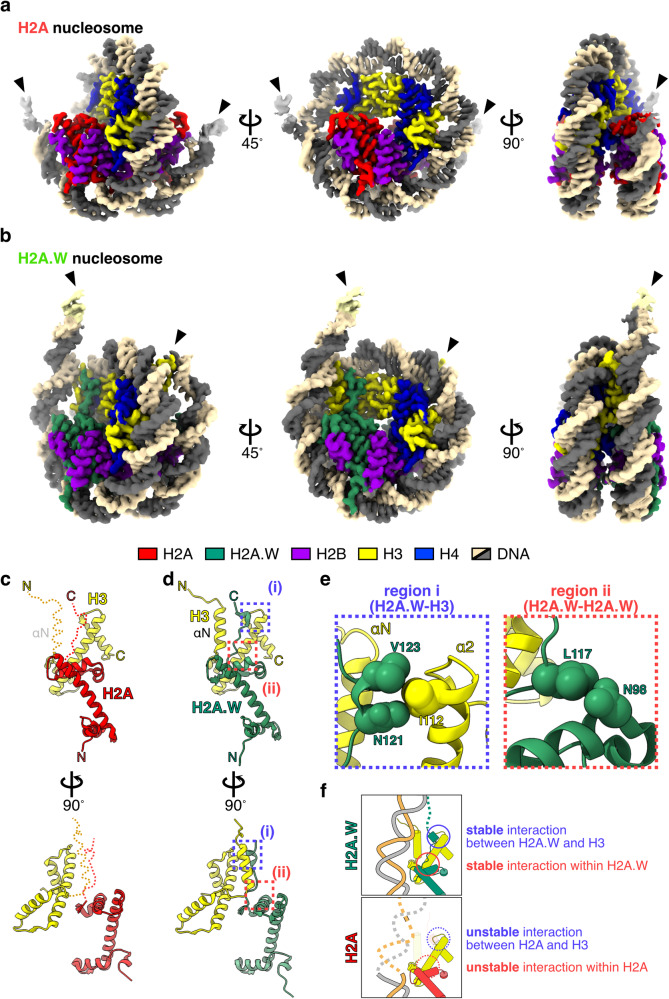
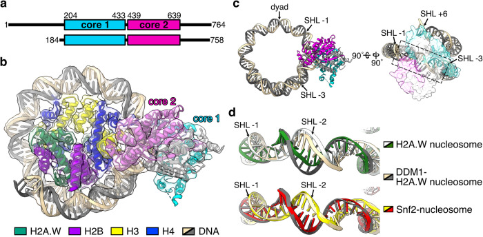
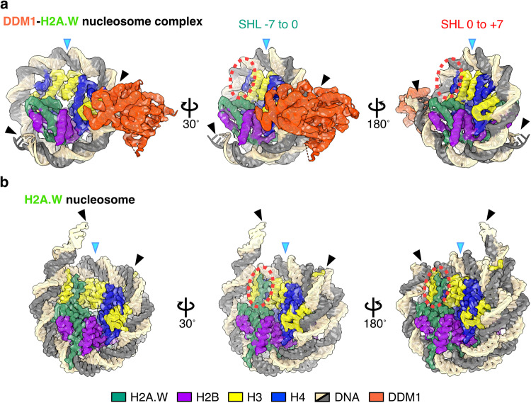
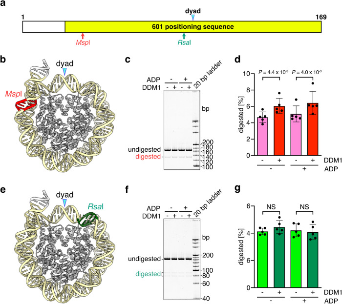
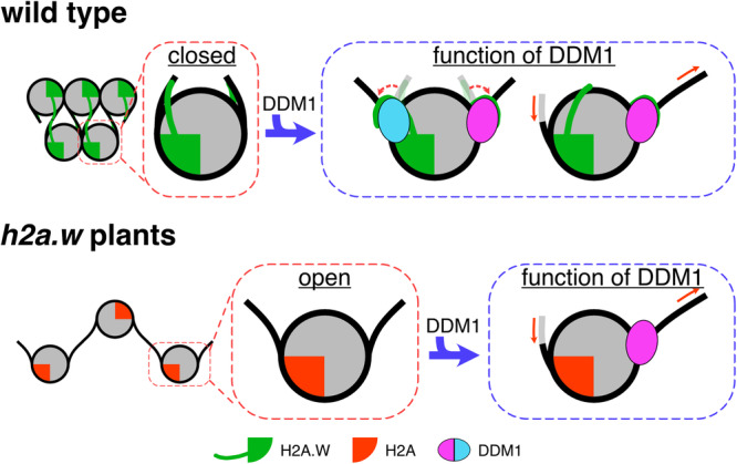
The histone H2A variant H2A.W occupies transposons and thus prevents access to them in Arabidopsis thaliana. H2A.W is deposited by the chromatin remodeler DDM1, which also promotes the accessibility of chromatin writers to heterochromatin by an unknown mechanism. To shed light on this question, we solve the cryo-EM structures of nucleosomes containing H2A and H2A.W, and the DDM1-H2A.W nucleosome complex. These structures show that the DNA end flexibility of the H2A nucleosome is higher than that of the H2A.W nucleosome. In the DDM1-H2A.W nucleosome complex, DDM1 binds to the N-terminal tail of H4 and the nucleosomal DNA and increases the DNA end flexibility of H2A.W nucleosomes. Based on these biochemical and structural results, we propose that DDM1 counters the low accessibility caused by nucleosomes containing H2A.W to enable the maintenance of repressive epigenetic marks on transposons and prevent their activity.


Conflict of interest statement

The authors declare no competing interests.

Figures

Fig. 1
Fig. 1. Cryo-EM structures of nucleosomes containing H2A and H2A.W.
Cryo-EM structures of nucleosome containing AtH2A (a) and AtH2A.W (b). Arrowheads indicate the terminal DNA detected by cryo-EM density maps. Structures of AtH2A (c) and AtH2A.W (d) complexed with H3. Red and yellow dashed lines indicate the disordered regions in AtH2A nucleosomes (c). The two contact sites discussed in panel e are enclosed in dashed boxes (i) and (ii). e Close-up views of regions (i) and (ii) from panel d, where AtH2A.W and H3 are colored green and yellow, respectively. Map-to-density figures of region (i) in the nucleosomes containing AtH2A and AtH2A.W are shown in Supplementary Fig. 4. f Graphical summary for the flexibility of the entry/exit nucleosomal DNA ends in the nucleosomes containing H2A.W (upper) and H2A (lower).
Fig. 2
Fig. 2. Cryo-EM structure of the AtDDM1-nucleosome complex.
a Schematic representation of the full-length AtDDM1 (upper) and the AtDDM1 fragment observed by cryo-EM (lower). The ATPase core domains 1 and 2 are colored cyan and magenta, respectively. The amino acid sequence alignment between AtDDM1 and Saccharomyces cerevisiae Snf2 is presented in Supplementary Fig. 8. b Cryo-EM structure of the AtDDM1-nucleosome complex. The atomic structure model of the AtDDM1-nucleosome complex is fitted to the transparent cryo-EM density map. The ATPase core domains 1 and 2 of AtDDM1 are colored cyan and magenta, respectively. c Structure of nucleosomal DNA bound by AtDDM1. The dashed box corresponds to the nucleosomal DNA around SHL-2, where DNA distortion was observed. d Structural comparison of nucleosomal DNAs bound by AtDDM1 (light orange and gray), ScSnf2 in the absence of ADP (yellow and red, PDB ID: 5X0Y (Snf2-nucleosome complex)), or H2A.W nucleosome (white and green). Map-to-density figures of the nucleosomal DNA around SHL-2 of the H2A.W nucleosome and the DDM1-H2A.W nucleosome complex are shown in Supplementary Fig. 7.
Fig. 3
Fig. 3. Structural comparison of AtDDM1-bound and AtDDM1-free nucleosomes.
Cryo-EM structures of the AtDDM1-AtH2A.W nucleosome complex (a) and the AtH2A.W nucleosome (b). Structures of nucleosomal DNA are compared at SHL −7 to 0 (middle) and SHL 0 to +7 (right). Blue arrowheads indicate the dyad axis. Black arrowheads indicate the terminal DNA detected by cryo-EM density maps. Dashed red circles indicate the AtH2A.W docking domain and the H3 αN helix disordered in the AtDDM1-AtH2A.W nucleosome complex.
Fig. 4
Fig. 4. Analyses of the nucleosomal DNA end flexibility by the restriction enzyme susceptibility assay.
a Graphical presentation of the relevant restriction enzyme recognition sites within the DNA fragment used in this experiment. The sequence of the DNA fragment is shown in Supplementary Fig. 1a. Cryo-EM structures of the AtH2A.W nucleosome with the locations of the MspI (b) and RsaI (e) sites. Native-PAGE analyses of DNA fragments after the restriction enzyme susceptibility assays with MspI (c) and RsaI (f). Graphical presentations of the restriction enzyme susceptibility assay results with MspI (d) and RsaI (g). Means and error bars represent SD from five independent experiments. The statistical significance (P) was determined using a t-test. NS not significant. Source data are provided as a Source Data file.
Fig. 5
Fig. 5. Analyses of nucleosomal DNA end flexibility by the FRET assay.
a Graphical summary of the FRET assay in this study. The emission of fluorescein is inhibited by nucleosome formation because the neighboring BHQ-1 quenches fluorophores, resulting in low fluorescence signals. When the entry/exit nucleosomal DNA ends are unwrapped, fluorescence signals can be detected. b Graphical presentation of the FRET assay results with DDM1. Means and error bars represent SD from four independent experiments. Source data are provided as a Source Data file.
Fig. 6
Fig. 6. C-terminal tail of AtH2A.W binds DDM1.
a Schematic representation (left) and sequence information (right) for the results obtained by crosslinking mass spectrometry of the DDM1-bound nucleosomes containing AtH2A.W. Two contacts between the H2A.W C-terminal tail and DDM1 are shown. Other crosslinks of the DDM1-bound nucleosomes containing AtH2A.W in the top 25% of ld-scores are shown in Supplementary Fig. 10. b Graphical summary of the contacts between the H2A.W C-terminal tail and DDM1, identified by crosslinking mass spectrometry. The yellow circle represents the 115.35 Å radius, corresponding to residues 113–140 of H2A.W (the central point is the Cα atom of His113 of H2A.W), which indicates the possible crosslinking area of H2A.W Lys140 by DSS-H12/D12. The residues of DDM1 contacting the H2A.W C-terminal tail are shown in red with orange circles.
Fig. 7
Fig. 7. Nucleosome sliding assay.
a Native-PAGE analyses of the nucleosomes containing AtH2A and AtH2A.W after the sliding assay. b, e Graphical presentation of the nucleosome sliding assay results. Means and error bars represent SD from three independent experiments. The efficiency of the remodeled nucleosome was calculated by the ratio of the intensity of each band and normalized to the ratio obtained at 0 min. Source data are provided as a Source Data file. c Overall structures of the AtDDM1-AtH2A.W nucleosome (middle) and the AtH2A.W nucleosome (right). The calculated electrostatic potential of the atomic surfaces of AtDDM1 and the H4 N-terminal tail (residues 19–24) molecules are presented (left). The dashed red circle indicates the disordered regions of the H4 N-terminal tail (residues 19–24) in the AtH2A.W nucleosome. Map to density figures of the H4 N-terminal tail of the AtDDM1-AtH2A.W nucleosome and the AtH2A.W nucleosome are shown in Supplementary Fig. 13. d Native-PAGE analyses of the nucleosomes after the sliding assay with nucleosomes containing AtH2A.W, with or without the N-terminal tail of AtH4.
Fig. 8
Fig. 8. Model of DDM1 activity for the maintenance of repressive marks.
The pericentromeric heterochromatin is occupied with H2A.W, which forms a condensed structure caused by the extended C-terminal tail of H2A.W interacting with the linker DNA, and the H3 αN and α2 helices. In the absence of H2A.W, the pericentromeric heterochromatin is occupied with H2A. The nucleosome containing H2A forms an open structure, caused by the loss of the interactions with the H3 αN and α2 helices. In the presence of DDM1, the C-terminal tail of H2A.W might dissociate from the linker DNA and H3 by interacting with DDM1. In addition, DDM1 slides nucleosomes containing H2A.W and H2A with identical efficiency. These activities might promote the increased accessibility of the heterochromatin to DNA methyltransferases.

Similar articles

Cited by

References

    1. Cosby RL, Chang N-C, Feschotte C. Host-transposon interactions: conflict, cooperation, and cooption. Genes Dev. 2019;33:1098–1116. doi: 10.1101/gad.327312.119. - DOI - PMC - PubMed
    1. Bourque G, et al. Ten things you should know about transposable elements. Genome Biol. 2018;19:199. doi: 10.1186/s13059-018-1577-z. - DOI - PMC - PubMed
    1. Wicker T, et al. A unified classification system for eukaryotic transposable elements. Nat. Rev. Genet. 2007;8:973–982. doi: 10.1038/nrg2165. - DOI - PubMed
    1. Gagnier L, Belancio VP, Mager DL. Mouse germ line mutations due to retrotransposon insertions. Mob. DNA. 2019;10:15. doi: 10.1186/s13100-019-0157-4. - DOI - PMC - PubMed
    1. Kidwell MG, Lisch D. Transposable elements as sources of variation in animals and plants. Proc. Natl Acad. Sci. USA. 1997;94:7704–7711. doi: 10.1073/pnas.94.15.7704. - DOI - PMC - PubMed

MeSH terms

LinkOut - more resources