. 2024 Dec;45(12):2487-2496.
doi: 10.1038/s41401-024-01339-z. Epub 2024 Jul 11.

Carotid artery transplantation of brain endothelial cells enhances neuroprotection and neurorepair in ischaemic stroke rats

Affiliations

Carotid artery transplantation of brain endothelial cells enhances neuroprotection and neurorepair in ischaemic stroke rats

Yi-Ting Du et al. Acta Pharmacol Sin. 2024 Dec.

Abstract

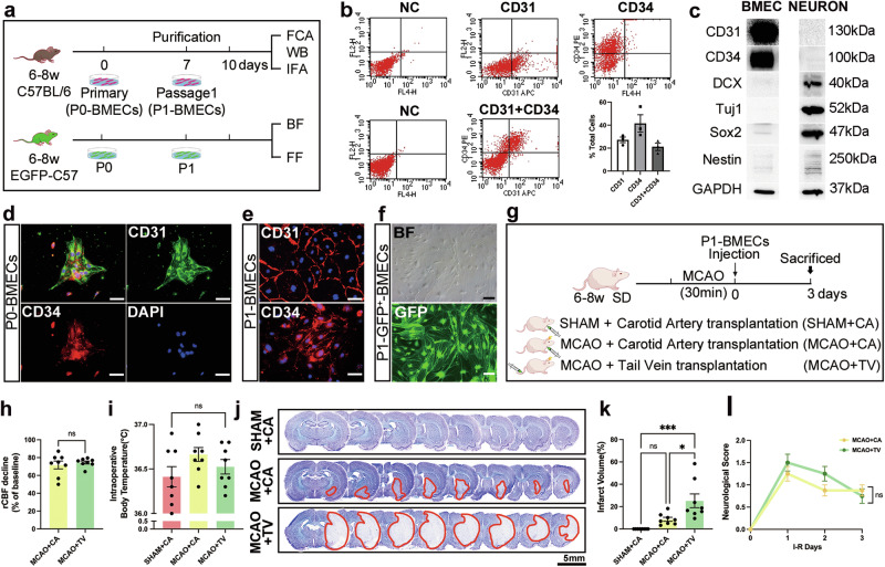
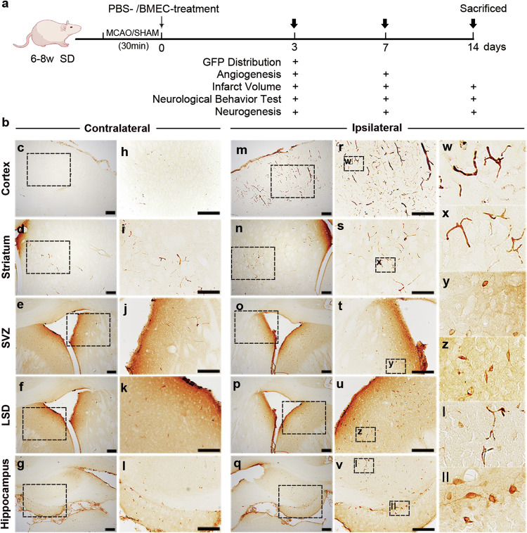
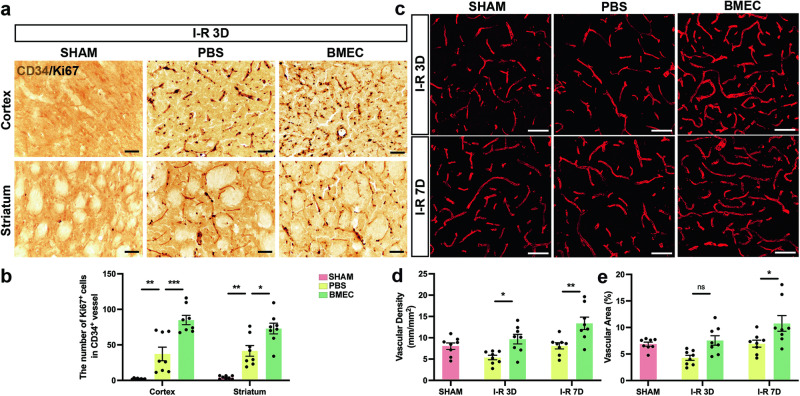
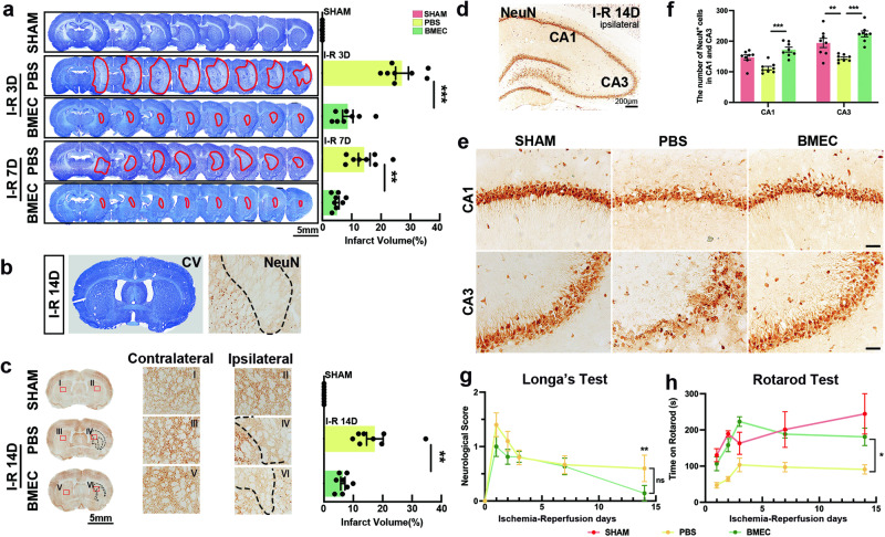
Brain microvascular endothelial cells (BMECs), an important component of the neurovascular unit, can promote angiogenesis and synaptic formation in ischaemic mice after brain parenchyma transplantation. Since the therapeutic efficacy of cell-based therapies depends on the extent of transplanted cell residence in the target tissue and cell migration ability, the delivery route has become a hot research topic. In this study, we investigated the effects of carotid artery transplantation of BMECs on neuronal injury, neurorepair, and neurological dysfunction in rats after cerebral ischaemic attack. Purified passage 1 endothelial cells (P1-BMECs) were prepared from mouse brain tissue. Adult rats were subjected to transient middle cerebral artery occlusion (MCAO) for 30 min. Then, the rats were treated with 5 × 10⁵ P1-BMECs through carotid artery infusion or tail vein injection. We observed that carotid artery transplantation of BMECs produced more potent neuroprotective effects than caudal injection in MCAO rats, including reducing infarct size and alleviating neurological deficits in behavioural tests. Carotid artery-transplanted BMECs displayed a wider distribution in the ischaemic rat brain. Immunostaining for endothelial progenitor cells and the mature endothelial cell markers CD34 and RECA-1 showed that carotid artery transplantation of BMECs significantly increased angiogenesis. Carotid artery transplantation of BMECs significantly increased the number of surviving neurons, decreased the cerebral infarction volume, and alleviated neurological deficits. In addition, we found that carotid artery transplantation of BMECs significantly enhanced ischaemia-induced hippocampal neurogenesis, as measured by doublecortin (DCX) and Ki67 double staining within 2 weeks after ischaemic injury. We conclude that carotid artery transplantation of BMECs can promote cerebral angiogenesis, neurogenesis, and neurological function recovery in adult rats after ischaemic stroke. Our results suggest that carotid injection of BMECs may be a promising new approach for treating acute brain injuries.

Keywords: brain stroke; carotid artery transplantation; endothelial cell therapy; neurorepair; neurovascular unit.


Conflict of interest statement

Competing interests: The authors declare no competing interests.

Figures

Fig. 1
Fig. 1. Carotid transplantation of mouse P1-BMECs expressing endothelial cell and stem cell markers reduces the infarct volume more than tail vein transplantation after MCAO.
a BMECs isolated from adult GFP-expressing or C57 mouse brains were cultured, purified after one passage, and harvested at 80% confluence. b, c P1-BMECs were identified by WB and FCA. WB analysis of the expression of endothelial cell markers (CD31 and CD34), neuron markers (DCX and Tuj1), a transcription factor marker (Sox2) and a stem cell marker (Nestin) in BMECs. d The cell morphology of P0-BMECs immunofluorescently labelled for CD31 (green), CD34 (red), and DAPI (blue). e Representative images of P1-BMECs showing different morphometrics for CD31, CD34, and DAPI (blue). f The efficiency of GFP expression in cultured BMECs can be observed in bright fields and fluorescent fields. Scale bars: 100 μm. g Schematic of the experimental procedure for BMEC transplantation into rat brains via the carotid artery or tail vein. h Statistical analysis of the proportion of the decrease in the rCBF when the monofilament was inserted revealed no significant difference in the decrease in the CBF between the CA and TV groups. i Cumulative data showing the intraoperative body temperature during MCAO. j Representative whole-section images of CV staining in the SHAM + CA, MCAO + CA, and MCAO + TV groups at 3 days after surgery. The blue areas are the normal brain tissue, while the light-coloured areas indicate ischaemic tissue and are marked with red lines. Scale bars: 5 mm. k Quantitative analysis of infarct volume calculated as a percentage of contralateral hemisphere volume. l The neurological performance of the CA group and TV group was assessed and scored according to Longa’s method (n = 8). ***P < 0.001; *P < 0.05; ns, not significant according to Student’s t test or one-way ANOVA with Tukey’s post hoc test. The data are presented as the mean ± SEM.
Fig. 2
Fig. 2. Schematic of the carotid artery transplantation procedure and spatial dynamics of infused GFP+ BMEC accumulation in the rat brain at 3 days after transplantation.
a The purified GFP+ BMECs (5 × 105 cells in 50 μl of PBS) were transplanted into SD rats at 3, 7, and 14 days after MCAO or Sham surgery for cell fate tracing analysis. The distribution, angiogenesis, infarct volume, behaviour test, and neurogenesis were observed at separate time points. b Representative immunohistochemistry images of GFP in the contralateral cl and ipsilateral (mz, I, II) cerebral cortex (c, h, m, r, w), striatum (d, i, n, s, x), SVZ (e, j, o, t, y), LSD (f, k, p, u, z), and hippocampus (g, l, q, v, I, II) 3 days after MCAO. h-l and r-v are magnified images of c-g and m-q, respectively. w-z, I, and II are magnified images of r-v, respectively. Scale bars: 100 μm.
Fig. 3
Fig. 3. BMEC transplantation promotes angiogenesis and increases the number of mature vessels in the ipsilateral periinfarct areas after stroke.
a Representative immunohistochemical images of CD34 (brown) and Ki67 (black) expression in the ipsilateral cortex and striatum of the SHAM, PBS, and BMEC groups at 3 days after stroke. Scale bars: 50 μm. b Cumulative data showing that the number of Ki67+ cells colocalized with CD34+ vessels increased after BMEC transplantation. c Representative images of RECA1+ vessels from the SHAM, PBS, and BMEC groups at 3 and 7 days after MCAO. Scale bars: 50 μm. d, e Quantification of the RECA1+ microvascular length and percentage of the area occupied by vascular structures in each group (n = 8). ***P < 0.001, **P < 0.01, *P < 0.05 by two-way ANOVA with Bonferroni’s multiple comparison test. The data are presented as the mean ± SEM.
Fig. 4
Fig. 4. Transplantation of BMECs attenuates neuronal loss and improves the recovery of neurological function after MCAO.
a Representative whole-section images of CV staining in the SHAM, PBS, and BMEC groups at 3 days and 7 days after surgery. Quantitative analysis of infarct volume calculated as a percentage of contralateral hemisphere volume (n = 8). b CV staining cannot clearly show ischaemic tissue, whereas representative immunohistochemical images for NeuN clearly show unstained ischaemic tissue at 14 days after MCAO. c Representative whole-section images and magnified images (I-VI) of NeuN in the ipsilateral (II, IV, VI) and contralateral (I, III, V) hemispheres in each group. The stained areas indicate normal brain tissue, while the blank unstained areas indicate ischaemic tissue marked with black lines. The change in the NeuN area was quantified as the infarct volume (n = 8). ***P < 0.001, **P < 0.01 by one-way ANOVA with Tukey’s post hoc test. Scale bars: 5 mm. d The illustration shows immunostaining for NeuN in the ischaemic hippocampus. Scale bars: 200 μm. e Representative immunohistochemical images of NeuN (brown) in the CA1 and CA3 regions of the ipsilateral hippocampus in the SHAM, PBS, and BMEC groups at 14 days after stroke. Scale bars: 50 μm. f Cumulative data showing that the number of NeuN+ cells increased after BMEC transplantation (n = 8). ***P < 0.001, **P < 0.01 by two-way ANOVA with Bonferroni’s multiple comparison test. g The neurological performance of the rats in the PBS group and BMEC group was assessed and scored according to Longa’s method (n = 8). h Rotarod test of the three groups. After three days of preoperative training, latency was scored at 1, 2, 3, 7, and 14 days after surgery (n = 8). **P < 0.01, *P < 0.05, ns, not significant by two-way ANOVA with Bonferroni’s multiple comparison test. The data are presented as the mean ± SEM.
Fig. 5
Fig. 5. BMEC transplantation promotes the proliferation of neural precursor cells in the granular layer of the dentate gyrus of the ipsilateral hippocampus after stroke.
a Representative whole-section image of the ipsilateral DG showing the field of view (red box). b Representative immunofluorescence images of DCX (green), Ki67 (red), and DAPI (blue) from sham-operated (SHAM) rats. c Representative images of DCX-Ki67 double immunostaining in rats transplanted with PBS or BMECs. Scale bars: 50 μm. d The number of DCX+ cells in the field of view was counted. e The number of DCX+–Ki67+ cells was calculated as the percentage of the total number of DCX+ cells in each group (n = 8). **P < 0.01, ns, not significant by two-way ANOVA with Bonferroni’s multiple comparison test. The data are presented as the mean ± SEM.

References

    1. GBD Stroke Collaborators. Global, regional, and national burden of stroke and its risk factors, 1990–2019: a systematic analysis for the Global Burden of Disease Study 2019. Lancet Neurol. 2021;20:795–820. - PMC - PubMed
    1. Powers WJ, Rabinstein AA, Ackerson T, Adeoye OM, Bambakidis NC, Becker K, et al. Guidelines for the early management of patients with acute ischemic stroke: 2019 update to the 2018 guidelines for the early management of acute ischemic stroke: a guideline for healthcare professionals from the American Heart Association/American Stroke Association. Stroke. 2019;50:e344–e418. - PubMed
    1. Qiu S, Xu Y. Guidelines for acute ischemic stroke treatment. Neurosci Bull. 2020;36:1229–32. - PMC - PubMed
    1. Campbell BCV, Ma H, Ringleb PA, Parsons MW, Churilov L, Bendszus M, et al. Extending thrombolysis to 4.5-9 h and wake-up stroke using perfusion imaging: a systematic review and meta-analysis of individual patient data. Lancet. 2019; 394: 139–47. - PubMed
    1. Nogueira RG, Jadhav AP, Haussen DC, Bonafe A, Budzik RF, Bhuva P, et al. Thrombectomy 6 to 24 h after stroke with a mismatch between deficit and infarct. N Engl J Med. 2018;378:11–21. - PubMed

LinkOut - more resources