. 2024 Jul 11;7(1):830.
doi: 10.1038/s42003-024-06492-z.

Nuclear actin assembly is an integral part of decidualization in human endometrial stromal cells

Affiliations

Nuclear actin assembly is an integral part of decidualization in human endometrial stromal cells

Isao Tamura et al. Commun Biol. .

Abstract

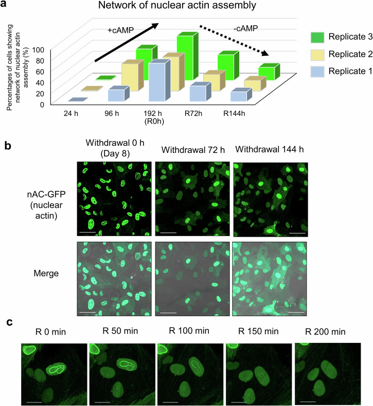
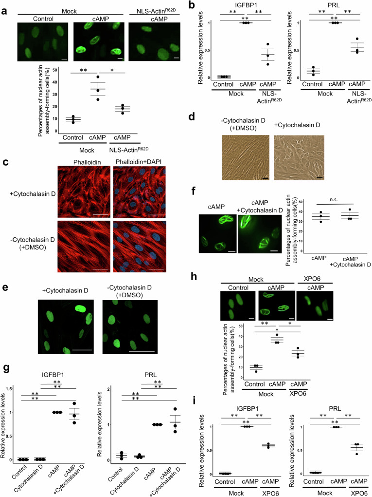
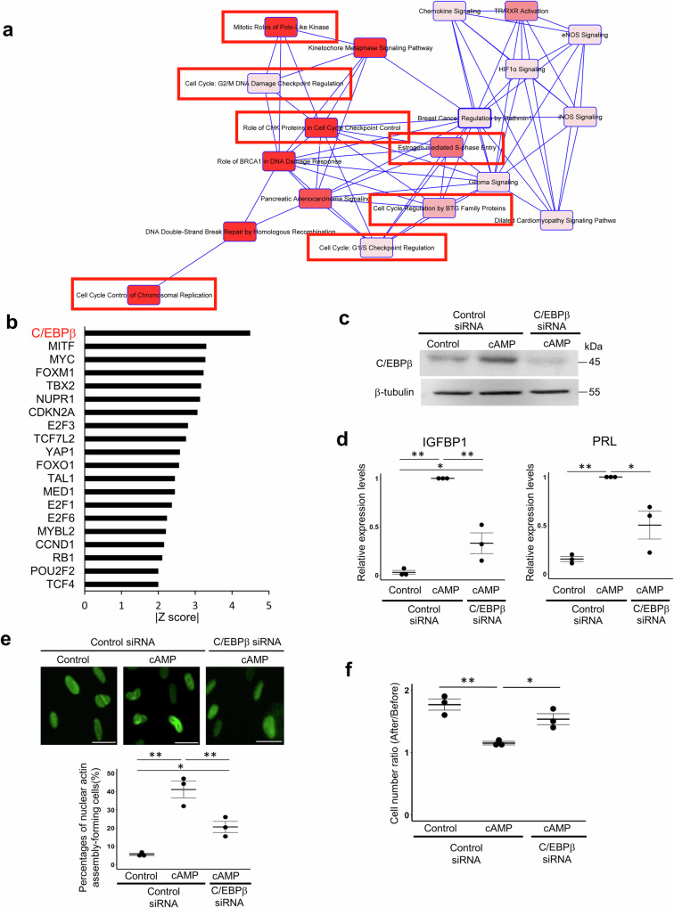
Decidualization of the human endometrium is critical for establishing pregnancy and is entailed by differentiation of endometrial stromal cells (ESCs) into decidual cells. During decidualization, the actin cytoskeleton is dynamically reorganized for the ESCs' morphological and functional changes. Although actin dynamically alters its polymerized state upon external stimuli not only in the cytoplasm, but also in the nucleus, nuclear actin dynamics during decidualization have not been elucidated. Here, we show that nuclear actin was specifically assembled during decidualization of human ESCs. This decidualization-specific formation of nuclear actin filaments was disassembled following the withdrawal of the decidualization stimulus, suggesting its reversible process. Mechanistically, RNA-seq analyses revealed that the forced disassembly of nuclear actin resulted in the suppression of decidualization, accompanied with the abnormal upregulation of cell proliferation genes, leading to incomplete cell cycle arrest. CCAAT/enhancer-binding protein beta (C/EBPβ), an important regulator for decidualization, was responsible for downregulation of the nuclear actin exporter, thus accelerating nuclear actin accumulation and its assembly for decidualization. Taken together, we demonstrate that decidualization-specific nuclear actin assembly induces cell cycle arrest for establishing the decidualized state of ESCs. We propose that not only the cytoplasmic actin, but also nuclear actin dynamics profoundly affect decidualization process in humans for ensuring pregnancy.


Conflict of interest statement

The authors declare no competing interests.

Figures

Fig. 1
Fig. 1. Nuclear actin is assembled after cAMP-mediated decidualization of hESCs.
a A representative image of a hESC that showed the network of nuclear actin assembly after transient expression of nuclear actin chromobody (nAC)-GFP that probes nuclear actin. hESCs were treated with cAMP for 96 h. The control represents hESCs that were not treated with cAMP as shown in Supplementary Fig. 1a. Scale bars, 5 μm. b A schematic diagram for observing the formation of nuclear actin assembly during decidualization of hESCs stably expressing nAC-GFP. c Percentages of cells that showed nuclear actin assembly. Cells were established from three different patients (Rep. 1–3) and were treated with or without cAMP for 96 h. Cells that showed nuclear actin assembly and network of nuclear actin assembly (Supplementary Fig. 2) were both counted as being positive for the formation of nuclear actin assembly. Hours after cAMP supplementation are indicated. Left, data of each case. Right, mean ± SE of three cases. Each data point is indicated as a dot. *P < 0.05 (Tukey-Kramer test). n.s. represents not significant. d, e Representative images that capture the formation of nuclear actin assembly during decidualization. Images were taken from 57 h after cAMP supplementation (d). As a control, no cAMP-supplemented control cells cultured in DMEM with 2% FBS were also captured by confocal microscopy under the same setting (e). Scale bars, 20 μm.
Fig. 2
Fig. 2. Nuclear actin is disassembled by reversing the decidualized state of hESCs.
a Percentages of cells that showed the network of nuclear actin assembly. Cells were established from three different patients (Rep. 1–3). Cells that showed the network of nuclear actin assembly (Supplementary Fig. 2) were counted. Hours after cAMP supplementation are indicated. After 192 h of culture with cAMP, cells were cultured without cAMP for recovery (R0h). Cells were counted at 72 h (R72h) and 144 h (R144h) after the removal of cAMP. Data of 24 h and 96 h are same as Fig. 1c. b Representative confocal images of decidualized hESC cells expressing nAC-GFP after withdrawal of cAMP. Times after cAMP withdrawal are indicated above (Recovery). Merged images of GFP channels and DIC are shown (Merge). Scale bars, 50 μm. c Representative images that follow a process of nuclear actin disassembly after withdrawal of cAMP. Images were taken from 73 h after cAMP withdrawal. Recovery times are shown. Scale bars, 20 μm.
Fig. 3
Fig. 3. Nuclear actin assembly is required for decidualization of hESCs.
a The effect of overexpressing NLS-ActinR62D on the formation of nuclear actin assembly during decidualization. hESCs stably expressing nAC-GFP were overexpressed with mCherry (as a mock control) or NLS-ActinR62D, and were treated with or without cAMP for 96 h. Representative images and percentages of cells that showed nuclear actin assembly are indicated. Mean ± SE of three independent experiments. Each data point is indicated as a dot. **P < 0.01; *P < 0.05 (Tukey-Kramer test). Scale bars, 10 μm. b RT-qPCR analyses of decidualization markers (IGFBP1 and PRL) after overexpressing NLS-ActinR62D. hESCs were overexpressed with mCherry (as a mock control) or NLS-ActinR62D, and were treated with or without cAMP for 96 h. Relative expression levels to cAMP-treated mock hESCs are shown. Mean ± SE of three independent experiments. Each data point is indicated as a dot. **P < 0.01 (Tukey-Kramer test). c Endogenous F-actin stained with phalloidin in hESCs treated with Cytochalasin D or DMSO (vehicle control). DNA was visualized by DAPI. Scale bars, 50 μm. d A bright filed image of hESCs after 4 days of Cytochalasin D treatment. As a control, hESCs were cultured in DMEM with 2% FBS with DMSO. Scale bar, 50 μm. e Representative images of hESCs expressing nAC-GFP treated with or without Cytochalasin D for 4 days. Scale bars, 50 μm. f Representative images of cAMP-treated hESCs expressing nAC-GFP, co-cultured with or without Cytochalasin D for 4 days. Scale bars, 10 μm. Percentages of cells that showed nuclear actin assembly are indicated. Mean ± SE of three independent experiments. Each data point is indicated as a dot. n.s. represents not significant (Student’s t test). g RT-qPCR analyses of decidualization markers (IGFBP1 and PRL) in cAMP-treated hESCs co-cultured with or without Cytochalasin D for 4 days. Relative expression levels to cAMP-treated hESCs are shown. Mean ± SE of three independent experiments. Each data point is indicated as a dot. **P < 0.01 (Tukey-Kramer test). h The effect of overexpressing XPO6 on the formation of nuclear actin assembly during decidualization. hESCs stably expressing nAC-GFP were overexpressed with mCherry (as a mock control) or XPO6, and were treated with or without cAMP for 96 h. Representative images and percentages of cells that showed nuclear actin assembly are indicated. Mean ± SE of three independent experiments. Each data point is indicated as a dot. **P < 0.01; *P < 0.05 (Tukey-Kramer test). Scale bars, 10 μm. i RT-qPCR analyses of decidualization markers (IGFBP1 and PRL) after overexpressing XPO6 in cAMP-treated hESCs. hESCs were overexpressed with mCherry (as a mock control) or XPO6, and were treated with or without cAMP for 96 h. Relative expression levels to cAMP-treated mock hESCs are shown. Mean ± SE of three independent experiments. Each data point is indicated as a dot. **P < 0.01 (Tukey-Kramer test).
Fig. 4
Fig. 4. Incomplete downregulation of decidualization-related genes after forced disassembly of nuclear actin in hESCs undergoing decidualization, as revealed by RNA-seq analyses.
a A schematic diagram for RNA-seq analyses. Three different types of samples were collected (i–iii). b Ven diagrams show the number of total and overlapping genes. Up- or down-regulated genes were identified (p adj < 0.05) by comparing samples i vs ii or i vs iii, as shown in Fig. 4a. Genes that are downregulated by cAMP treatment, but are not downregulated by overexpressing NLS-ActinR62D are termed as “nuclear actin assembly-regulated decidualization genes”. c GO and pathway analyses of nuclear actin assembly-regulated decidualization genes, identified in Fig. 4b. d A heatmap shows expression levels of 60 genes related to cellular proliferation, selected from nuclear actin assembly-regulated decidualization genes.
Fig. 5
Fig. 5. Cell cycle arrest of hESCs after cAMP stimulation is inhibited by forced disassembly of nuclear actin.
a hESCs were overexpressed with mCherry (as a mock control) or NLS-ActinR62D. Cell numbers were counted before and after 96 h of incubation with or without cAMP, and the ratios were compared. NLS-ActinR62D overexpression increased cell numbers that were normally unchanged after cAMP stimulation. Mean ± SE of three independent experiments. Each data point is indicated as a dot. **P < 0.01 (Tukey-Kramer test). b hESCs were overexpressed with mCherry (as a mock control) or NLS-ActinR62D. Percentages of Ki67-positive cells were counted after 96 h of incubation with or without cAMP. NLS-ActinR62D overexpression increased Ki67-positive proliferating cells, when compared to the mock control with cAMP. Mean ± SE of three independent experiments. Each data point is indicated as a dot. **P < 0.01; *P < 0.05 (Tukey-Kramer test). Scale bars, 100 μm.
Fig. 6
Fig. 6. C/EBPβ contributes to nuclear actin assembly in decidualized hESCs.
a Canonical pathways predicted by IPA using nuclear actin assembly-regulated decidualization genes. Many cell cycle-related terms are found (red boxes). Terms with strong significance are indicated as intense red. b Upstream regulators for nuclear actin assembly-regulated decidualization genes are predicated by IPA. Among them, top 20 transcription regulators based on the absolute values of z-score are shown. c Representative images of western blot analyses of cAMP-treated hESCs with or without siRNA-mediated knockdown of C/EBPβ. hESCs were transfected with siRNA against C/EBPβ or control siRNA, and were treated with or without cAMP for 96 h. Control represents hESCs not treated with cAMP. Antibodies against C/EBPβ and β-tubulin are used. d RT-qPCR analyses of decidualization markers (IGFBP1 and PRL) in cAMP-treated hESCs transfected with siRNA. Relative expression levels to the cAMP-treated hESCs transfected with control siRNA are shown. Mean ± SE of three independent experiments. Each data point is indicated as a dot. **P < 0.01; *P < 0.05 (Tukey-Kramer test). e The effect of C/EBPβ knockdown on the formation of nuclear actin assembly during decidualization. Representative images and proportions of cells that showed nuclear actin assembly are indicated. Mean ± SE of three independent experiments. Each data point is indicated as a dot. **P < 0.01; *P < 0.05 (Tukey-Kramer test). Scale bars, 50 μm. f The effect of C/EBPβ knockdown on cell cycle arrest during decidualization. Cell numbers were counted before and after 96 h of incubation with or without cAMP, and the ratios were compared. Mean ± SE of three independent experiments. Each data point is indicated as a dot. **P < 0.01; *P < 0.05 (Tukey-Kramer test).
Fig. 7
Fig. 7. The increase of nuclear actin levels by XPO6 downregulation is associated with nuclear actin assembly during decidualization.
a Representative images of western blot analyses of isolated hESC nuclei and cytoplasm against β-actin, cofilin, β-tubulin, and histone H2B. hESCs were treated with or without cAMP (control) for 48 h. Band intensities of actin and cofilin were quantified and the relative intensities of cAMP-treated samples to the control are shown in the graph. Mean ± SE. Each data point is indicated as a dot. *P < 0.05 (Student’s t test). n.s. represents not significant. Three independent experiments were repeated. b Representative images of western blot analyses of whole hESC lysates against XPO6 and β-tubulin. hESCs were treated with or without cAMP (control) for 96 h. Band intensities were quantified and the relative intensities of cAMP-treated samples to the control are shown in the graph. Mean ± SE. Each data point is indicated as a dot. **P < 0.05 (Student’s t test). Three independent experiments were repeated. c Accumulation of cofilin in nuclei after induction of cAMP-stimulated decidualization of hESCs. Representative immunofluorescence images of hESCs stably expressing nAC-GFP treated with cAMP for 96 h. As a control, hESCs without cAMP addition were examined (control). DNA was stained with DAPI. Scale bar, 10 μm. d The effect of C/EBPβ knockdown on the increase of nuclear actin and cofilin during decidualization. Representative images of western blot analyses of isolated hESCs nuclei against β-actin, cofilin, β-tubulin, and histone H2B are shown. Band intensities were quantified and the relative intensities to no-treated hESCs transfected with control siRNA are shown (control). Mean ± SE of three independent experiments. Each data point is indicated as a dot. **P < 0.01 (Tukey-Kramer test). n.s. represents not significant. e The effect of C/EBPβ knockdown on the nuclear accumulation of cofilin after induction of cAMP-stimulated decidualization of hESCs. hESCs were transfected with siRNA against C/EBPβ or control siRNA, and were treated with or without cAMP. Representative immunofluorescence images are shown. As a control, hESCs without cAMP addition were examined (control). DNA was stained with DAPI. Scale bar, 50 μm. f The effect of C/EBPβ knockdown on XPO6 expression during decidualization. Representative images of western blot analyses of isolated hESCs nuclei against XPO6 and β-tubulin are shown. Band intensities were quantified and the relative intensities to control cAMP-untreated hESCs transfected with control siRNA (control) are shown. Mean ± SE of three independent experiments. Each data point is indicated as a dot. **P < 0.01; *P < 0.05 (Tukey-Kramer test).

Similar articles

Cited by

  • Recent advances in nuclear actin research.
    Szabó A, Borkúti P, Kovács Z, Kristó I, Vilmos P. Szabó A, et al. Nucleus. 2025 Dec;16(1):2498643. doi: 10.1080/19491034.2025.2498643. Epub 2025 May 4. Nucleus. 2025. PMID: 40320716 Free PMC article. Review.

References

    1. Salker M, et al. Natural selection of human embryos: impaired decidualization of endometrium disables embryo-maternal interactions and causes recurrent pregnancy loss. PLoS One. 2010;5:e10287. doi: 10.1371/journal.pone.0010287. - DOI - PMC - PubMed
    1. Zhang Q, Yan J. Update of Wnt signaling in implantation and decidualization. Reprod. Med. Biol. 2016;15:95–105. doi: 10.1007/s12522-015-0226-4. - DOI - PMC - PubMed
    1. Fukui Y, et al. Uterine receptivity, embryo attachment, and embryo invasion: Multistep processes in embryo implantation. Reprod. Med. Biol. 2019;18:234–240. doi: 10.1002/rmb2.12280. - DOI - PMC - PubMed
    1. Dimitriadis E, Menkhorst E, Salamonsen LA, Paiva P. Review: LIF and IL11 in trophoblast-endometrial interactions during the establishment of pregnancy. Placenta. 2010;31:S99–S104. doi: 10.1016/j.placenta.2009.12.027. - DOI - PubMed
    1. Gellersen B, Brosens J. Cyclic decidualization of the human endometrium in reproductive health and failure. Endocr. Rev. 2014;35:851–905. doi: 10.1210/er.2014-1045. - DOI - PubMed

Publication types