

Modified ventriculoperitoneal shunt applied to temporary external ventricular drainage
- PMID: 38992174
- PMCID: PMC11239655
- DOI: 10.1038/s41598-024-66917-x
Modified ventriculoperitoneal shunt applied to temporary external ventricular drainage
Abstract
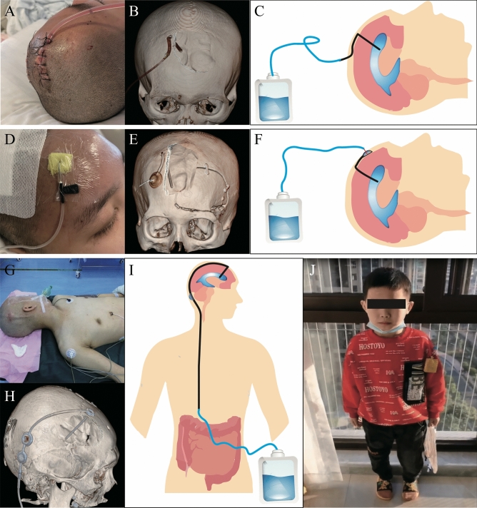
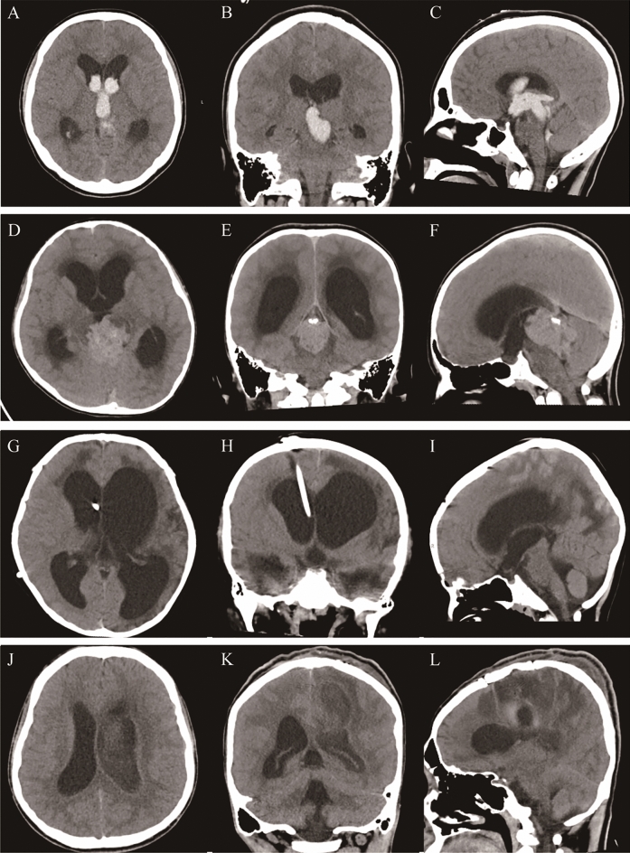
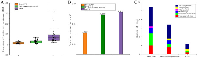
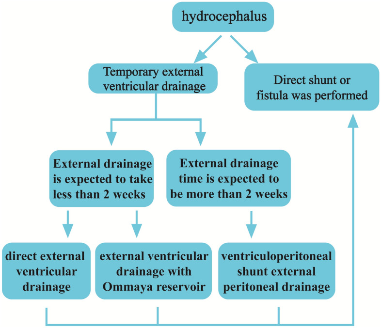
External ventricular drainage (EVD) is a common procedure in neurosurgical practice. Presently, the three methods used most often include direct EVD (dEVD), long-tunneled external ventricular drains (LTEVDs), and EVD via the Ommaya reservoir (EVDvOR). But they possess drawbacks such as limited duration of retention, vulnerability to iatrogenic secondary infections, and challenges in regulating drainage flow. This study aimed to explore the use of a modified ventriculoperitoneal shunt (mVPS)-the abdominal end of the VPS device was placed externally-as a means of temporary EVD to address the aforementioned limitations. This retrospective cohort study, included 120 cases requiring EVD. dEVD was performed for 31 cases, EVDvOR for 54 cases (including 8 cases with previously performed dEVD), and mVPS for 35 cases (including 6 cases with previously performed EVDvOR). The one-time success rate (no need for further other EVD intervention) for dEVD, EVDvOR, and mVPS were 70.97%, 88.89%, and 91.42%, dEVD vs EVDvOR (P < 0.05), dEVD vs mVPS (P < 0.05), EVDvOR vs mVPS (P > 0.05). Puncture needle displacement or detachment was observed in nearly all cases of EVDvOR, while no such complications have been observed with mVPS. Apart from this complication, the incidence of postoperative complications was 35.48%, 14.81%, and 8.5%, dEVD vs EVDvOR (P < 0.05), dEVD vs mVPS (P < 0.05), EVDvOR vs mVPS (P > 0.05). Mean postoperative retention for EVD was 14.68 ± 9.50 days, 25.96 ± 15.14 days, and 82.43 ± 64.45 days, respectively (P < 0.001). In conclusion, mVPS significantly extends the duration of EVD, which is particularly beneficial for patients requiring long-term EVD.
Keywords: External ventricular drainage; Hydrocephalus; Modified ventriculoperitoneal shunt; Ommaya reservoir.
© 2024. The Author(s).
Conflict of interest statement
The authors declare no competing interests.
Figures
Similar articles
-
Indications for pediatric external ventricular drain placement and risk factors for conversion to a ventriculoperitoneal shunt.Pediatr Neurosurg. 2012;48(6):342-7. doi: 10.1159/000353608. Epub 2013 Aug 13. Pediatr Neurosurg. 2012. PMID: 23941907
-
External ventricular drain related complications-whether continuous csf drainage via ommaya reservoir is the answer?Neurol India. 2020 Mar-Apr;68(2):458-461. doi: 10.4103/0028-3886.284354. Neurol India. 2020. PMID: 32415024
-
Predictors for delayed ventriculoperitoneal shunt placement after external ventricular drain removal in patients with subarachnoid hemorrhage.Br J Neurosurg. 2015 Apr;29(2):219-24. doi: 10.3109/02688697.2014.967753. Epub 2014 Oct 9. Br J Neurosurg. 2015. PMID: 25299790
-
The role of external ventricular drainage for the management of posterior cranial fossa tumours: a systematic review.Neurosurg Rev. 2021 Jun;44(3):1243-1253. doi: 10.1007/s10143-020-01325-z. Epub 2020 Jun 3. Neurosurg Rev. 2021. PMID: 32494987
-
Rapid Versus Gradual Weaning of External Ventricular Drain: A Systematic Literature Review and Meta-analysis.Neurocrit Care. 2023 Aug;39(1):250-259. doi: 10.1007/s12028-023-01766-6. Epub 2023 Jun 12. Neurocrit Care. 2023. PMID: 37308728 Free PMC article.
Cited by
-
Correct understanding of the definition and management strategies for refractory hydrocephalus.Chin Neurosurg J. 2025 Aug 18;11(1):16. doi: 10.1186/s41016-025-00403-9. Chin Neurosurg J. 2025. PMID: 40826122 Free PMC article. Review.
References
MeSH terms
LinkOut - more resources
Full Text Sources
