. 2024 Jul 12;14(1):16083.
doi: 10.1038/s41598-024-66779-3.

ActivinA modulates B-acute lymphoblastic leukaemia cell communication and survival by inducing extracellular vesicles production

Affiliations

ActivinA modulates B-acute lymphoblastic leukaemia cell communication and survival by inducing extracellular vesicles production

Eugenia Licari et al. Sci Rep. .

Abstract

Extracellular vesicles (EVs) are a new mechanism of cellular communication, by delivering their cargo into target cells to modulate molecular pathways. EV-mediated crosstalk contributes to tumor survival and resistance to cellular stress. However, the role of EVs in B-cell Acute Lymphoblastic Leukaemia (B-ALL) awaits to be thoroughly investigated. We recently published that ActivinA increases intracellular calcium levels and promotes actin polymerization in B-ALL cells. These biological processes guide cytoskeleton reorganization, which is a crucial event for EV secretion and internalization. Hence, we investigated the role of EVs in the context of B-ALL and the impact of ActivinA on this phenomenon. We demonstrated that leukemic cells release a higher number of EVs in response to ActivinA treatment, and they can actively uptake EVs released by other B-ALL cells. Under culture-induced stress conditions, EVs coculture promoted cell survival in B-ALL cells in a dose-dependent manner. Direct stimulation of B-ALL cells with ActivinA or with EVs isolated from ActivinA-stimulated cells was even more effective in preventing cell death. This effect can be possibly ascribed to the increase of vesiculation and modifications of EV-associated microRNAs induced by ActivinA. These data demonstrate that ActivinA boosts EV-mediated B-ALL crosstalk, improving leukemia survival in stress conditions.

Keywords: ActivinA; B-cell acute lymphoblastic leukaemia; Cell survival; Extracellular vesicles; Intercellular communication; Microenvironment.


Conflict of interest statement

The authors declare no competing interests.

Figures
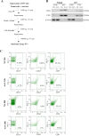
Figure 1
Figure 1
Characterization of EVs. (A) Isolation protocol for EVs. After 24 or 48 h of culture in serum-free medium, cell supernatants were centrifuged three times with increasing speed. Then, the samples were ultracentrifuged at 110,000×g for 97 min to pellet total EVs. (B) Enrichment of EV marker. Western blot analysis of CD81 and CD9 tetraspanins and β-actin on 697 and Nalm6 cell lysates (cells) and EVs isolated after 24 h of culture in serum-free medium alone (NS) or in presence of ActivinA (ActA). The blots are representative of two independent experiments. (C) Flow cytometry evaluation of CD9 and CD63 tetraspanins and B-cell marker CD19 on EVs isolated from 697 cells stimulated or not with ActivinA for 24 or 48 h. The plots are representative of three independent experiments (CD19/CD63) and two independent experiments for CD9.
Figure 2
Figure 2
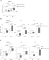
ActivinA upregulates EV release from B-ALL cells. (A) 697 cells were pretreated or not with ActivinA 50 ng/ml or 200 ng/ml, for 24 and 48 h. The concentration (particle/ml) of EVs in the supernatant was determined by means of NTA. Fold change (FC) was calculated as number of particle/ml in the supernatant of ActivinA-stimulated cells (ActA)/not stimulated (NS) cells. Each box plot shows the median, the mean (+) and extends from the lowest to the highest value (n ≥ 8 independent experiments per condition). Two-tailed one sample t test: FC ActA/NS vs 1; Mann–Whitney two-tailed test: ActivinA 200 ng/ml vs ActivinA 50 ng/ml. (B,C) Box plot graphs representing the concentration of total EVs, sEV and lEV released by 697 (B) or Nalm6 cells (C) pretreated or not with ActivinA 200 ng/ml for 24 and 48 h. Each box plot shows the median, the mean (+) and extends from the lowest to the highest value (n = 7 for 697 cells or n ≥ 5 for Nalm6 cells independent experiments per condition). Wilcoxon matched-pairs two-tailed test.
Figure 3
Figure 3
B-ALL cells can crosstalk through the internalization of released EVs. EVs were isolated from 697 cells stimulated (EV-ActA) or not (EV-NS) with ActivinA (200 ng/ml). EV-NS and EV-ActA were stained with CFSE (green fluorescent dye) or PBS (EV-PBS, negative control) and cocultured with 697 cells for 24 h. (A) Representative confocal 3D maximum intensity reconstruction of 697 cells after internalization of EV-PBS, EV-NS and EV-ActA. The green fluorescent dots around the cell nucleus (DAPI staining, blue) indicate internalized EVs. (B) Confocal microscopy quantification of CFSE Integrated Fluorescence Intensity. Each box plot shows the median and the mean (+) and extends from the lowest to the highest value (n = 3 independent experiments). Two-way ANOVA with Tukey’s correction for multiple comparisons. (C) Representative overlay histogram showing CFSE fluorescence evaluated by flow cytometry in 697 cells cultured with CFSE-stained EV-NS (gray) or EV-ActA (red). Unstained 697 cells were used as negative control (dashed gray line). (D) Flow cytometry quantification of CFSE MFI in 697 cells cultured with CFSE-labeled EV-NS and EV-ActA. Each box plot shows the median, the mean (+) and extends from the lowest to the highest value (n = 5 independent experiments). Wilcoxon matched-pairs two-tailed test.
Figure 4
Figure 4
B-ALL cell crosstalk mediated by EV release contributes to cell viability modulation. (A) EV-NS were isolated from the supernatant of 25 × 106 (EV-NS_25 × 106, black empty square) or 80 × 106 (EV-NS_80 × 106, green empty square) unstimulated 697 cells and added on day 0, 3 and 6 (black arrows) to 697 cells that were kept in culture for 12 days without changing medium. PBS-stimulated 697 cells were used as unstimulated control (NS, black square). The panel shows the growth curves obtained by counting the number of viable cells by an automated cell counter in three wells for each condition (mean ± sd). One representative experiment out of three independent cultures performed is shown. *p: NS vs EV-NS_25 × 106, #p: EV-NS_25 × 106 vs EV-NS_80 × 106, $p: NS vs EV-NS_80 × 106, two-way ANOVA with Tukey’s correction for multiple comparison, all the actual p values are reported in Supplementary Table 1. (B) 697 cells were stimulated or not with ActivinA 200 ng/ml (red square) or with PBS (NS, black square) on day 0, 3 and 6 (black arrows). EVs were isolated from the supernatant of 80 × 106 697 cells unstimulated (EV-NS_80 × 106, black empty square) or stimulated with ActivinA (EV-ActA_80 × 106, red empty square) and added on day 0, 3 and 6 (black arrows) to 697 cells that were kept in culture for 12 days without changing medium. The panel shows the growth curves obtained by counting the number of viable cells by an automated cell counter in three wells for each condition (mean ± sd). One representative experiment out of three independent cultures performed is shown. *p: NS vs ActA (50 ng/mL), $p: NS vs EV-NS_80 × 106, #p: EV-NS_80 × 106 vs EV-ActA_80 × 106, §p: NS vs EV-ActA_80 × 106, two-way ANOVA with Tukey’s correction for multiple comparison, all the actual p values are reported in Supplementary Table 1.
Figure 5
Figure 5
ActivinA-stimulated B-ALL 697 cells secrete EVs enriched in distinctive miRNA species. Volcano plots comparing EV-associated miRNAs from ActivinA-stimulated (EV-ActA) and not stimulated (EV-NS) 697 cells for 24 h and 48 h. The X-axis represents the log2 transformed fold change of each miRNA expression between ActivinA-derived and non-stimulated samples. The Y-axis shows the −log10 of the p-value. Different colored dots show the differential regulation of miRNAs: in red the up-regulated and in green the down-regulated.

Similar articles

Cited by

References

    1. Hunger SP, Mullighan CG. Acute lymphoblastic leukemia in children. N. Engl. J. Med. 2015;373:1541–1552. doi: 10.1056/NEJMra1400972. - DOI - PubMed
    1. Chiarini F, et al. Advances in understanding the acute lymphoblastic leukemia bone marrow microenvironment: From biology to therapeutic targeting. Biochim. Biophys. Acta-Mol. Cell Res. 2016;1863:449–463. doi: 10.1016/j.bbamcr.2015.08.015. - DOI - PubMed
    1. Colmone A, et al. Leukemic cells create bone marrow niches that disrupt the behavior of normal hematopoietic progenitor cells. Science. 2008;322:1861–1865. doi: 10.1126/science.1164390. - DOI - PubMed
    1. Portale F, et al. ActivinA: A new leukemia-promoting factor conferring migratory advantage to B-cell precursor-acute lymphoblastic leukemic cells. Haematologica. 2019;104:533–545. doi: 10.3324/haematol.2018.188664. - DOI - PMC - PubMed
    1. Bashir M, Damineni S, Mukherjee G, Kondaiah P. Activin-A signaling promotes epithelial–mesenchymal transition, invasion, and metastatic growth of breast cancer. npj Breast Cancer. 2015;1:1–13. doi: 10.1038/npjbcancer.2015.7. - DOI

MeSH terms

LinkOut - more resources