. 2024 Jul;41(7):1475-1491.
doi: 10.1007/s11095-024-03733-y. Epub 2024 Jul 12.

Duo photoprotective effect via silica-coated zinc oxide nanoparticles and Vitamin C nanovesicles composites

Affiliations

Duo photoprotective effect via silica-coated zinc oxide nanoparticles and Vitamin C nanovesicles composites

Soha M Kandil et al. Pharm Res. 2024 Jul.

Abstract

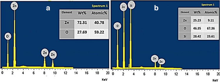
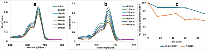
Objective: Zinc Oxide nanoparticles (ZnO NPs) are used widely in nowadays personal care products, especially sunscreens, as a protector against UV irradiation. Yet, they have some reports of potential toxicity. Silica is widely used to cage ZnO NPs to reduce their potential toxicity. Vitamin C derivative, Magnesium Ascorpyl Phosphate (MAP), is a potent antioxidant that can efficiently protect human skin from harmful impacts of UV irradiation and oxidative stress. The combination of silica coated ZnO NPs and MAP nanovesicles could have potential synergistic protective effect against skin photodamage.

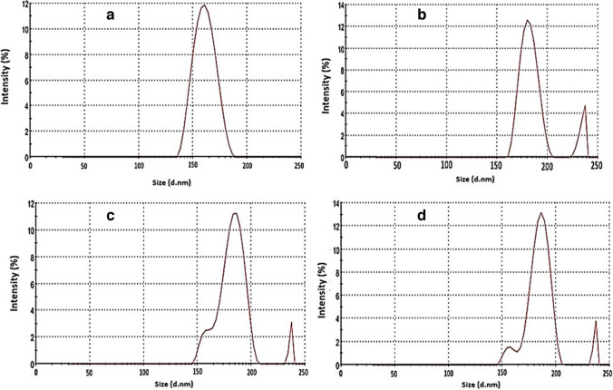
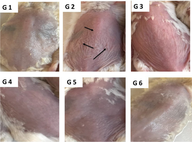
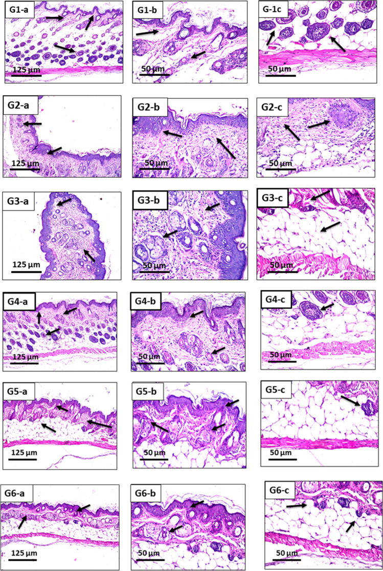
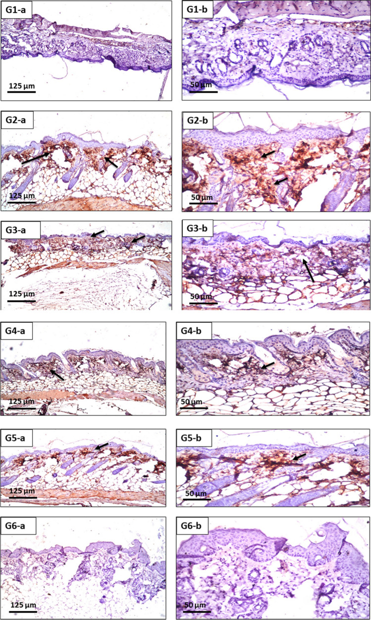
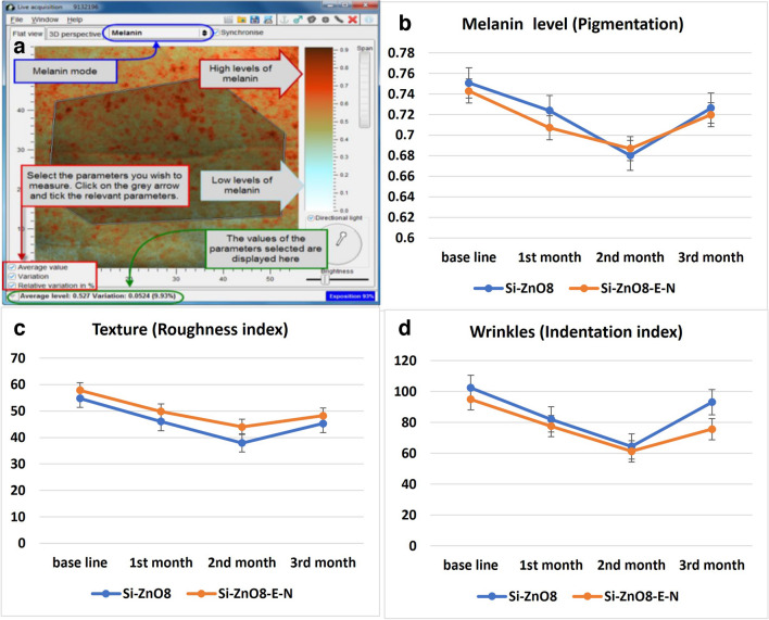
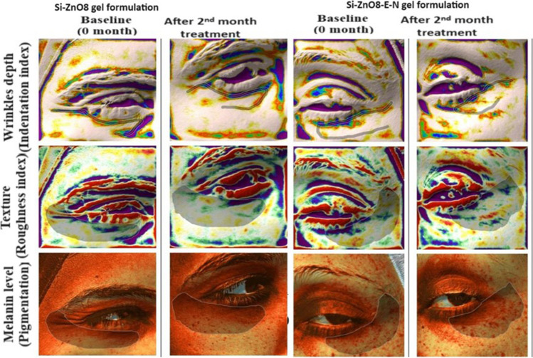
Methods: Silica coated ZnO NPs and MAP nanovesicles (ethosomes and niosomes) were synthesized, formulated, and evaluated as topical gels. These gel formulations were evaluated in mice for their photoprotective effect against UV irradiation through histopathology and immuno-histochemistry study. Split-face clinical study was conducted to compare the effect of application of silica coated ZnO NPs either alone or combined with MAP nanovesicles. Their photoprotective action was evaluated, using Antera 3D® camera, for melanin level, roughness index and wrinkles depth.

Results: Silica coated ZnO NPs when combined with MAP nanovesicles protected mice skin from UV irradiation and decreased the expression of the proinflammatory cytokines, NF-κB. Clinically, silica coated ZnO NPs, alone or combined with MAP nanovesicles, could have significant effect to decrease melanin level, roughness index and wrinkles depth with higher effect for the combination.

Conclusion: A composite of silica coated ZnO NPs and MAP nanovesicles could be a promising cosmetic formulation for skin protection against photodamage signs such as hyperpigmentation, roughness, and wrinkles.

Keywords: NF-κB; UV irradiation; Zno; antera 3D® camera; ethosomes; magnesium ascorbyl phosphate (MAP); niosomes; photodamage; silica.


Conflict of interest statement

The authors declare that they have no financial interests or personal relationships that could have appeared to influence the work reported in this paper.

Figures

Fig. 1
Fig. 1
The optical absorption of ZnO NPs (a) and photoluminescence emission spectra of ZnO, Si-ZnO4, Si-ZnO6 and Si-ZnO8 NPs (b).
Fig. 2
Fig. 2
The X- ray diffractometers of the prepared samples.
Fig. 3
Fig. 3
The TEM images of ZnO (a, b), Si-ZnO4 (c, d), Si-ZnO6 (e, f) and Si-ZnO8 (g, h).
Fig. 4
Fig. 4
Zeta potentials (ZP) of ZnO (a), Si-ZnO4 (b), Si-ZnO6 (c) and Si-ZnO8 (d) NPs.
Fig. 5
Fig. 5
Dynamic light scattering (DLS) of ZnO (a), Si-ZnO4 (b), Si-ZnO6 (c) and Si-ZnO8 (d) NPs.
Fig. 6
Fig. 6
Elemental analysis with energy dispersive X-ray spectroscopy (EDX) of ZnO NPs (a) and Si-ZnO8 NPs (b).
Fig. 7
Fig. 7
Photocatalytic activity assessment of ZnO NPs (a), Si-ZnO8 NPs (b) and their UV shielding performance (c).
Fig. 8
Fig. 8
The typical appearance of hair-removed mouse skin after UV irradiation. G1: Normal control, G2: Photodamage model (UV), G3: photodamage model + control (ZnO8 NPs), G4: photodamage model + (Si-ZnO8-E), G5: photodamage model + (Si-ZnO8-N), G6: photodamage model + (Si-ZnO8-E-N).
Fig. 9
Fig. 9
G1-a (× 16): skin of mice of G1, showing normal histological structure of the epidermis, dermis with hair follicle and sebaceous glands followed by subcutaneous tissue and musculature. G1-b (× 40): skin of mice of G1, showing the magnification of Fig. 1a to identify the normal histology of epidermal layer with underlying dermis containing hair follicles. G1-c (× 40): skin of mice of G1, showing the magnification of Fig. 1a to identify the subcutaneous tissue and musculature. G2-a (× 16): skin of mice of G2, showing focal acanthosis was detected in the epidermis while the underlying dermis showed inflammatory cells infiltration, hyperplasia in the sebaceous glands and necrosis in some of the hair follicles. G2-b (× 40): skin of mice of G2, showing the magnification of Fig. 2a to identify the focal acanthosis in the epidermis with hyperplasia of sebaceous glands and inflammatory cells infiltration in the dermis. G2-c (× 40): skin of mice of G2, showing the magnification of Fig. 2a to identify the necrosis of some hair follicles and inflammatory cells infiltration in the dermis and subcutaneous adipose tissue. G3-a (× 16): skin of mice of G3, showing acanthosis in the epidermis with inflammatory cells infiltration in the dermis. G3-b (× 40): skin of mice of G3, showing the magnification of Fig. 3a to identify the acanthosis in the epidermis and inflammatory cells infiltration in the dermis. G3-c (× 40): skin of mice of G3, showing the magnification of Fig. 3a to identify the normal histological structure of subcutaneous adipose tissue and muscles. G4-a (× 16): skin of mice of G4, showing mild acanthosis in the epidermis with few inflammatory cells infiltration in the dermis with intact underlying subcutaneous adipose tissue and muscles. G4-b (× 40): skin of mice of G4, showing the magnification of Fig. 4a to identify the mild acanthosis in the epidermis with few inflammatory cells infiltration in the dermis. G4-c (× 40): skin of mice of G4, showing the magnification of Fig. 4a to identify the normal histopathological structure of subcutaneous adipose tissue and muscles. G5-a (× 16): skin of mice of G5, showing mild acanthosis in the epidermis with few inflammatory cells infiltration in the dermis with intact underlying subcutaneous adipose tissue and muscles. G5-b (× 40): skin of mice of G5, showing the magnification of Fig. 5a to identify the mild acanthosis in the epidermis with few inflammatory cells infiltration in the dermis. G5-c (× 40): skin of mice of G5, showing the magnification of Fig. 5a to identify the normal histological structure of subcutaneous adipose tissue and muscles. G6-a (× 16): skin of mice of G6, showing normal histological structure of the epidermis, dermis, subcutaneous tissue and musculature. G6-b (× 40): skin of mice of G6, showing the magnification of Fig. 6a to identify the normal histology of epidermis and dermis. G6-c (× 40): skin of mice of G6, showing the magnification of Fig. 6a to identify the normal histology of subcutaneous tissue and musculature.
Fig. 10
Fig. 10
G1-a (× 16): skin of mice of G1, showing nil immunoexpression of NF-κB. G1-b (× 40): skin of mice of G1, showing the magnification of Fig. 1a. G2-a (× 16): skin of mice of G1, showing severe immunoexpression of NF-κB. G2-b (× 40): skin of mice of G1, showing the magnification of Fig. 2a. G3-a (× 16): skin of mice of G1, showing moderate immunoexpression of NF-κB. G3-b (× 40): skin of mice of G1, showing the magnification of Fig. 3a. G4-a (× 16): skin of mice of G1, showing mild immunoexpression of NF-κB. G4-b (× 40): skin of mice of G1, showing the magnification of Fig. 4a. G5-a (× 16): skin of mice of G1, showing mild immunoexpression of NF-κB. G5-b (× 40): skin of mice of G1, showing the magnification of Fig. 5a. G6-a (× 16): skin of mice of G1, showing nil immunoexpression of NF-κB. G6-b (× 40): skin of mice of G1, showing the magnification of Fig. 6a.
Fig. 11
Fig. 11
a Representative model of collecting data from Antera 3D® Camera, b: Effect of Si-ZnO8 and Si-ZnO8-E-N gel formulations on melanin levels, c: Effect of Si-ZnO8 and Si-ZnO8-E-N gel formulations on texture, d: Effect of Si-ZnO8 and Si-ZnO8-E-N gel formulations on wrinkles.
Fig. 12
Fig. 12
Representative examples of the involved patients’ photos taken by Antera 3D® camera for the melanin level, roughness index and wrinkles depth at different treatment times.

Similar articles

Cited by

References

    1. Singh O, Singh MP, Kohli N, Singh RC. Effect of pH on the morphology and gas sensing properties of ZnO nanostructures. Sens Actuators B. 2012;166–167(402):438–43. 10.1016/j.snb.2012.02.085.10.1016/j.snb.2012.02.085 - DOI
    1. Cross SE, Innes B, Roberts MS, Tsuzuki T, Robertson TA, McCormick P. Human skin penetration of sunscreen nanoparticles: in-vitro assessment of a novel micronized zinc oxide formulation Skin. Pharmacol Physiol. 2007;20(3):148–54. 10.1159/000098701.10.1159/000098701 - DOI - PubMed
    1. Smijs TG, Pavel S. Titanium dioxide and zinc oxide nanoparticles in sunscreens: focus on their safety and effectiveness. Nanotechnol Sci Appl. 2011;4:95–112. 10.2147/NSA.S19419. 10.2147/NSA.S19419 - DOI - PMC - PubMed
    1. Kim YR, Park JI, Lee EJ, Park SH, Seong NW, Kim JH. Toxicity of 100 nm zinc oxide nanoparticles: a report of 90-day repeated oral administration in Sprague Dawley rats. Int J Nanomedicine. 2014;9(Suppl 2):109–26. 10.2147/IJN.S57928. 10.2147/IJN.S57928 - DOI - PMC - PubMed
    1. Oberdörster G, Oberdörster E, Oberdörster J. Nanotoxicology: an emerging discipline evolving from studies of ultrafine particles. Environ Health Perspect. 2005;113(7):823–39. 10.1289/ehp.7339. 10.1289/ehp.7339 - DOI - PMC - PubMed

MeSH terms

LinkOut - more resources