. 2024 Oct;17(5):1019-1028.
doi: 10.1016/j.mucimm.2024.07.001. Epub 2024 Jul 9.

Western diet reduces small intestinal intraepithelial lymphocytes via FXR-Interferon pathway

Affiliations

Western diet reduces small intestinal intraepithelial lymphocytes via FXR-Interferon pathway

Chen-Ting Hung et al. Mucosal Immunol. 2024 Oct.

Abstract

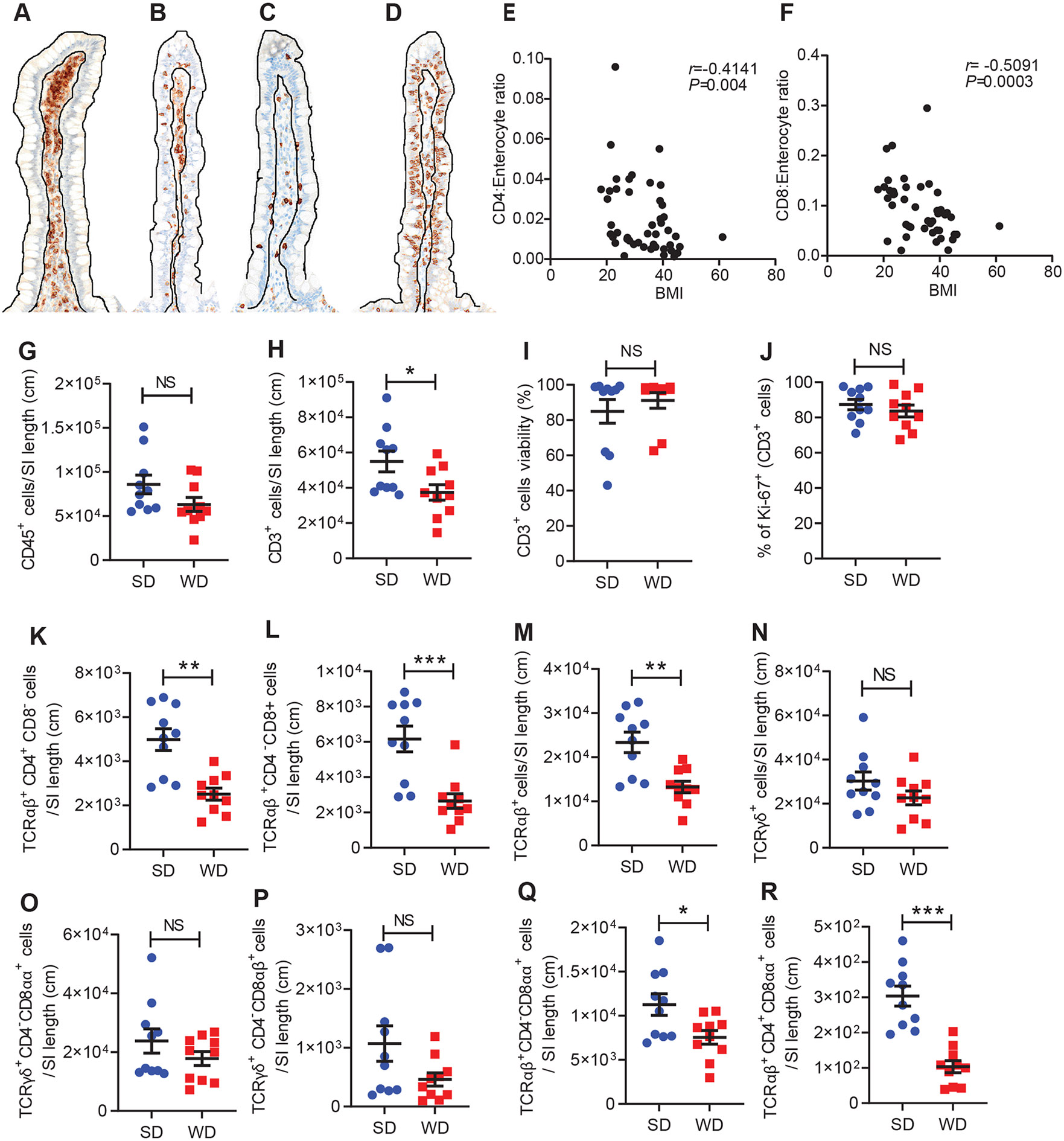
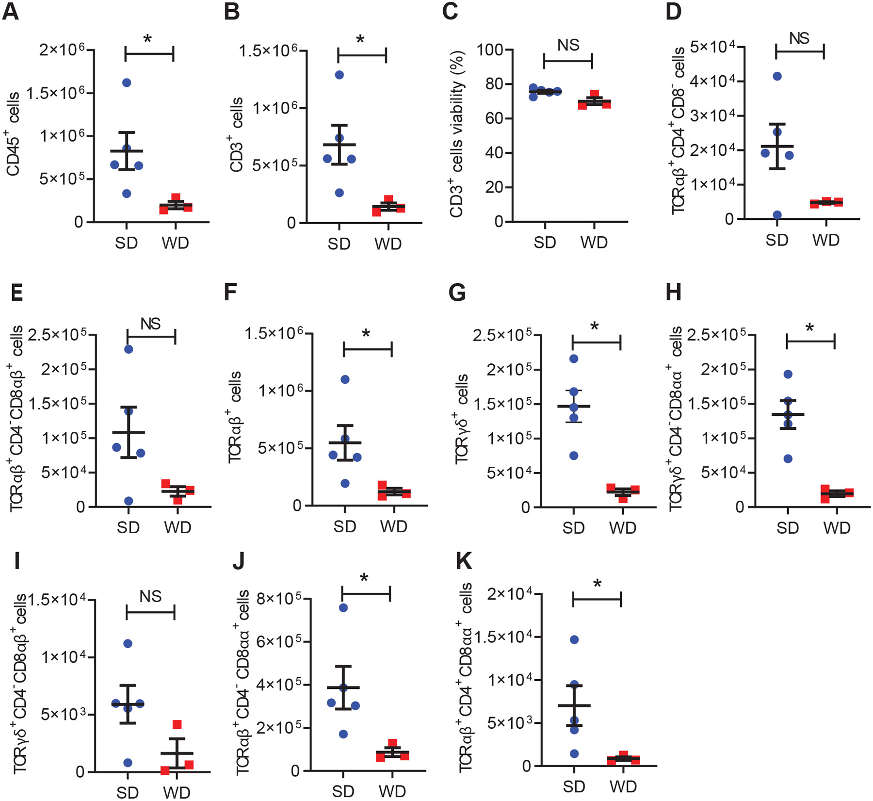
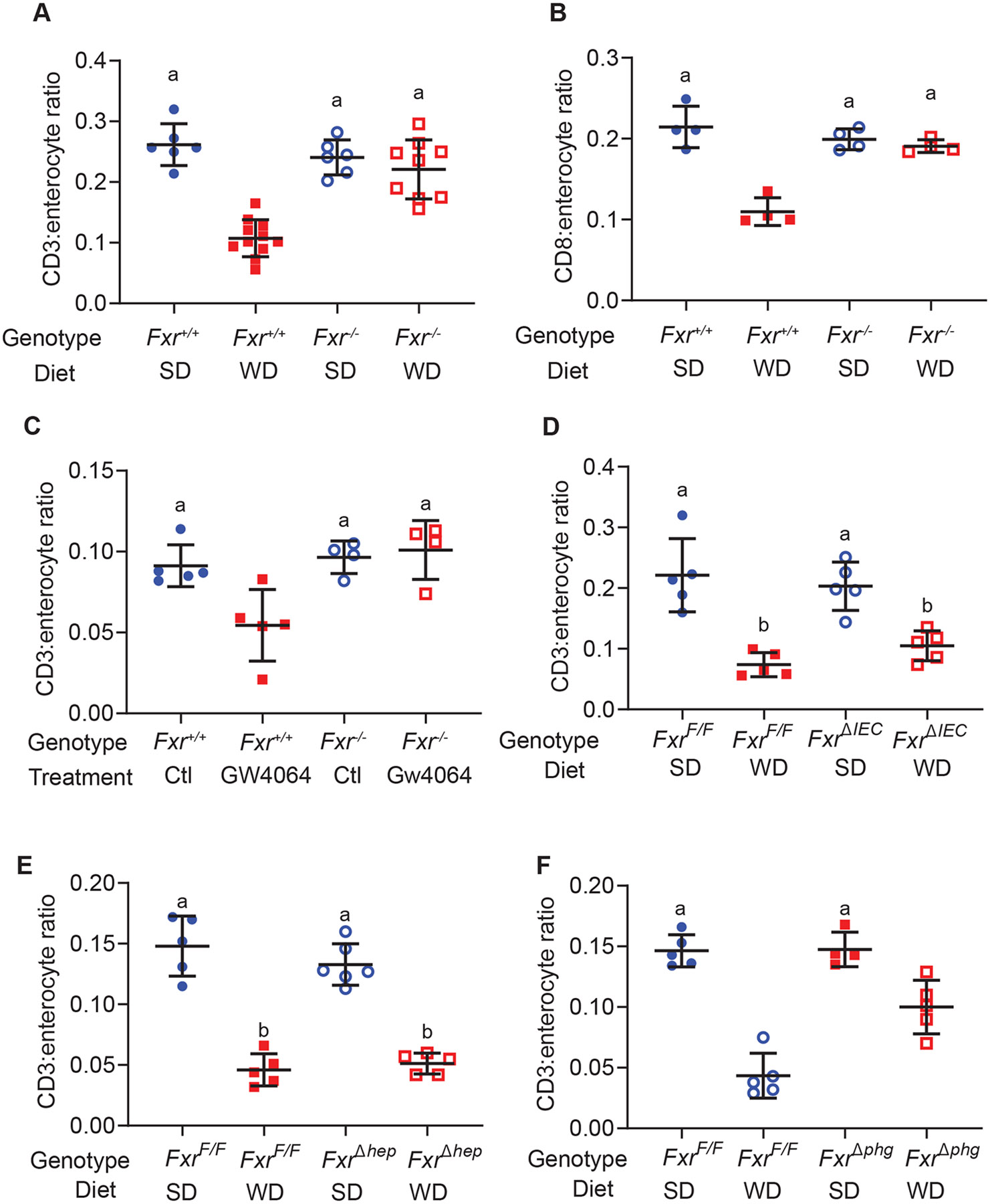
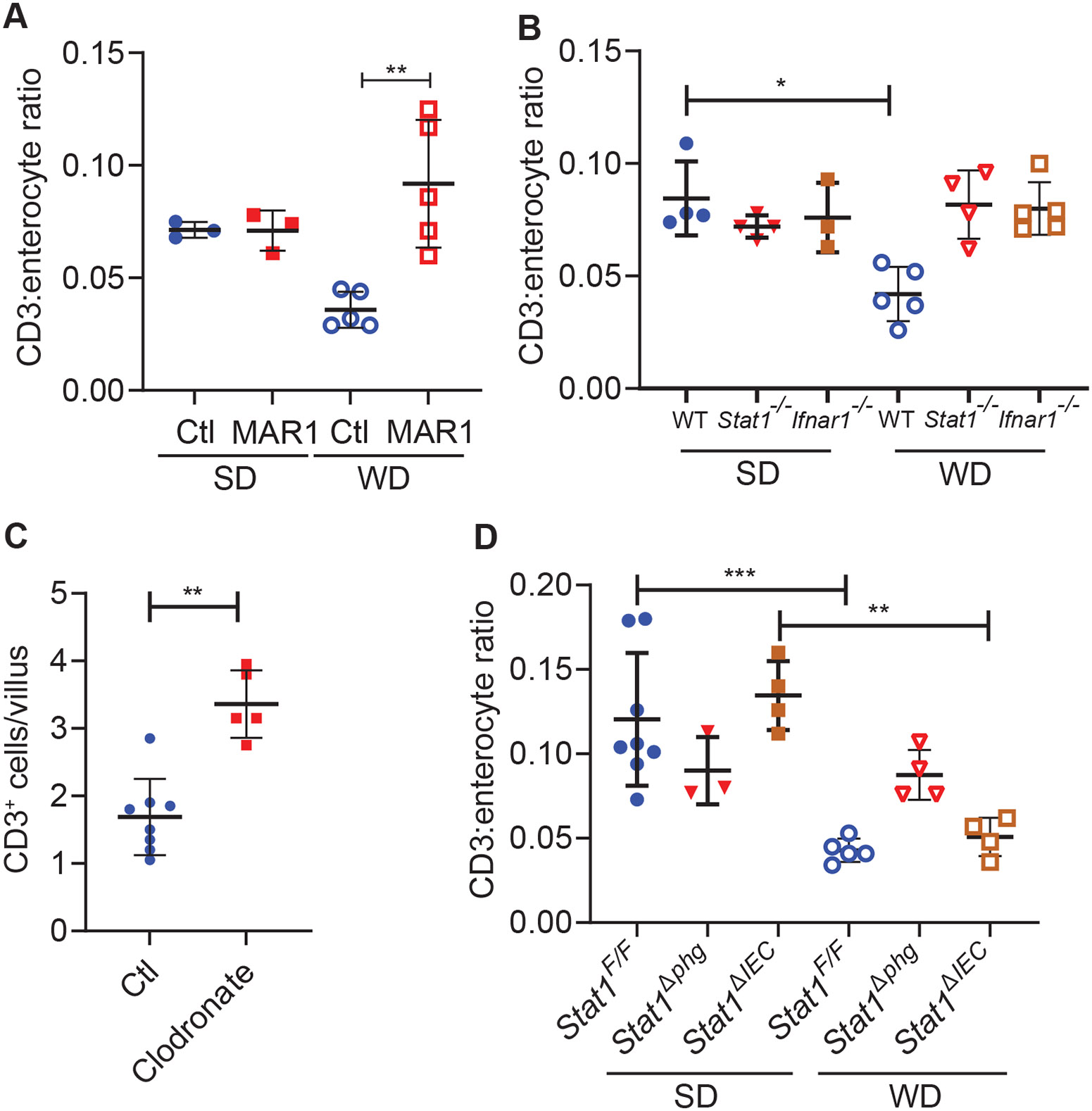
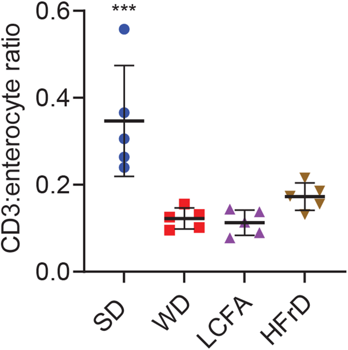
The prevalence of obesity in the United States has continued to increase over the past several decades. Understanding how diet-induced obesity modulates mucosal immunity is of clinical relevance. We previously showed that consumption of a high fat, high sugar "Western" diet (WD) reduces the density and function of small intestinal Paneth cells, a small intestinal epithelial cell type with innate immune function. We hypothesized that obesity could also result in repressed gut adaptive immunity. Using small intestinal intraepithelial lymphocytes (IEL) as a readout, we found that in non-inflammatory bowel disease (IBD) subjects, high body mass index correlated with reduced IEL density. We recapitulated this in wild type (WT) mice fed with WD. A 4-week WD consumption was able to reduce IEL but not splenic, blood, or bone marrow lymphocytes, and the effect was reversible after another 2 weeks of standard diet (SD) washout. Importantly, WD-associated IEL reduction was not dependent on the presence of gut microbiota, as WD-fed germ-free mice also showed IEL reduction. We further found that WD-mediated Farnesoid X Receptor (FXR) activation in the gut triggered IEL reduction, and this was partially mediated by intestinal phagocytes. Activated FXR signaling stimulated phagocytes to secrete type I IFN, and inhibition of either FXR or type I IFN signaling within the phagocytes prevented WD-mediated IEL loss. Therefore, WD consumption represses both innate and adaptive immunity in the gut. These findings have significant clinical implications in the understanding of how diet modulates mucosal immunity.

Keywords: Adaptive immunity; Fatty acid; Macrophages; Obesity.


Conflict of interest statement

Declaration of Competing Interest The authors declare the following financial interests/personal relationships which may be considered as potential competing interests: T. Stappenbeck advises Janssen, Boehringer Ingelheim, Kallyope, Takada, and Roche. T.C. Liu has research contracts with Denali Therapeutics and Interline Therapeutics. All other authors declare no relevant competing interests.

Figures

Fig. 1.
Fig. 1.. Small intestinal intraepithelial lymphocytes (IELs) are reduced in obese humans and wild type mice fed with western diet (WD).
Representative photomicrographs of (A) low CD4+ IEL; (B) high CD4+ IEL; (C) low CD8+ IEL; (D) high CD8+ IEL immunohistochemistry from non-IBD patient ileal resection samples. BMI has inverse correlation with the densities of (E) CD4+ and (F) CD8+ T cells. n=47. In WT mice, those fed with WD showed (G) no changes in CD45+ cells, (H) reduced CD3+ cells, but no changes in CD3+ (I) viability or (J) proliferation. There was reduced (K) TCRαβ+CD4+CD8, (L) TCRαβ+CD4CD8+, (M) TCRαβ+ IELs and not (N) TCRγδ+ IELs. Both (O) TCRγδ+CD4CD8αα+ and (P) TCRγδ+CD4CD8αβ+ cells were not changed. (Q) TCRαβ+CD4 CD8αα+ and (R) TCRαβ+CD4+CD8αα+ cells were reduced. (G-R) performed by flow cytometry with n=10/group. Statistical analysis was performed using Spearman correlation (E, F), and Mann-Whitney test (G-R). Error bars represent SEM. *: P < 0.05; **: P < 0.01; ***: P < 0.001. SI: small intestine.
Fig. 2.
Fig. 2.. Time course and reversibility of WD-induced IEL reduction.
WT mice were fed with SD or WD for 1, 2, or 4 weeks, followed by SD for another 2 weeks. IEL samples at these time points were analyzed by flow cytometry with n=5-10/group. Data presented as relative value between WD to SD groups. (A) CD3+ cells; (B) TCRαβ+CD4+CD8 cells; (C) TCRαβ+CD4CD8+ cells; (D) TCRαβ+ cells; (E) TCRαβ+CD4 CD8αα+ cells; and (F) TCRαβ+CD4+CD8αα+ cells. Statistical analysis was performed using 2-way ANOVA. Error bars represent SEM. *: P < 0.05; **: P < 0.01; ***: P < 0.001.
Fig. 3.
Fig. 3.. WD consumption reduced small intestinal IEL numbers in germ-free mice.
WD consumption in germ-free mice resulted in reduction of the total numbers of (A) CD45+ cells, (B) CD3+ cells, with (C) no changes in CD3+ viability. WD consumption did not significantly reduce (D) TCRαβ+CD4+CD8 or (E) TCRαβ+CD4CD8+ cells, but reduced (F) TCRαβ+ IELs and (G) TCRγδ+ IELs. WD also (H) depleted TCRγδ+CD4CD8αα+ cells but not (I) TCRγδ+CD4CD8αβ+ cells. (J) TCRαβ+CD4 CD8αα+ and (K) TCRαβ+CD4+CD8αα+ cells were also reduced by WD. n=3-5/group. Statistical analysis was performed using Mann-Whitney test. Error bars represent SEM. *: P < 0.05.
Fig. 4.
Fig. 4.. FXR activation in phagocytes drives WD-induced IEL reduction.
Fxr+/+ and Fxr−/− mice were fed with SD or WD for 4 weeks. Immunohistochemistry studies showed that only WD-fed Fxr+/+ mice developed depletion in (A) CD3+ and (B) CD8+ IELs. n=6-11/group. (C) FXR agonist GW4064 triggered CD3+ IEL reduction in Fxr+/+ but not Fxr−/− mice. n=4-5/group. (D) FxrΔIEC mice and (E) FxrΔhep mice were not protected from WD-induced CD3+ IEL reduction. In contrast, (F) FxrΔphg mice did not develop WD-associated CD3+ IEL reduction. (C-F) n=5/group. Statistical analysis was performed using 2-way ANOVA. Error bars represent SD. The groups labeled with the same alphabets indicate no statistically significance from each other.
Fig. 5.
Fig. 5.. Phagocyte type I IFN signaling mediates WD-associated IEL reduction.
Immunohistochemistry studies showed that: (A) Monoclonal antibody to type I IFN MAR1 prevented WD-induced IEL reduction in WT mice. n=3-5/group. (B) WD-induced IEL reduction was not observed in either Stat1−/− or Ifnar−/− mice. n=3-5/group. (C) Clodronate reduction of phagocyte rescued WD-mediated IEL reduction in WT mice. n=5-8/group. Type I IFN signaling in (D) phagocytes (n=3-6/group) rather than (E) intestinal epithelial cells (n=5/group) were responsible for WD-induced IEL reduction. Statistical analysis was performed using 2-way ANOVA (A, B) or Mann-Whitney test (C-E). Error bars represent SD. *: P < 0.05; **: P < 0.01.
Fig. 6.
Fig. 6.. High dietary fructose and LCFA could trigger IEL depletion.
WT mice fed with WD, or high fructose diet (HFrD; no increase in fat content), or LCFA-predominant high fat diet (no fructose) all showed similar degrees of IEL depletion by immunohistochemistry. n=5/group. Statistical analysis was performed using Kruskal-Wallis test. Error bars represent SD. ***: P < 0.001.
Fig. 7.
Fig. 7.. Proposed mechanism of WD-induced IEL reduction.
WD consumption activates FXR pathway, which triggers type I IFN secretion from phagocytes such as macrophages. This then reduces IEL density in the small intestine.

References

    1. Hales CM, Carroll MD, Fryar CD, Ogden CL. Prevalence of Obesity Among Adults and Youth: United States, 2015-2016. NCHS Data Brief 2017; (288): 1–8. - PubMed
    1. Wu H, Ballantyne CM. Skeletal muscle inflammation and insulin resistance in obesity. The Journal of clinical investigation 2017; 127(1): 43–54. - PMC - PubMed
    1. Gruchala-Niedoszytko M, Malgorzewicz S, Niedoszytko M, Gnacinska M, Jassem E. The influence of obesity on inflammation and clinical symptoms in asthma. Adv Med Sci 2013; 58(1): 15–21. - PubMed
    1. Reilly SM, Saltiel AR. Adapting to obesity with adipose tissue inflammation. Nat Rev Endocrinol 2017; 13(11): 633–643. - PubMed
    1. Liu TC, Kern JT, Jain U, Sonnek NM, Simpson KF, VanDussen KL et al. Western diet induces Paneth cell defects through microbiome alterations and Farnesoid X Receptor and type I interferon activation. Cell host & microbe 2021; 29(6): 998–1001. - PMC - PubMed

MeSH terms

Substances