. 2024 Aug;300(8):107541.
doi: 10.1016/j.jbc.2024.107541. Epub 2024 Jul 9.

Eta-secretase-like processing of the amyloid precursor protein (APP) by the rhomboid protease RHBDL4

Affiliations

Eta-secretase-like processing of the amyloid precursor protein (APP) by the rhomboid protease RHBDL4

Ylauna Christine Mégane Penalva et al. J Biol Chem. 2024 Aug.

Abstract

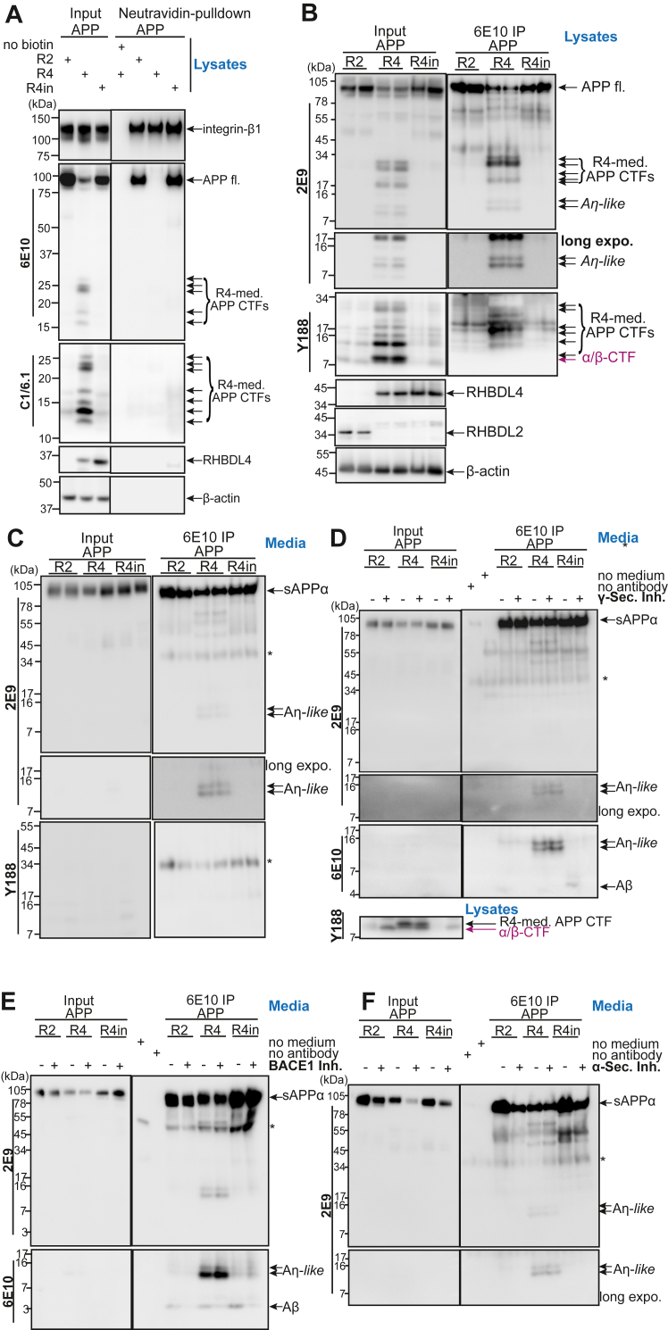
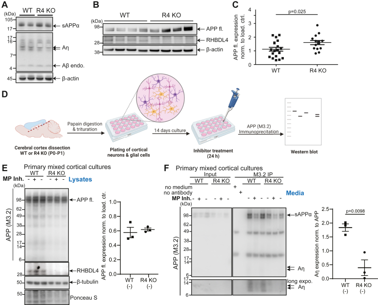
The amyloid precursor protein (APP) is a key protein in Alzheimer's disease synthesized in the endoplasmic reticulum (ER) and translocated to the plasma membrane where it undergoes proteolytic cleavages by several proteases. Conversely, to other known proteases, we previously elucidated rhomboid protease RHBDL4 as a novel APP processing enzyme where several cleavages likely occur already in the ER. Interestingly, the pattern of RHBDL4-derived large APP C-terminal fragments resembles those generated by the η-secretase or MT5-MMP, which was described to generate so-called Aη fragments. The similarity in large APP C-terminal fragments between both proteases raised the question of whether RHBDL4 may contribute to η-secretase activity and Aη-like fragments. Here, we identified two cleavage sites of RHBDL4 in APP by mass spectrometry, which, intriguingly, lie in close proximity to the MT5-MMP cleavage sites. Indeed, we observed that RHBDL4 generates Aη-like fragments in vitro without contributions of α-, β-, or γ-secretases. Such Aη-like fragments are likely generated in the ER since RHBDL4-derived APP-C-terminal fragments do not reach the cell surface. Inherited, familial APP mutations appear to not affect this processing pathway. In RHBDL4 knockout mice, we observed increased cerebral full-length APP in comparison to wild type (WT) in support of RHBDL4 being a physiologically relevant protease for APP. Furthermore, we found secreted Aη fragments in dissociated mixed cortical cultures from WT mice, however significantly fewer Aη fragments in RHBDL4 knockout cultures. Our data underscores that RHBDL4 contributes to the η-secretease-like processing of APP and that RHBDL4 is a physiologically relevant protease for APP.

Keywords: Alzheimer’s disease; Aη; MMP24; MT5-MMP; RHBDD1; RHBDL4; amyloid precursor protein (APP); eta-secretase; rhomboid protease.


Conflict of interest statement

Conflict of interest The authors declare that they have no conflicts of interest with the contents of this article

Figures

Figure 1
Figure 1
Identification of RHBDL4 cleavage sites in APP. A, identification of RHBDL4 cleavage sites by mass spectrometry. Immunoprecipitation of N-terminally myc-tagged APP fragments after co-transfection with either active or inactive (inac.) RHBDL4 (R4) in HEK293T cells. Samples were digested with LysC and analyzed by electrospray ionization mass spectrometry (ESI-MS). Representative extracted ion chromatograms showing retention times for different identified peptides. The table lists the identified retention time per peak, the peptide mass per charge (m/z), and peptide sequences along with APP695 amino acid numbering for fragments or cleavage sites. Due to subtle differences in the automated injections, the retention time of the complete LysC peptide differs between the samples containing inactive RHBDL4 (55.7 min) and active RHBDL4 (56.4 min). B, schematic representation of the identified RHBDL4 cleavage sites in APP created with BioRender.com. The previously identified η-secretase cleavage site, as well as conventional APP processing enzymes, are indicated, scheme is not to scale. Antibody binding sites for 6E10, M3.2, 2E9, 22C11, Y188 and C1/6.1 antibodies are indicated. C–E, analysis of RHBDL4-mediated (R4-med.) cleavage of the APP deletion (APPΔ) mutant. Amino acid stretches comprising two amino acids N- and C-terminal of both identified cleavage sites were deleted (as shown in C). Comparison of RHBDL4 cleavage pattern for APP WT and APPΔ upon co-transfection. Different gel systems were used to optimally analyse the fragments, 4 to 12% bis-tris (D), 8% tris-glycine (upper panel E) and 10 to 20% tris-tricine (lower panel E). Blue arrows indicate novel bands in the APPΔ samples. Detection of APP full length (APP fl.) and APP ectodomain (APP ecto.) with 22C11, CTFs with 6E10 and Y188; RHBDL4 with anti-myc antibody. β-actin or tubulin as loading controls. A representative Western blot of three individual experiments is shown. F, schematic representation of the luciferase constructs used for the RHBDL4 activity assay. All constructs are N-terminally tagged with a Flag sequence. GLuc-APP-KDEL and GLuc-KDEL contain the ER-retention motif KDEL at their C terminus. GLuc-APP-KDEL contains the APP sequence with the RHBDL4 cleavage sites. G and H, luciferase activity measured in the cell culture supernatant (luciferase released from ER, (G) or in cell lysates (H). GLuc-APP-KDEL (green bars) only yields extracellular luciferase activity when co-expressed with RHBDL4 (R4), but not with RHBDL1 (R1) or inactive RHBDL4. GLuc-KDEL (dark grey) is not cleaved by RHBDL4 and yields only luciferase activity in the lysate (ER retained). GLuc (light grey) is constitutively secreted and serves as a positive control. R1 + GLuc luminescence signal (G) or R4 + GLuc-KDEL (H) was set to one for normalization to plot other conditions as a fold change between biological replicates. Mean ± SEM is displayed, n = 4 to 5, one-way ANOVA (p < 0.0001) with Tukey’s multiple comparison test. Selected statistical differences are indicated. I, detection of GLuc constructs with mouse anti-flag antibody by Western blot; RHBDL4 and RHBDL1 with direct antibodies and β-actin as a loading control. A representative Western blot of three individual experiments is shown.
Figure 2
Figure 2
Familial APP mutations do not affect the RHBDL4-mediated processing of APP.A and B, RHBDL4-mediated (R4-med.) processing of familial AD mutants of APP. Co-transfection of various familial APP mutants with active RHBDL4. Detection of APP full length (APP fl.) and APP ectodomain (APP ecto.) with 22C11, CTFs with 6E10 and Y188; RHBDL4 with anti-myc antibody. β-actin as loading controls. APP-CTFs were quantified and normalized first to β-actin and then the fold change compared to WT was calculated. WT was always set to one in each individual experiment (green dashed line). Mean ± SEM is displayed, n = 3 to 6, p values for Bonferroni-corrected one sample t-tests are reported.
Figure 3
Figure 3
RHBDL4 generates Aη-like peptides in vitro.A, investigation of RHBDL4-mediated APP-CTFs at the cell surface using cell surface biotinylation. Co-transfection of APP and RHBDL2 (R2), active RHBDL4 (R4), or inactive RHBDL4 (R4in). The input consists of lysates without neutravidin to serve as loading controls (left panels). Biotinylated cell surface proteins were pulled down using neutravidin (right panels). Integrin-β1 is a positive control for successful pulldown of plasma membrane proteins. Detection of APP fl. with 6E10, CTFs with 6E10 and C1/6.1; RHBDL4 with rabbit anti-RHBDL4 antibody; Integrin-β1 with rabbit anti-integrin-β1 antibody. Representative Western blot of three individual experiments is shown. B and C, immunoprecipitation of Aη species from cell culture supernatant and lysate. Total cell culture lysates or supernatant are used as input (left panels) while immunoprecipitation (IP) was performed using the 6E10 antibody (right panels). Detection of APP full length (fl.), sAPP⍺ and Aη with 2E9, CTFs with Y188; RHBDL4 with rabbit-anti-RHBDL4 antibody; RHBDL2 with mouse-anti-flag antibody and β-actin as a loading control. A representative Western blot of three individual experiments is shown. D–F, RHBDL4-mediated Aη generation is independent of canonical processing by ⍺-, β- or γ-secretases. Cells were treated with either ⍺-secretase inhibitor (⍺-Sec. Inh.), BACE-1 inhibitor (BACE1 Inh.) or γ-secretase inhibitor (γ-sec. Inh). Total cell culture supernatant is used as input (left panels) while immunoprecipitation (IP) was performed using the 6E10 antibody (right panels). Detection of sAPP⍺ and Aη with 2E9 and 6E10. Representative western blots of each inhibitor experiment are shown, n = 3 per inhibitor. Asterix indicates signals derived from the antibody used in the immunoprecipitation.
Figure 4
Figure 4
RHBDL4 knockout affects APP levels and Aη production in vivo.A, Aη extraction was performed according to (18), extraction of soluble proteins upon DEA extraction from brain tissue homogenates of WT and RHBDL4 KO mice at 10 to 11 months of age. sAPPα and Aη were detected with M3.2 antibody (specific for mouse Aβ), β-actin as loading control. Representative Western blot for n = 8 brain samples (WT and KO, each). B and C, full-length APP levels in brain tissue lysates of 10 to 11 months old RHBDL4 knockout (R4 KO) mice as compared to age-matched wild-type (WT) mice. Equal amounts of protein were loaded per lane. Detection of APP full length (fl) with 22C11, endogenous RHBDL4 with rabbit anti-RHBDL4 antibody, and β-actin as a loading control. Quantification with ImageJ, normalized to β-actin, mean ± SEM, n = 14 to 19, p-value for unpaired two-tailed t test is reported. D, schematic representation of cortical dissociation and primary cell culture procedure followed by Aη immunoprecipitation from cell medium and downstream analysis via Western blot. Created using BioRender. E, full-length APP expression from primary cortical cell culture lysates prepared from WT or RHBDL4 knockout brains. Representative Western blot of three individual experiments is shown; APP quantification of untreated condition normalized to ponceau S with ImageJ, mean ± SEM, unpaired two-tailed t test performed. Detection of APP full length (fl.) with M3.2, endogenous RHBDL4 with rabbit-anti-RHBDL4 antibody, β-tubulin, and ponceau S as loading controls. F, Immunoprecipitation of Aη species from primary cortical cell culture supernatant prepared from WT or RHBDL4 knockout mouse brains. Input consists of total cell culture supernatant (left panels) while immunoprecipitation (IP) was performed using the M3.2 antibody (right panels). sAPP and Aη detection using M3.2 antibody. Representative Western blot of three individual experiments is shown; quantification of untreated condition with ImageJ normalized to full-length APP from lysates, mean ± SEM, the p-value for unpaired two-tailed t test is reported. Treatment with metalloprotease inhibitor (MP Inh.) TIMP2.
Figure 5
Figure 5
Scheme of APP processing and Aη formation in different compartments. In the absence of RHBDL4, full-length APP traffics to the cell surface where MT5-MMP as well as α- or β-secretases will process APP to generate Aη at the cell surface (left panels). In the presence of RHBDL4, APP will be cleaved by RHBDL4 in the ER, and RHBDL4-derived large APP C-terminal fragments do not reach the cell surface. RHBDL4-derived Aη-like peptides are directly generated in the ER (right panels).

References

    1. Duran-Aniotz C., Hetz C. Glucose metabolism: a sweet relief of Alzheimer's disease. Curr. Biol. 2016;26:R806–R809. - PubMed
    1. Iturria-Medina Y., Sotero R.C., Toussaint P.J., Mateos-Perez J.M., Evans A.C., Weiner M.W., et al. Early role of vascular dysregulation on late-onset Alzheimer's disease based on multifactorial data-driven analysis. Nat. Commun. 2016;7 - PMC - PubMed
    1. Puglielli L., Tanzi R.E., Kovacs D.M. Alzheimer's disease: the cholesterol connection. Nat. Neurosci. 2003;6:345–351. - PubMed
    1. Spillantini M.G., Goedert M. Tau pathology and neurodegeneration. Lancet Neurol. 2013;12:609–622. - PubMed
    1. Winblad B., Amouyel P., Andrieu S., Ballard C., Brayne C., Brodaty H., et al. Defeating Alzheimer's disease and other dementias: a priority for European science and society. Lancet Neurol. 2016;15:455–532. - PubMed

Substances

LinkOut - more resources