. 2024 Jul 11;15(1):208.
doi: 10.1186/s13287-024-03806-0.

Modified mesenchymal stromal cells by in vitro transcribed mRNA: a therapeutic strategy for hepatocellular carcinoma

Affiliations

Modified mesenchymal stromal cells by in vitro transcribed mRNA: a therapeutic strategy for hepatocellular carcinoma

María José Cantero et al. Stem Cell Res Ther. .

Abstract

Background: Mesenchymal stromal cells (MSCs) tropism for tumours allows their use as carriers of antitumoural factors and in vitro transcribed mRNA (IVT mRNA) is a promising tool for effective transient expression without insertional mutagenesis risk. Granulocyte-macrophage colony-stimulating factor (GM-CSF) is a cytokine with antitumor properties by stimulating the specific immune response. The aim of this work was to generate modified MSCs by IVT mRNA transfection to overexpress GM-CSF and determine their therapeutic effect alone or in combination with doxorubicin (Dox) in a murine model of hepatocellular carcinoma (HCC).

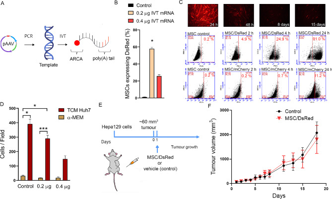
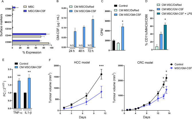
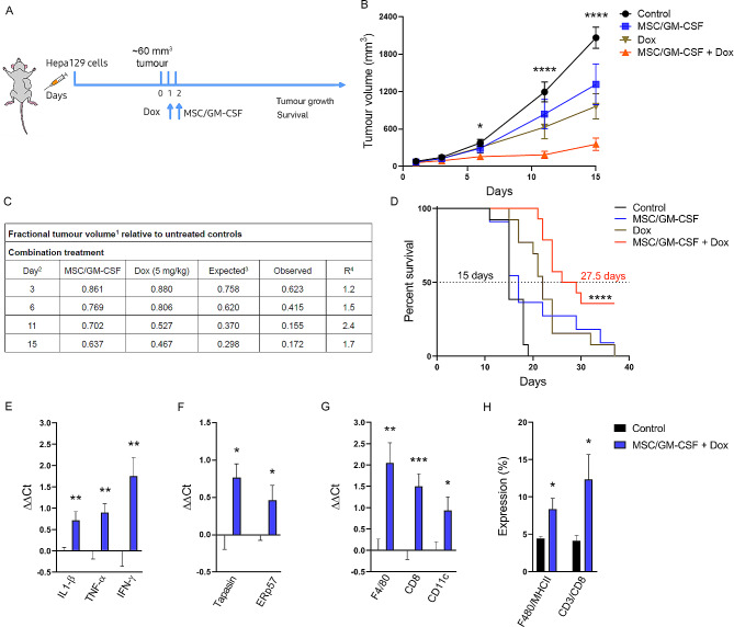
Methods: DsRed or GM-CSF IVT mRNAs were generated from a cDNA template designed with specific primers followed by reverse transcription. Lipofectamine was used to transfect MSCs with DsRed (MSC/DsRed) or GM-CSF IVT mRNA (MSC/GM-CSF). Gene expression and cell surface markers were determined by flow cytometry. GM-CSF secretion was determined by ELISA. For in vitro experiments, the J774 macrophage line and bone marrow monocytes from mice were used to test GM-CSF function. An HCC model was developed by subcutaneous inoculation (s.c.) of Hepa129 cells into C3H/HeN mice. After s.c. injection of MSC/GM-CSF, Dox, or their combination, tumour size and mouse survival were evaluated. Tumour samples were collected for mRNA analysis and flow cytometry.

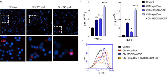
Results: DsRed expression by MSCs was observed from 2 h to 15 days after IVT mRNA transfection. Tumour growth remained unaltered after the administration of DsRed-expressing MSCs in a murine model of HCC and MSCs expressing GM-CSF maintained their phenotypic characteristic and migration capability. GM-CSF secreted by modified MSCs induced the differentiation of murine monocytes to dendritic cells and promoted a proinflammatory phenotype in the J774 macrophage cell line. In vivo, MSC/GM-CSF in combination with Dox strongly reduced HCC tumour growth in C3H/HeN mice and extended mouse survival in comparison with individual treatments. In addition, the tumours in the MSC/GM-CSF + Dox treated group exhibited elevated expression of proinflammatory genes and increased infiltration of CD8 + T cells and macrophages.

Conclusions: Our results showed that IVT mRNA transfection is a suitable strategy for obtaining modified MSCs for therapeutic purposes. MSC/GM-CSF in combination with low doses of Dox led to a synergistic effect by increasing the proinflammatory tumour microenvironment, enhancing the antitumoural response in HCC.

Keywords: Granulocyte-macrophage colony-stimulating factor; Hepatocellular carcinoma; Immunogenic cell death; Immunotherapy; In vitro transcribed mRNA; Mesenchymal stromal cell.


Conflict of interest statement

The authors declare no competing interests.

Figures

Fig. 1
Fig. 1
Transfection with IVT mRNA is a suitable strategy for obtaining engineered MSCs. A Scheme of IVT mRNA design. The coding sequence (CDS) of DsRed was amplified via PCR from the pAAV plasmid to generate the template. In vitro transcription (IVT) requires a mixture of nucleosides triphosphate (ATP, GTP, methylcytidine-5’-triphosphate (MeCTP), pseudouridine-5’-triphosphate (PseudoUTP)) and 3´-O-Me-m7G (5´) ppp(5´)G RNA cap structure analog (ARCA) to obtain a complete IVT mRNA. B Expression of DsRed was analysed by flow cytometry in MSCs transfected with different amounts of IVT mRNA (0.2–0.4 µg). Dunn’s multiple comparisons test, *p < 0.05 vs. control. C Representative images by fluorescence microscopy of MSCs transfected with 0.2 µg of IVT mRNA of DsRed at 24 h, 48 h, 8 days and 15 days (top panel). Kinetics of IVT mRNA expression determined by flow cytometry (middle panel) in comparison with the plasmid transfection method (bottom panel) at 2 h, 4 h and 24 h. D Untransfected MSCs (control) or MSC/DsRed transfected with 0.2–0.4 µg of IVT mRNA were evaluated for their ability to migrate toward tumour conditioned medium derived from Huh7 cells (TCM Huh7) or α-MEM in a modified Boyden chamber. Dunn’s multiple comparisons test, *p < 0.05 and **p < 0.01 vs. α-MEM (basal migration). E Experimental model: C3H/HeN mice were subcutaneously (s.c.) injected with 1 × 106 syngeneic Hepa129 cells, and when the tumour reached ~60 mm3 (day 0), 2 × 105 MSC/DsRed or saline (control) was s.c. injected (day 1). F In vivo tumour growth of Hepa129 tumour-bearing mice with vehicle (control) or MSC/DsRed. Two-way ANOVA and Sidak’s multiple comparisons test
Fig. 2
Fig. 2
MSCs engineered to overexpress GM-CSF by IVT mRNA. A Percentage of expression of surface markers in untransfected MSCs (MSC) and MSCs transfected with 0.2 µg IVT mRNA / 4 × 104 cells (MSC/GM-CSF) analysed by flow cytometry. Sidak’s multiple comparisons test was not significant for any marker. B GM-CSF production in the conditioned media of MSC/DsRed (CM MSC/DsRed) or MSC/GM-CSF (CM MSC/GM-CSF) analysed by ELISA. C In vitro proliferation of murine splenocytes cultured with ConA plus CM MSC/GM-CSF, CM MSC/DsRed or RPMI (control). Cell proliferation was evaluated by [3H]-thymidine incorporation assay and results are expressed as counts per minute (CPM). Dunn’s multiple comparisons test, *p < 0.05 vs. control. D Analysis of DCs surface markers (CD11c, MHCII and CD86) in murine bone marrow cells cultured with CM MSC/DsRed, CM MSC/GM-CSF or CM MSC/GM-CSF plus LPS. Dunn’s multiple comparisons test, *p < 0.05 vs. CM MSC/DsRed. E mRNA expression of TNF-α and IL-1β in J774 cells stimulated with CM from MSC/GM-CSF or RPMI as control determined by RT-qPCR. Mann-Whitney test, **p < 0,001 vs. control. F Tumour growth in the HCC mouse model (left) or colorectal carcinoma murine model (right). When the tumours reached ~60 mm3 peritumoral injection of 2 × 105 MSC/GM-CSF or PBS (control) was administered. Two-way ANOVA and Sidak’s comparison test, *p < 0.05 and ***p < 0.001 vs. control
Fig. 3
Fig. 3
Effects of MSC/GM-CSF combined with a low dose of Dox. A Immunofluorescence of calreticulin (CRT, red) in Hepa129 cells after incubation for 48 h with 20 or 30 µM Dox or without treatment (control). Scale bar, 100 μm. B Analysis of TNF-α and IL-1β mRNA expression in J774 cells after 30 h of incubation with CM Hepa/Dox, CM MSC/GM-CSF, CM Hepa/Dox + CM MSC/GM-CSF or RPMI as control. ANOVA and Tukey’s post test, *p < 0,05, ***p < 0,0001 vs. control. C Representative flow cytometry image showing CD86 expression in J774 cells after 30 h of incubation with CM Hepa/Dox, CM MSC/GM-CSF, CM Hepa/Dox + CM MSC/GM-CSF or RPMI as control
Fig. 4
Fig. 4
Synergistic inhibition of tumour growth by combination treatment of MSC/GM-CSF with a low dose of Dox. A Scheme of treatment for in vivo experiments. C3H/HeN mice were subcutaneously (s.c.) injected with 1 × 106 syngeneic Hepa129 cells, and when the tumour reached ~60 mm3 (day 0), mice received Dox (5 mg/kg, day 1), 2 × 105 MSC/GM-CSF (day2), both treatments (MSC/GM-CSF + Dox) or saline (control). B Tumour growth of Hepa129 tumour-bearing mice. Two-way ANOVA, *p < 0.05 and ****p < 0.0001 vs. control group. C Analysis of the in vivo interaction between MSC/GM-CSF and Dox by the fractional product method (FTV) in the HCC model. 1FTV (experimental mean tumour volume) / (control mean tumour volume); 2Day after treatment onset; 3(MSC/GM mean FTV) x (Dox mean FTV); 4R = [Expected FTV/Observed FTV]. A ratio > 1 indicates a synergistic effect, and a ratio < 1 indicates a less than additive effect. D Survival Kaplan-Meier curve, ****p < 0.0001 vs. control (log rank test). Analysis of mRNA expression of IL-1β, TNF-α and IFN-γ (E), tapasin and ERp57 (F) or F4/80, CD8 and CD11c (G) in mouse tumours 7 days after combination treatment (MSC/GM-CSF + Dox) or vehicle administration (control). Unpaired t test, *p < 0.05, **p < 0.01 and ***p < 0.001 vs. control. H Quantification of F4/80+/MHCII+ and CD3+/CD8+ cells by flow cytometry. Unpaired t test, *p < 0.05 vs. control

References

    1. Dominici M, Le Blanc K, Mueller I, Slaper-Cortenbach I, Marini FC, Krause DS et al. Minimal criteria for defining multipotent mesenchymal stromal cells. The International Society for Cellular Therapy position statement. Cytotherapy [Internet]. 2006;8:315–7. 10.1080/14653240600855905 - PubMed
    1. Bernardo ME, Fibbe WE. Mesenchymal stromal cells: Sensors and switchers of inflammation. Cell Stem Cell [Internet]. 2013;13:392–402. 10.1016/j.stem.2013.09.006 - PubMed
    1. Di Valfrè L, Ferrero I, Cravanzola C, Mareschi K, Rustichell D, Novo E, et al. Human mesenchymal stem cells as a two-edged sword in hepatic regenerative medicine: Engraftment and hepatocyte differentiation versus profibrogenic potential. Gut. 2008;57:223–31. doi: 10.1136/gut.2006.111617. - DOI - PubMed
    1. Hass R, Kasper C, Böhm S, Jacobs R. Different populations and sources of human mesenchymal stem cells (MSC): a comparison of adult and neonatal tissue-derived MSC. Cell Communication Signal. 2011;9:1–14. doi: 10.1186/1478-811X-9-12. - DOI - PMC - PubMed
    1. Sarugaser R, Lickorish D, Baksh D, Hosseini MM, Davies JE. Human umbilical cord Perivascular (HUCPV) cells: a source of mesenchymal progenitors. Stem Cells. 2005;23:220–9. doi: 10.1634/stemcells.2004-0166. - DOI - PubMed

Publication types

MeSH terms

Substances