

Right ventricular diastolic adaptation to pressure overload in different rat strains
- PMID: 38993022
- PMCID: PMC11239975
- DOI: 10.14814/phy2.16132
Right ventricular diastolic adaptation to pressure overload in different rat strains
Abstract
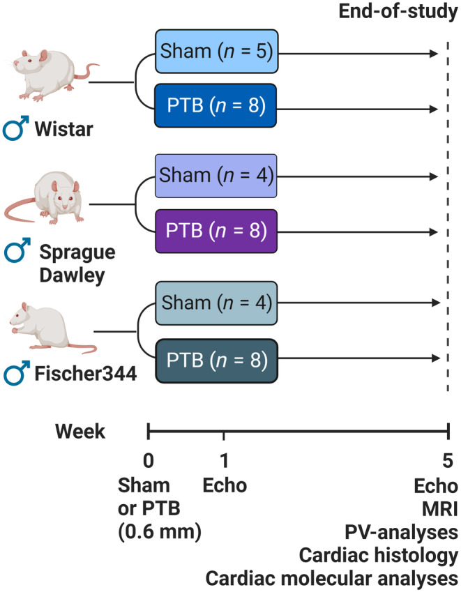
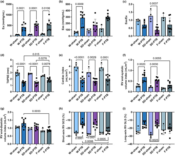
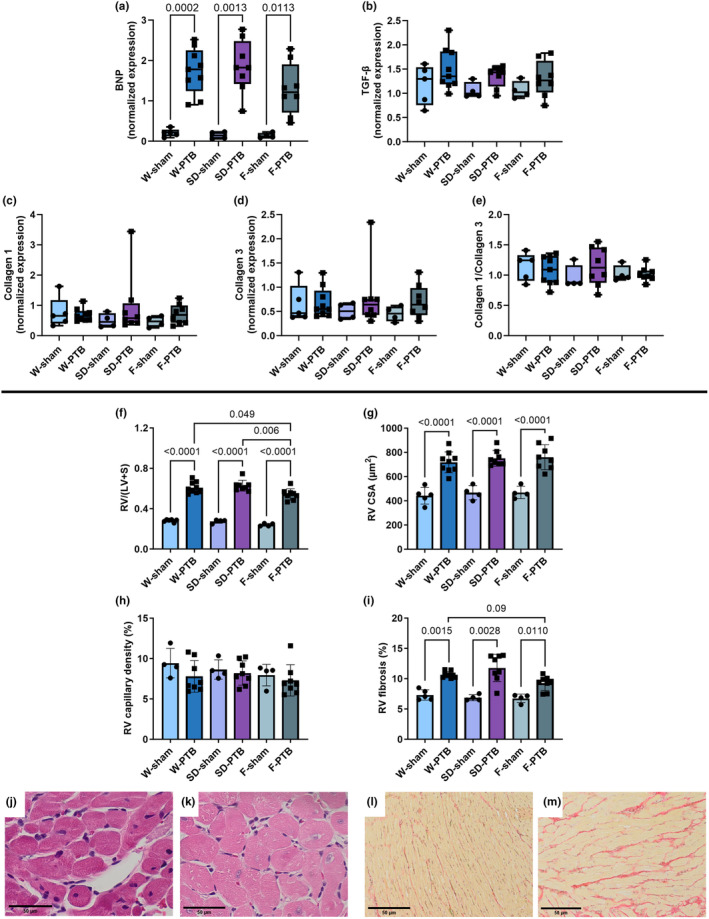
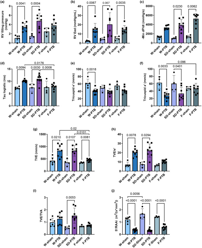
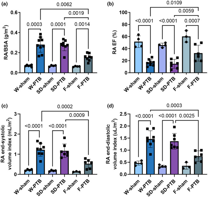
Different rat strains are used in various animal models of pulmonary hypertension and right ventricular (RV) failure. No systematic assessment has been made to test differences in RV response to pressure overload between rat strains. We compared RV adaptation to pulmonary trunk banding (PTB) in Wistar (W), Sprague Dawley (SD), and Fischer344 (F) rats by hemodynamic profiling focusing on diastolic function. Age-matched male rat weanlings were randomized to sham surgery (W-sham, n = 5; SD-sham, n = 4; F-sham, n = 4) or PTB (W-PTB, n = 8; SD-PTB, n = 8; F-PTB, n = 8). RV function was evaluated after 5 weeks by echocardiography, cardiac MRI, and invasive pressure-volume measurements. PTB caused RV failure and increased RV systolic pressures four-fold in all three PTB groups compared with sham. W- and SD-PTB had a 2.4-fold increase in RV end-systolic volume index compared with sham, while F-PTB rats were less affected. Diastolic and right atrial impairment were evident by increased RV end-diastolic elastance, filling pressure, and E/e' in PTB rats compared with sham, again F-PTB the least affected. In conclusions, PTB caused RV failure with signs of diastolic dysfunction. Despite a similar increase in RV systolic pressure, F-PTB rats showed less RV dilatation and a more preserved diastolic function compared with W- and SD-PTB.
Keywords: diastolic dysfunction; pulmonary trunk banding; rat strains; right ventricular failure.
© 2024 The Author(s). Physiological Reports published by Wiley Periodicals LLC on behalf of The Physiological Society and the American Physiological Society.
Conflict of interest statement
Prof.dr de Man has received research grant support from Janssen and BIAL. Asger Andersen has received research grant support from BIAL and teaching honorariums from EPS vascular, ABBOTT, Angiodynamics, Inari Medical, Gore Medical, and Janssen. All other authors have declared that they have no conflict of interest, financial, or otherwise.
Figures
References
-
- Alaa, M. , Abdellatif, M. , Tavares‐Silva, M. , Oliveira‐Pinto, J. , Lopes, L. , Leite, S. , Leite‐Moreira, A. F. , & Lourenço, A. P. (2016). Right ventricular end‐diastolic stiffness heralds right ventricular failure in monocrotaline‐induced pulmonary hypertension. American Journal of Physiology Heart and Circulatory Physiology, 311, H1004–H1013. - PubMed
-
- Andersen, S. , Schultz, J. G. , Holmboe, S. , Axelsen, J. B. , Hansen, M. S. , Lyhne, M. D. , Nielsen‐Kudsk, J. E. , & Andersen, A. (2018). A pulmonary trunk banding model of pressure overload induced right ventricular hypertrophy and failure. Journal of Visualized Experiments: JoVE, 141, 1–7. - PubMed
-
- Axell, R. G. , Hoole, S. P. , Hampton‐Till, J. , & White, P. A. (2015). RV diastolic dysfunction: Time to re‐evaluate its importance in heart failure. Heart Failure Reviews, 20, 363–373. - PubMed
-
- Bochnowicz, S. , Osborn, R. R. , Luttmann, M. A. , Louden, C. , Hart, T. , Hay, D. W. , & Underwood, D. C. (2000). Differences in time‐related cardiopulmonary responses to hypoxia in three rat strains. Clinical and Experimental Hypertension (New York, NY: 1993), 22, 471–492. - PubMed
-
- Boehm, M. , Lawrie, A. , Wilhelm, J. , Ghofrani, H. A. , Grimminger, F. , Weissmann, N. , Seeger, W. , Schermuly, R. T. , & Kojonazarov, B. (2017). Maintained right ventricular pressure overload induces ventricular–arterial decoupling in mice. Experimental Physiology, 102, 180–189. - PubMed
MeSH terms
Grants and funding
- 20-R140-A9671-22167/Hjerteforeningen (Heart Foundation)
- 2017-1064/96/The Danish Medical Research Grant
- 20-L-0184/A.P. Moeller Fonden
- DC472123-004-lek/Helga og Peter Kornings Fond (Helga and Peter Korning's Fund)
- 25/11-2021/Soester og Verner Lipperts Fond
- J167/1/Snedkermester Sophus Jacobsen og Hustru Astrid Jacobsens Fond
- CVON-2018-29/The Netherlands Cardiovascular Research Initiative
- CVON-2017-10/The Netherlands Cardiovascular Research Initiative
- NWO-VIDI: 917.18.338/The Netherlands organization for scientific research
- 2018t059/The Dutch Heart Foundation dekker senior postdoc grant
LinkOut - more resources
Full Text Sources
