. 2025 Nov 1;20(11):3273-3286.
doi: 10.4103/NRR.NRR-D-24-00068. Epub 2024 Jul 10.

Exosomes originating from neural stem cells undergoing necroptosis participate in cellular communication by inducing TSC2 upregulation of recipient cells following spinal cord injury

Affiliations

Exosomes originating from neural stem cells undergoing necroptosis participate in cellular communication by inducing TSC2 upregulation of recipient cells following spinal cord injury

Shiming Li et al. Neural Regen Res. .

Abstract

JOURNAL/nrgr/04.03/01300535-202511000-00030/figure1/v/2024-12-20T164640Z/r/image-tiff We previously demonstrated that inhibiting neural stem cells necroptosis enhances functional recovery after spinal cord injury. While exosomes are recognized as playing a pivotal role in neural stem cells exocrine function, their precise function in spinal cord injury remains unclear. To investigate the role of exosomes generated following neural stem cells necroptosis after spinal cord injury, we conducted single-cell RNA sequencing and validated that neural stem cells originate from ependymal cells and undergo necroptosis in response to spinal cord injury. Subsequently, we established an in vitro necroptosis model using neural stem cells isolated from embryonic mice aged 16-17 days and extracted exosomes. The results showed that necroptosis did not significantly impact the fundamental characteristics or number of exosomes. Transcriptome sequencing of exosomes in necroptosis group identified 108 differentially expressed messenger RNAs, 104 long non-coding RNAs, 720 circular RNAs, and 14 microRNAs compared with the control group. Construction of a competing endogenous RNA network identified the following hub genes: tuberous sclerosis 2 ( Tsc2 ), solute carrier family 16 member 3 ( Slc16a3 ), and forkhead box protein P1 ( Foxp1 ). Notably, a significant elevation in TSC2 expression was observed in spinal cord tissues following spinal cord injury. TSC2-positive cells were localized around SRY-box transcription factor 2-positive cells within the injury zone. Furthermore, in vitro analysis revealed increased TSC2 expression in exosomal receptor cells compared with other cells. Further assessment of cellular communication following spinal cord injury showed that Tsc2 was involved in ependymal cellular communication at 1 and 3 days post-injury through the epidermal growth factor and midkine signaling pathways. In addition, Slc16a3 participated in cellular communication in ependymal cells at 7 days post-injury via the vascular endothelial growth factor and macrophage migration inhibitory factor signaling pathways. Collectively, these findings confirm that exosomes derived from neural stem cells undergoing necroptosis play an important role in cellular communication after spinal cord injury and induce TSC2 upregulation in recipient cells.


Conflict of interest statement

Conflicts of interest: The authors declare that they have no competing financial interests or personal relationships that could have appeared to influence the work reported in this paper.

Figures

Figure 1
Figure 1
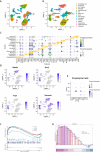
Identification of NSCs mainly derived from ependymal cells and undergoing necroptosis. (A) UMAP plot of single-cell type clusters in SCI. (B) Annotated UMAP plot of single-cell type clusters in SCI. (C) Bubble plot of cell type marker gene expression in SCI. (D) Expression of NSC-related marker genes as determined by single-cell RNA-sequencing. (E) The expression of key genes and ligand receptor–related genes associated with necroptosis in ependymal cells was examined in the sham group and 1, 3, 7 days following SCI. (F) GSEA of PCD in ependymal cells (3 days post-SCI vs. control). (G) Enrichment of key genes and ligand receptor–related genes associated with necroptosis as assessed by GSEA. GSEA: Gene set enrichment analysis; NSC: neural stem cell; PCD: programmed cell death; SCI: spinal cord injury; UMAP: uniform manifold approximation and projection.
Figure 2
Figure 2
Validation of the necroptosis model and characterization of NSCs and exosomes. (A) Characterization of NSC neurospheres. The NSCs displayed a typical spherical neurosphere morphology. Scale bars: 500 μm (upper) and 100 μm (lower). (B) Representative immunofluorescence images of SOX2 (green, Alexa Fluor 488) and Nestin (red, Alexa Fluor 594) expression in NSCs. Both neurospheres and individual NSCs expressed SOX2 and Nestin. Cell nuclei were counterstained with DAPI (blue). Scale bar: 100 μm. (C) NSCs were treated with DMSO or TSZ for 8 hours, after which cell death was analyzed by flow cytometry. (D) Exosome morphology as assessed by transmission electron microscopy. The exosomes in both the NSC-control and NSC-TSZ groups exhibited a typical one-sided semi-concave bilayer membrane structure. Scale bars: 200 nm. (E, F) Nanoflow cytometry detection of particle size distribution, concentration, and exosome surface marker expression. DAPI: 4′,6-Diamidino-2-phenylindole; DMSO: dimethyl sulfoxide; NSC: neural stem cell; SOX2: SRY-box transcription factor 2; TSZ: tumor necrosis factor α, SMAC mimetic, zVAD-fmk.
Figure 3
Figure 3
WTS network construction and the identification and validation of hub genes. (A, B) Construction of the WTS regulatory network, which was subsequently divided into two ceRNA networks (down-up-down/up-down-up). In these networks, circles represent miRNAs, squares represent mRNAs, and triangles represent lncRNAs and circRNAs. Red indicates up-regulation, while green indicates down-regulation. (C) Bidirectional hierarchical clustering maps of hub genes between the control and TSZ groups. (D) Expression of hub genes at different time points following SCI, as assessed by single-cell RNA-sequencing. ceRNA: Competing endogenous RNA; circRNA: circular RNA; lncRNA: long non-coding RNA; miRNA: microRNA; SCI: spinal cord injury; WTS: whole-transcriptome sequencing.
Figure 4
Figure 4
Western blot and immunofluorescence analysis of the expression of proteins encoded by hub genes in the spinal cord 3 days following injury. (A, B) Western blot and semi-quantitative analyses of the expression of proteins encoded by hub genes in the spinal cord 3 days after SCI. Data are expressed as mean ± SD (n = 3), and were analyzed by two-tailed unpaired Student’s t-test. (C) Immunofluorescence staining for the NSC marker SOX2 (green, Alexa Fluor 488) and the hub gene product TSC2 (red, Alexa Fluor 594). Compared with the sham group, TSC2 and SOX2 expression levels were up-regulated in the SCI group, and the fluorescence signals from both proteins overlapped. Cell nuclei were counterstained with DAPI. Scale bars: 250 μm (left) and 50 μm (middle and right). DAPI: 4′,6-Diamidino-2-phenylindole; dpi: day post-injury; ns: not significant; NSC: neural stem cell; SCI: spinal cord injury; SOX2: SRY-box transcription factor 2; TSC2: tuberous sclerosis 2.
Figure 5
Figure 5
Western blot and immunofluorescence analyses of TSC2 expression after uptake of exosomes derived from TSZ-treated NSCs in vitro. (A, B) Western blot and semi-quantitative analyses of TSC2 expression at different time points after uptake of exosomes derived from TSZ-treated NSCs in vitro. Data are expressed as mean ± SD (n = 3), and were analyzed by one-way analysis of variance followed by Bonferroni’s post hoc test. (C) Immunofluorescence staining for TSC2 (green, Alexa Fluor 488) and DID-labeled exosomes derived from TSZ-treated NSCs (red) 12 hours after uptake in vitro. When the NSC-34 cells took up the exosomes, there was a significant increase in TSC2 expression. Cell nuclei were counterstained with DAPI. Scale bars: 50 μm (left) and 20 μm (middle and right). DAPI: 4′,6-Diamidino-2-phenylindole; DID: 1,1-dioctadecyl-3,3,3,3-tetramethylindodicarbocyanine; TSC2: tuberous sclerosis 2; TSZ: tumor necrosis factor α, SMAC mimetic, zVAD-fmk.
Figure 6
Figure 6
Ependymal cell communication in SCI and cellular communication involving hub genes. (A) Ependymal cell communication with other cell types after SCI. (B) Cellular communication through the TNF signaling pathway after SCI. (C) Interaction network of hub genes and receptor–ligand genes after SCI, constructed using data from the STRING database. (D, E) Cellular communication involving the interaction of hub genes with receptors and ligand genes. The line thickness signifies the strength of the intercellular communication; the line color corresponds to the color of the cell from which the ligand originates. dpi: Day(s) post-injury; SCI: spinal cord injury; TNF: tumor necrosis factor.

Similar articles

Cited by

References

    1. Aran D, Looney AP, Liu L, Wu E, Fong V, Hsu A, Chak S, Naikawadi RP, Wolters PJ, Abate AR, Butte AJ, Bhattacharya M. Reference-based analysis of lung single-cell sequencing reveals a transitional profibrotic macrophage. Nat Immunol. 2019;20:163–172. - PMC - PubMed
    1. Browaeys R, Saelens W, Saeys Y. NicheNet: modeling intercellular communication by linking ligands to target genes. Nat Methods. 2020;17:159–162. - PubMed
    1. Ceto S, Sekiguchi KJ, Takashima Y, Nimmerjahn A, Tuszynski MH. Neural stem cell grafts form extensive synaptic networks that integrate with host circuits after spinal cord injury. Cell Stem Cell. 2020;27:430–440.e5. - PMC - PubMed
    1. Chen G, Tong K, Li S, Huang Z, Liu S, Zhu H, Zhong Y, Zhou Z, Jiao G, Wei F, Chen N. Extracellular vesicles released by transforming growth factor-beta 1-preconditional mesenchymal stem cells promote recovery in mice with spinal cord injury. Bioact Mater. 2024;35:135–149. - PMC - PubMed
    1. Chen N, Zhou P, Liu X, Li J, Wan Y, Liu S, Wei F. Overexpression of rictor in the injured spinal cord promotes functional recovery in a rat model of spinal cord injury. FASEB J. 2020;34:6984–6998. - PubMed

LinkOut - more resources