. 2025 Jun 1;20(6):1525-1540.
doi: 10.4103/NRR.NRR-D-24-00231. Epub 2024 Jul 10.

Single-cell and spatial omics: exploring hypothalamic heterogeneity

Affiliations

Single-cell and spatial omics: exploring hypothalamic heterogeneity

Muhammad Junaid et al. Neural Regen Res. .

Abstract

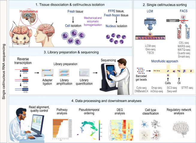
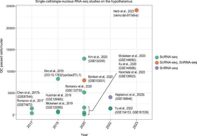
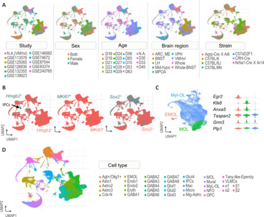
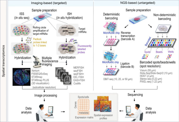
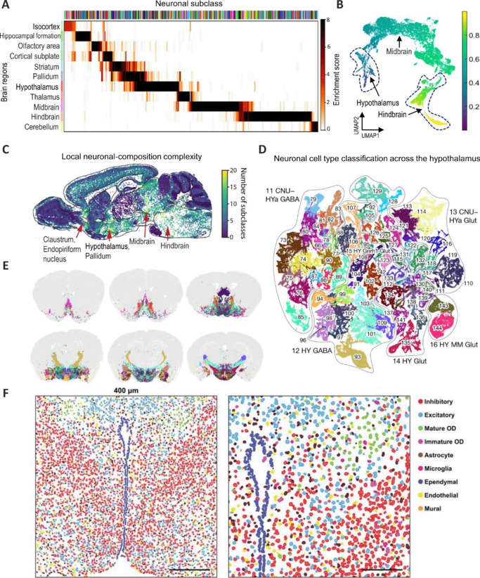
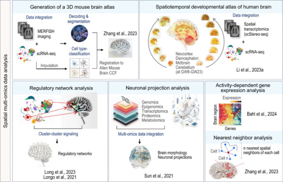
Elucidating the complex dynamic cellular organization in the hypothalamus is critical for understanding its role in coordinating fundamental body functions. Over the past decade, single-cell and spatial omics technologies have significantly evolved, overcoming initial technical challenges in capturing and analyzing individual cells. These high-throughput omics technologies now offer a remarkable opportunity to comprehend the complex spatiotemporal patterns of transcriptional diversity and cell-type characteristics across the entire hypothalamus. Current single-cell and single-nucleus RNA sequencing methods comprehensively quantify gene expression by exploring distinct phenotypes across various subregions of the hypothalamus. However, single-cell/single-nucleus RNA sequencing requires isolating the cell/nuclei from the tissue, potentially resulting in the loss of spatial information concerning neuronal networks. Spatial transcriptomics methods, by bypassing the cell dissociation, can elucidate the intricate spatial organization of neural networks through their imaging and sequencing technologies. In this review, we highlight the applicative value of single-cell and spatial transcriptomics in exploring the complex molecular-genetic diversity of hypothalamic cell types, driven by recent high-throughput achievements.


Conflict of interest statement

Conflicts of interest: The authors declare no conflicts of interest.

Figures

Figure 1
Figure 1
Schematic representation of single-cell and single-nucleus RNA sequencing. (1) The hypothalamic brain tissue is collected from mice and humans. Single cells are mostly lysed from fresh frozen tissue, while single nuclei mostly require cell lysis and elimination of cytoplasmic RNA material from FFPE tissue. (2) Subsequently, various emerging sequencing technologies based on FACS, LCM, and microfluidics utilize their unique methods to capture and analyze single cells/nuclei. (3) Then, sorted cells undergo reverse transcription followed by library amplification and quantification to build the sequencing library for downstream analyses. (4) Lastly, the generated sequence data undergoes data processing, including read alignment, pathway analyses, and DEG analyses between cells/samples/groups to identify rare cell-type populations. Created with Midjourney, and Microsoft PowerPoint. DEG: Differentially expressed gene; FACS: fluorescence-activated cell sorting; FFPE: formalin-fixed paraffin-embedded; LCM: laser capture microdissection; TSCS: topographic single-cell sequencing.
Figure 2
Figure 2
Distribution of QC-passed single-cell/nuclei in the hypothalamic RNA-seq studies. The dot plot depicts the distribution of QC passed single-cell/nuclei by various single-cell/single-nuclei RNA-seq studies in hypothalamic research. Each dot is colored by a unique single-cell/single-nuclei RNA-seq approach, with the X-axis representing the year of the study conducted between 2017 and 2023, and the Y-axis indicating the QC passed cells/nuclei used in these studies. Each dot is labeled by the accession ID and author’s name related to these studies. Created with RStudio (ggplot2 package) and Microsoft PowerPoint. QC: Quality control; ScRNA-seq: single-cell RNA sequencing; SnRNA-seq: single-nucleus RNA sequencing.
Figure 3
Figure 3
Integration of scRNA-seq studies from postnatal hypothalamic regions. (A) UMAP plots showing the curated cell-level attributes of the integrated dataset after integration, cells are grouped by study, age, brain region, sex, and mouse strain. (B) UMAP plots showing positive and negative expression of Mki67/Hmgb2/Sox2 genes across all cell types vs. IPCs in the young adult hypothalamus. (C) UMAP plot (left) and relative expression (right) of oligodendrocyte subtype-specific genes showing the heterogeneity of MO clusters. Reprinted with permission from Junaid et al. (2023), Copyright © 2023, The Author(s) (D) UMAP plot showing annotations of 36 cell types encompassing all major and minor neural and non-neuronal cell populations from the integrated dataset. Created with RStudio (Seurat package) and Microsoft PowerPoint. Agt+/Olig1+: Astrocyte + oligodendrocytes; Astro 1/2/3: astrocytes; Cck-N: cholecystokinin neurons; EMOL: early mature oligodendrocytes; Endo 1/2: endothelial cell; Eryth: erythrocytes; GABA 1–8: GABAergic neurons; Glut 1–4: glutamatergic neurons; IPCs: intermediate progenitor cells; Mac: macrophages; Micro: microglia; Mig-Astro: migrating astrocytes; MO: myelinating oligodendrocytes; MyL-OL: myelin-forming oligodendrocytes; NFO: newly formed oligodendrocytes; OPCs: oligodendrocytes progenitor cells; ScRNA-seq: single-cell RNA sequencing; Tany-like-Ependy: tanycyte-like ependymal cells; UMAP: uniform manifold approximation and projection; VLMCs: vascular leptomeningeal cells; α1/2: tanycytes alpha; β1/2: tanycytes beta.
Figure 4
Figure 4
Schematic overview of spatial transcriptomics technologies used in brain research. Image-based spatial transcriptomics relies on two methods. One is the in situ sequencing (ISS) technique, which amplifies mRNA. The other is in situ hybridization (ISH), which detects target sequences by hybridization of complementary fluorescent probes. The output data of each technique is visualized using multiple fluorescence-based imaging techniques, providing subcellular resolution of mRNA localization. Each image capturing the mRNA is further processed for data analysis to extract the expression matrix data on the gene expression pattern. Next-generation sequencing-based (NGS) spatial transcriptomics techniques employ two types of barcoding. Deterministic barcoding utilizes microfluidic channel chips of 10 μm, 25 μm, and 50 μm resolution to deliver barcodes to the surface of a tissue slide by reverse transcription and ligation through crossflow. Barcoded samples then undergo sequencing for further analyses. In non-deterministic barcoding, a barcoded array is used to barcode transcripts according to their location in lattice spots. Barcoded samples are then sequenced to extract the spatial expression profile on the gene expression pattern. Created with Midjourney and Microsoft PowerPoint.
Figure 5
Figure 5
Neuronal cell type distribution in the hypothalamus and its neighborhood at adult age. (A) Heatmap showing the calculated enrichment score of each neuronal subclass in the hypothalamus and across the whole brain. (B) UMAP plots highlight the large-scale gradient of gene expression of neurons across the hypothalamus, midbrain, and hindbrain at adult age. (C) Spatial heatmap showing the local neuronal complexity of the hypothalamus, along with other regions of the brain. The local complexity of a single neuron is defined by measuring or counting different kinds of subclasses among its 50 near neighborhoods. (A, C) Reprinted and (B) adapted with permission from Zhang et al. (2023), Copyright © 2023, The Author(s).(D–E) UMAP plot and spatial coronal sections of the brain showing the six subclasses of both glutamatergic (Glut) and GABAergic (GABA) neurons in the hypothalamus and its neighborhood. Reprinted with permission from Yao et al. (2023), Copyright © 2023, The Author(s). (F) Enlarged image showing the spatial distribution of all major cell types in the hypothalamic preoptic area revealed by MERFISH. Reprinted with permission from Moffitt et al. (2018), Copyright © 2018, The American Association for the Advancement of Science. CNU: Cerebral nuclei; GABA: gamma-aminobutyric acid; Glut: glutamatergic; HY: hypothalamus; Hya: hypothalamus anterior; MERFISH: multiplexed error-robust fluorescence in situ hybridization; MM: medial mammillary nucleus; OD: oligodendrocytes; UMAP: uniform manifold approximation and projection.
Figure 6
Figure 6
Unveiling the multidimensional structural and functional complexity of the brain by integrating single-cell data with spatially resolved transcriptomics data. The generation of a three-dimensional (3D) mouse brain atlas is outlined, depicting the workflow to construct a 3D mouse brain cell atlas by integrating single-cell RNA sequencing (scRNA-seq) data with MERFISH imaging-based data. This process involves the imputation of transcriptome-wide expression profiles, decoding, segmentation, and cell type classification. Finally, MERFISH images are registered to the Allen Mouse Brain Common Coordinate Framework (CCF). Adapted with permission from Zhang et al. (2023), Copyright © 2023, The Author(s). The creation of a spatiotemporal developmental atlas of the human brain is represented, mapping various human brain regions from 6–23 gestational weeks (GWs) by integrating spatial transcriptomics data with scRNA-seq data. This provides comprehensive insights into regional specification in the developing human brain (Li et al., 2023a). The bottom three segments provide a comprehensive view of cluster-to-cluster signaling to map regulatory networks, followed by the integration of multi-omics data to explore brain morphology and neuronal projection patterns (Longo et al., 2021; Sun et al., 2021; Long et al., 2023). Furthermore, it highlights activity-dependent gene studies in line with the identification of local neural complexity of brain regions by implementing a nearest-neighbor analysis of the surrounding cell population (Zhang et al., 2023; Bahl et al., 2024).

Similar articles

References

    1. Alon S, et al. Expansion sequencing: spatially precise in situ transcriptomics in intact biological systems. Science. 2021;371:eaax2656. - PMC - PubMed
    1. Alvarez-Bolado G. Development of neuroendocrine neurons in the mammalian hypothalamus. Cell Tissue Res. 2019;375:23–39. - PubMed
    1. Amunts K, Knoll AC, Lippert T, Pennartz CMA, Ryvlin P, Destexhe A, Jirsa VK, D’Angelo E, Bjaalie JG. The human brain project-synergy between neuroscience, computing, informatics, and brain-inspired technologies. PLoS Biol. 2019;17:e3000344. - PMC - PubMed
    1. Athaya T, Ripan RC, Li X, Hu H. Multimodal deep learning approaches for single-cell multi-omics data integration. Brief Bioinform. 2023;24:bbad313. - PMC - PubMed
    1. Averbeck BB, Murray EA. Hypothalamic interactions with large-scale neural circuits underlying reinforcement learning and motivated behavior. Trends Neurosci. 2020;43:681–694. - PMC - PubMed

LinkOut - more resources