

Citronellal improves endothelial dysfunction by affecting the stability of the GCH1 protein
- PMID: 38993132
- PMCID: PMC11322867
- DOI: 10.3724/abbs.2024086
Citronellal improves endothelial dysfunction by affecting the stability of the GCH1 protein
Abstract
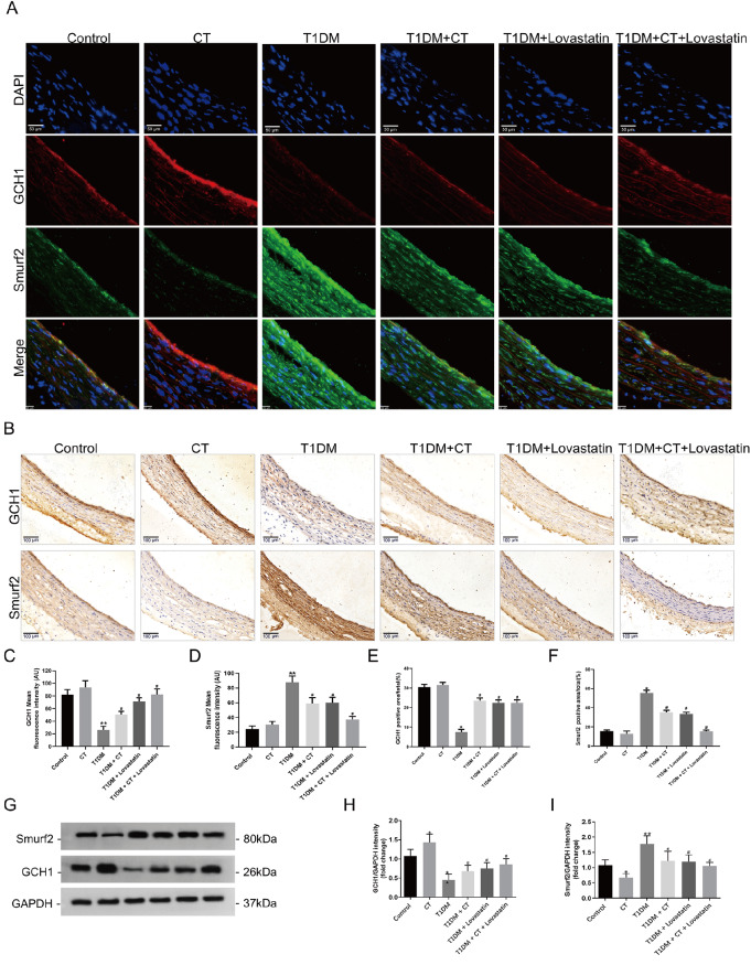
Endothelial dysfunction (ED) serves as the pathological basis for various cardiovascular diseases. Guanosine triphosphate cyclopyrrolone 1 (GCH1) emerges as a pivotal protein in sustaining nitric oxide (NO) production within endothelial cells, yet it undergoes degradation under oxidative stress, contributing to endothelial cell dysfunction. Citronellal (CT), a monoterpenoid, has been shown to ameliorate endothelial dysfunction induced by in atherosclerosis rats. However, whether CT can inhibit the degradation of GCH1 protein is not clear. It has been reported that ubiquitination may play a crucial role in regulating GCH1 protein levels and activities. However, the specific E3 ligase for GCH1 and the molecular mechanism of GCH1 ubiquitination remain unclear. Using data-base exploration analysis, we find that the levels of the E3 ligase Smad-ubiquitination regulatory factor 2 (Smurf2) negatively correlate with those of GCH1 in vascular tissues and HUVECs. We observe that Smurf2 interacts with GCH1 and promotes its degradation via the proteasome pathway. Interestingly, ectopic Smurf2 expression not only decreases GCH1 levels but also reduces cell proliferation and reactive oxygen species (ROS) levels, mostly because of increased GCH1 accumulation. Furthermore, we identify BH 4/eNOS as downstream of GCH1. Taken together, our results indicate that CT can obviously improve vascular endothelial injury in Type 1 diabetes mellitus (T1DM) rats and reverse the expressions of GCH1 and Smurf2 proteins in aorta of T1DM rats. Smurf2 promotes ubiquitination and degradation of GCH1 through proteasome pathway in HUVECs. We conclude that the Smurf2-GCH1 interaction might represent a potential target for improving endothelial injury.
Keywords: Smurf2; citronellal; degradation; endothelial dysfunction.
Conflict of interest statement
The authors declare that they have no conflict of interest.
Figures


Similar articles
-
The ubiquitination ligase SMURF2 reduces aerobic glycolysis and colorectal cancer cell proliferation by promoting ChREBP ubiquitination and degradation.J Biol Chem. 2019 Oct 4;294(40):14745-14756. doi: 10.1074/jbc.RA119.007508. Epub 2019 Aug 13. J Biol Chem. 2019. PMID: 31409643 Free PMC article.
-
The E3 ubiquitin ligase Smurf2 regulates PARP1 stability to alleviate oxidative stress-induced injury in human umbilical vein endothelial cells.J Cell Mol Med. 2020 Apr;24(8):4600-4611. doi: 10.1111/jcmm.15121. Epub 2020 Mar 13. J Cell Mol Med. 2020. PMID: 32167680 Free PMC article.
-
Citronellal alleviate macro- and micro-vascular damage in high fat diet / streptozotocin - Induced diabetic rats via a S1P/S1P1 dependent signaling pathway.Eur J Pharmacol. 2022 Apr 5;920:174796. doi: 10.1016/j.ejphar.2022.174796. Epub 2022 Feb 10. Eur J Pharmacol. 2022. PMID: 35151650
-
The E3 ubiquitin ligase SMAD ubiquitination regulatory factor 2 negatively regulates Krüppel-like factor 5 protein.J Biol Chem. 2011 Nov 18;286(46):40354-64. doi: 10.1074/jbc.M111.258707. Epub 2011 Sep 27. J Biol Chem. 2011. PMID: 21953463 Free PMC article.
-
Nedd8 targets ubiquitin ligase Smurf2 for neddylation and promote its degradation.Biochem Biophys Res Commun. 2016 May 20;474(1):51-56. doi: 10.1016/j.bbrc.2016.04.058. Epub 2016 Apr 13. Biochem Biophys Res Commun. 2016. PMID: 27086113
References
-
- Yang F, Yu J, Ke F, Lan M, Li D, Tan K, Ling J, et al. Curcumin alleviates diabetic retinopathy in experimental diabetic rats. Ophthalmic Res. . 2018;60:43–54. doi: 10.1159/000486574. - DOI - PubMed
-
- Xiao W, Yang Y, Shi J, Xu J, Zhu J, Wu Z. The diagnostic efficacy and predictive value of combined lipoprotein laboratory indexes for atherosclerosis. J Pak Med Assoc. 2020, 70 [Special Issue]: 115–119 - PubMed
-
- Zhao Y, Yang Z, Fang C, Xiao D, Shi Y, Lin Y, Zhai Q. A single-center observational study on the expression of circulating interleukin-20 levels and predicting outcomes in human chronic heart failure: a 2-year follow-up cohort study. Clinica Chim Acta. . 2020;510:5–10. doi: 10.1016/j.cca.2020.06.048. - DOI - PubMed
-
- Liu P, Liu J, Wu Y, Xi W, Wei Y, Yuan Z, Zhuo X. Zinc supplementation protects against diabetic endothelial dysfunction via GTP cyclohydrolase 1 restoration. Biochem Biophys Res Commun. . 2020;521:1049–1054. doi: 10.1016/j.bbrc.2019.11.046. - DOI - PubMed
MeSH terms
Substances
LinkOut - more resources
Full Text Sources
