. 2024 Aug;15(4):722-730.
doi: 10.14740/wjon1871. Epub 2024 Jul 5.

Hereditary Gastric Cancer Is Linked With Hereditary Breast and Ovarian Cancer

Affiliations

Hereditary Gastric Cancer Is Linked With Hereditary Breast and Ovarian Cancer

Takuma Hayashi et al. World J Oncol. 2024 Aug.

Abstract

Background: Helicobacter pylori (H. pylori), a bacterium which chronically infects the stomach of approximately half the world's population, is a risk factor for the development of gastric cancer (GC). However, the underlying mechanism whereby H. pylori infection induces GC development remains unclear. Intermittent injection of the H. pylori cytotoxin-associated gene A antigen (CagA) protein into its host cell inhibits nuclear translocation of BRCA1/BRCA2, DNA repair proteins involved in the development of breast cancer/ovarian cancer. Interestingly, hereditary breast and ovarian cancer (HBOC) syndrome is associated with GC development. Here, we aimed to clarify the molecular link between H. pylori infection, BRCA1/2 pathogenic variants (PVs), GC and higher GC incidence in HBOC families.

Methods: We retrospectively reviewed data from Japanese patients undergoing precision treatment using cancer genomic medicine.

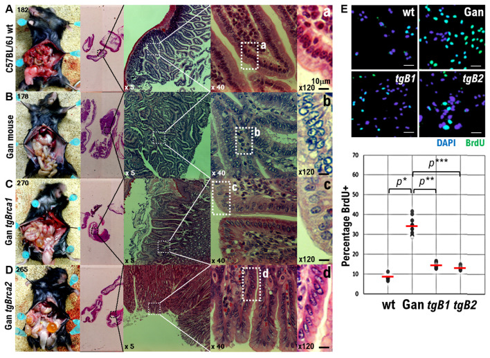
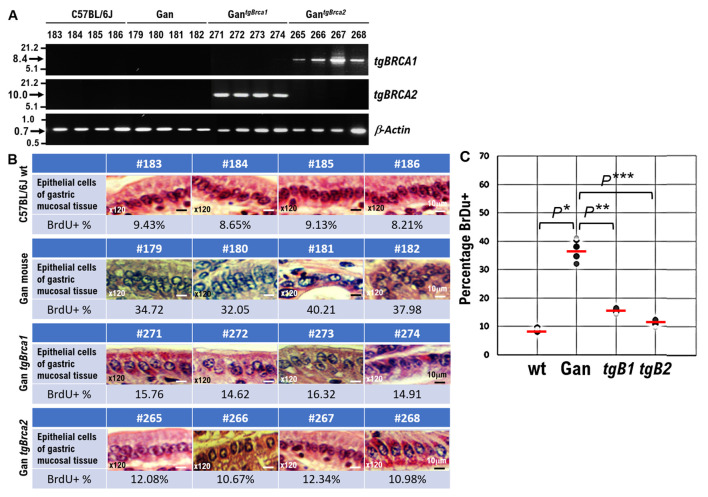
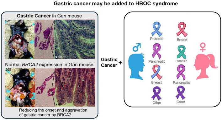
Results: We found a higher GC incidence in HBOC families having germline pathogenic variants (GPVs) of BRCA1/2 (2.95% vs. 0.78% in non-HBOC families). Next, we found that 96.1% of H. pylori-infected patients received cancer genomic medicine for advanced GC, and > 16% advanced GC patients had gBRCA2 PVs. Furthermore, expressing wild-type BRCA1/2 in Gan mice (a mouse model of human GC) inhibited GC development. Thus, gBRAC1/2 PVs and H. pylori infection synergistically increase the risk of GC development.

Conclusion: Our study highlights the need to investigate the potential of therapeutic agents against BRCA1/2 PVs to avoid the development of GC in HBOC families. In addition, our results suggest that poly (ADP-ribose) polymerase (PARP) inhibitors could potentially inhibit GC development and progression with gBRCA1/2 PVs.

Keywords: BRCA1; BRCA2; Gastric cancer; HBOC; Helicobacter pylori.


Conflict of interest statement

The authors state no competing interest.

Figures

Figure 1
Figure 1
Effects of wild-type BRCA1 and BRCA2 expression on gastric cancer (GC) development in Gan mice, a mouse model of GC. (A) Photographs showing surgical pathological findings of normal epithelial cells in gastric mucosal tissue of wild-type C57BL6J mice. (B) Photographs showing surgical pathological findings of highly atypical epithelial cells in gastric mucosal tissue developed in Gan mice. (C) Photographs showing surgical pathological findings of mildly atypical epithelial cells in the gastric mucosal tissue developed in GantgBrca1 mice constitutively expressing BRCA1. Decreased number of atypical epithelial cells in gastric mucosal tissue. (D) Photographs showing surgical pathological findings of epithelial cells in the gastric mucosal tissue in GantgBrca2 mice constitutively expressing BRCA2. Significantly less atypical epithelial cells can be observed in gastric mucosal tissue. The gastric mucosal tissue of GantgBrca2 mice pathologically resembles normal tissue. (a-d) Enlarged images of tissue areas showing pathological findings of epithelial cells in the gastric mucosal tissue of each mouse at × 40 magnification. In other words, these images have been further enlarged to × 120. Scalebars, 10 µm. (E) Each genetically modified mouse was inoculated with a BrdU solution, and proliferation of epithelial cells in the gastric mucosal tissue of each genetically modified mouse was examined by the number of BrdU-positive cells. The number of BrdU-positive cells in the epithelial cells of the gastric mucosal tissue of wild-type C57BL6J mice is extremely small. The number of BrdU-positive cells in the epithelial cells of the gastric mucosal tissue of the Gan mice was extremely large. The number of BrdU-positive cells in the epithelial cells of the gastric mucosal tissue of GantgBrca1 mice was reduced. The number of BrdU-positive cells in the epithelial cells of the gastric mucosal tissue of GantgBrca2 mice was significantly reduced. The percentage of number of BrdU-positive cells observed among epithelial cells of the gastric mucosal tissue of each genetically modified mouse is plotted in the graph. *P < 0.001, **P < 0.005, ***P < 0.002.
Figure 2
Figure 2
Effects of wild-type BRCA1 and BRCA2 expression on gastric cancer (GC) development in Gan mice, a mouse model of GC. (A) The results of reverse transcriptase polymerase chain reaction (RT-PCR) showing Brca 1 wild type or Brca2 wild type mRNA expression in GantgBrca1 or GantgBrca2 mice. (B) Surgical pathological findings of normal epithelial cells in gastric mucosal tissue of wild type C57BL6J mice (mouse ID number #183, #184, #185, #186). Surgical pathological findings of highly atypical epithelial cells in gastric mucosal tissue in Gan mice (mouse ID number #179, #180, #181, #182) and mildly atypical epithelial cells in the gastric mucosal tissue of GantgBrca1 mice (mouse ID number #271, #272, #273, #273) constitutively expressing BRCA1. The atypical epithelial cells in gastric mucosal tissue are reduced in this mouse. The surgical pathological findings of atypical epithelial cells in the gastric mucosal tissue of GantgBrca2 mice (mouse ID number #265, #266, #267, #268) constitutively expressing BRCA2 were significantly reduced. Pathologically, the gastric mucosal tissue of GantgBrca2 mice (mouse ID number #265, #266, #267, #268) resembles normal tissue. These images of the gastric mucosal tissue have been enlarged to × 120. Scalebar, 10 µm. (C) Each genetically modified mouse was inoculated with a BrdU solution, and the proliferation of epithelial cells in the gastric mucosal tissue of each genetically modified mouse was examined using the number of BrdU-positive cells as an indicator. The number of BrdU-positive cells in the epithelial cells of the gastric mucosal tissue of wild-type C57BL6J mice is extremely small. The number of BrdU-positive cells in the epithelial cells of the gastric mucosal tissue of the Gan mice was extremely large. The number of BrdU-positive cells in the epithelial cells of the gastric mucosal tissue of GantgBrca1 mice was reduced. The number of BrdU-positive cells in the epithelial cells of the gastric mucosal tissue of GantgBrca2 mice was significantly reduced The percentage of number of BrdU-positive cells observed among epithelial cells of the gastric mucosal tissue of each genetically modified mouse is plotted in the graph. *P < 0.001, **P < 0.005, ***P < 0.001.
Figure 3
Figure 3
The gastric cancer observed in some patients may be part of the hereditary breast and ovarian cancer (HBOC) syndrome. BRCA1/BRCA2 protect DNA replication forks by repairing damage (DNA double-strand breaks (DSBs)) during nuclear genomic DNA replication. In particular, in maintaining the stability of cellular genome (genome stability). BRCA1/2 genes are typical tumor suppressor genes, and loss-of-function mutations in BRCA1/2 significantly increase the risk of developing breast and ovarian cancers, as well as some prostate and pancreatic cancers. The present results indicate that BRCA1 or BRCA2 pathogenic variants may be involved in the malignant transformation of epithelial cells in the gastric mucosal tissue. Therefore, the gastric cancer observed in some patients could be part of the HBOC syndrome.

Similar articles

References

    1. Cancer Genome Atlas Research N. Comprehensive molecular characterization of gastric adenocarcinoma. Nature. 2014;513(7517):202–209. doi: 10.1038/nature13480. - DOI - PMC - PubMed
    1. Fiocca R, Villani L, Tenti P, Solcia E, Cornaggia M, Frigerio B, Capella C. Characterization of four main cell types in gastric cancer: foveolar, mucopeptic, intestinal columnar and goblet cells. An histopathologic, histochemical and ultrastructural study of "early" and "advanced" tumours. Pathol Res Pract. 1987;182(3):308–325. doi: 10.1016/S0344-0338(87)80066-3. - DOI - PubMed
    1. Huang B, Sun Z, Wang Z, Lu C, Xing C, Zhao B, Xu H. Factors associated with peritoneal metastasis in non-serosa-invasive gastric cancer: a retrospective study of a prospectively-collected database. BMC Cancer. 2013;13:57. doi: 10.1186/1471-2407-13-57. - DOI - PMC - PubMed
    1. Chu KM, Law S, Branicki FJ, Wong J. Extrahepatic biliary obstruction by metastatic gastric carcinoma. J Clin Gastroenterol. 1998;27(1):63–66. doi: 10.1097/00004836-199807000-00013. - DOI - PubMed
    1. Jung K, Park MI, Kim SE, Park SJ. Borrmann type 4 advanced gastric cancer: focus on the development of scirrhous gastric cancer. Clin Endosc. 2016;49(4):336–345. doi: 10.5946/ce.2016.057. - DOI - PMC - PubMed

LinkOut - more resources