. 2024 Jun 6;32(3):101279.
doi: 10.1016/j.omtm.2024.101279. eCollection 2024 Sep 12.

Mesenchymal stromal cells protect combined oncolytic and helper-dependent adenoviruses from humoral immunity

Affiliations

Mesenchymal stromal cells protect combined oncolytic and helper-dependent adenoviruses from humoral immunity

Ada Irmak Özcan et al. Mol Ther Methods Clin Dev. .

Abstract

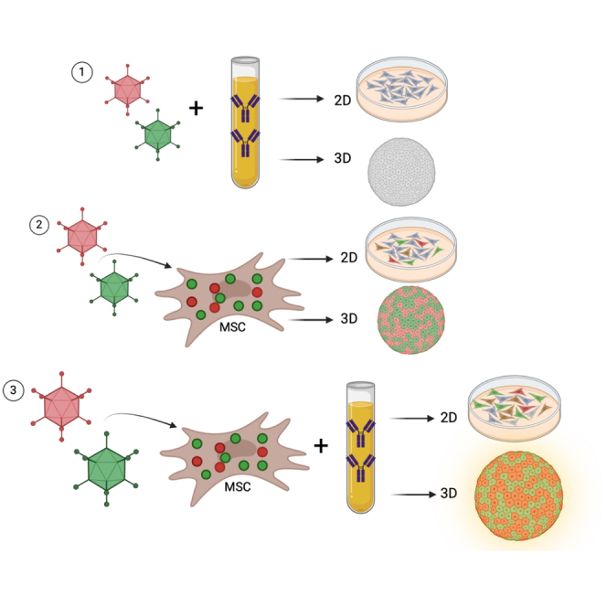
Systemic delivery of oncolytic and immunomodulatory adenoviruses may be required for optimal effects on human malignancies. Mesenchymal stromal cells (MSCs) can serve as delivery systems for cancer therapeutics due to their ability to transport and shield these agents while homing to tumors. We now use MSCs to deliver a clinically validated binary oncolytic and helper-dependent adenovirus combination (CAdVEC) to tumor cells. We show successful oncolysis and helper-dependent virus function in tumor cells even in the presence of plasma from adenovirus-seropositive donors. In both two- and three-dimensional cultures, CAdVEC function is eliminated even at high dilutions of seropositive plasma but is well sustained when CAdVEC is delivered by MSCs. These results provide a robust in vitro model to measure oncolytic and helper-dependent virus spread and demonstrate a beneficial role of using MSCs for systemic delivery of CAdVEC even in the presence of a neutralizing humoral response.

Keywords: helper-dependent adenovirus; mesenchymal stromal cells; oncolytic adenovirus; solid tumors; three-dimensional tumor spheroids.


Conflict of interest statement

M.K.B. serves on advisory boards for Marker Therapeutics, Allogene, Walking Fish, Abintus, Athenex-Kuur, Onk Therapeutics, Coya Therapeutics, Triumvira, Adaptimmune, Vor Therapeutics, and Tscan. M.K.B has equity in Allovir, Tessa Therapeutic Ltd, and Marker Therapeutics. M.K.B. has royalties from Takeda and Bellicum. M.S. received research funding from Tessa Therapeutic Ltd. and AstraZeneca. M.S. was a scientific consultant for Tessa Therapeutic Ltd.

Figures

None
Graphical abstract
Figure 1
Figure 1
Bone-marrow-derived MSCs are susceptible to CAdVEC infection (A) Bright-field and fluorescent microscopy images of MSCs infected with 100 vp/cell OAd.5 and 1,000 vp/cell HDAd.5/3 expressing RFP and GFP respectively 48 h post infection. (B) RFP expression (left axis) and GFP expression (right axis) of A549 cells incubated with 96 h CAdVEC MSC supernatant. Red object intensity (RCU) and green object intensity (GCU) measured by IncuCyte Live image analysis over time. (C) Supernatant was collected from CAdVEC MSCs 96 h post infection and added to A549 cells. Cell death measured by annexin V staining via flow cytometry of A549 tumor cells treated with dilutions of CAdVEC MSC supernatant (technical replicates) 96 h later to confirm OAd-mediated oncolysis. Error bars represent standard deviation.
Figure 2
Figure 2
Direct CAdVEC infection is neutralized by plasma proteins of healthy donors CAdVEC only or direct infection conditions are incubated with PBS as a control. (A) 2D A549 and H1650 tumor cells were infected with a high dose of 100 vp/cell OAd.5 and 1,000 vp/cell HDAd.5/3 expressing RFP (top) and GFP (bottom) measured by IncuCyte Live imaging over time. (B) IncuCyte images of A549 cells infected with a low-dose 10 vp/cell OAd.5 and 100 vp/cell HDAd.5/3 at 96 h with or without dilutions of plasma. (C) RFP (top) and GFP (bottom) expression in 2D A549 and H1650 tumor cells with low-dose infection over time. (D) High-dose CAdVEC infection in 3D tumor spheroids. RFP and GFP expressions were measured by IncuCyte Live imaging over time in both A549 and H1650 tumor spheroids with different dilutions of plasma. (E) IncuCyte images of 3D A549 tumor spheroids post infection of low-dose 10 vp/cell OAd.5 and 100 vp/cell HDAd.5/3 expressing RFP and GFP respectively at 96 h with different dilutions of plasma. (F) RFP and GFP expression in 3D A549 and H1650 tumor spheroids infected with low-dose CAdVEC. Scale bar represents 400 μM. p values are signified by asterisks: ∗p < 0.05, ∗∗p < 0.01, ∗∗ ∗p < 0.001 determined by unpaired Student's t test at 100 h; n = 9–12 replicates. Data are presented as means +/− SEM.
Figure 3
Figure 3
CAdVEC MSCs shield virus from neutralization in both 2D and 3D culture settings MSCs were infected with 100 vp/cell OAd.5_RFP and 1,000 vp/cell HDAd.5/3_GFP. 24 h later, CAdVEC MSCs were incubated in either PBS or diluted plasma for 1 h and then delivered to a tumor cultured in 2D and 3D settings. (A) Representative IncuCyte images of 2D culture of A549 cells at 96 h post addition. (B) Quantification of OAd.5_RFP (top) and HDAd.5/3_GFP (bottom) in 2D culture over time. Fluorescence was measured by object intensity within the IncuCyte software. (C) Images of A549 tumor spheroids 96 h after CAdVEC MSC delivery with different dilutions of neutralizing plasma. (D) Quantification of OAd.5_RFP (top) and HDAd.5/3_GFP (bottom) in 3D culture over time. Fluorescence measured by object integrated intensity within the IncuCyte software (n = 4 wells/condition). p value calculated at the 100-h time point. (E) Quantification of 96-h time point in 3D culture of A549 (left) and H1650 (right) comparing direct infection with CAdVEC MSC delivery (n = 3 MSC donors in quadruplicate) in the presence of 1:10 plasma dilution, indicated by “1:10.” Scale bar represents 400 μM. p values are signified by asterisks: ∗p < 0.05, ∗∗p < 0.01, ∗∗∗p < 0.001 determined by unpaired Student's t test. Data are presented as means +/− SEM.

References

    1. Wang D., Porter C.E., Lim B., Rosewell Shaw A., Robertson C.S., Woods M.L., Xu Y., Biegert G.G.W., Morita D., Wang T., et al. Ultralow-dose binary oncolytic/helper-dependent adenovirus promotes antitumor activity in preclinical and clinical studies. Sci. Adv. 2023;9 doi: 10.1126/sciadv.ade6790. - DOI - PMC - PubMed
    1. Farzad L., Cerullo V., Yagyu S., Bertin T., Hemminki A., Rooney C., Lee B., Suzuki M. Combinatorial treatment with oncolytic adenovirus and helper-dependent adenovirus augments adenoviral cancer gene therapy. Mol. Ther. Oncolytics. 2014;1 doi: 10.1038/mto.2014.8. - DOI - PMC - PubMed
    1. Rosewell Shaw A., Porter C.E., Watanabe N., Tanoue K., Sikora A., Gottschalk S., Brenner M.K., Suzuki M. Adenovirotherapy Delivering Cytokine and Checkpoint Inhibitor Augments CAR T Cells against Metastatic Head and Neck Cancer. Mol. Ther. 2017;25:2440–2451. doi: 10.1016/j.ymthe.2017.09.010. - DOI - PMC - PubMed
    1. Havunen R., Santos J.M., Sorsa S., Rantapero T., Lumen D., Siurala M., Airaksinen A.J., Cervera-Carrascon V., Tähtinen S., Kanerva A., Hemminki A. Abscopal Effect in Non-injected Tumors Achieved with Cytokine-Armed Oncolytic Adenovirus. Mol. Ther. Oncolytics. 2018;11:109–121. doi: 10.1016/j.omto.2018.10.005. - DOI - PMC - PubMed
    1. Ferguson M.S., Lemoine N.R., Wang Y. Systemic delivery of oncolytic viruses: hopes and hurdles. Adv. Virol. 2012;2012 doi: 10.1155/2012/805629. - DOI - PMC - PubMed

LinkOut - more resources