. 2017 May;2(5):17016.
doi: 10.1038/natrevmats.2017.16. Epub 2017 Apr 20.

Interplay between materials and microfluidics

Affiliations

Interplay between materials and microfluidics

Xu Hou et al. Nat Rev Mater. 2017 May.

Abstract

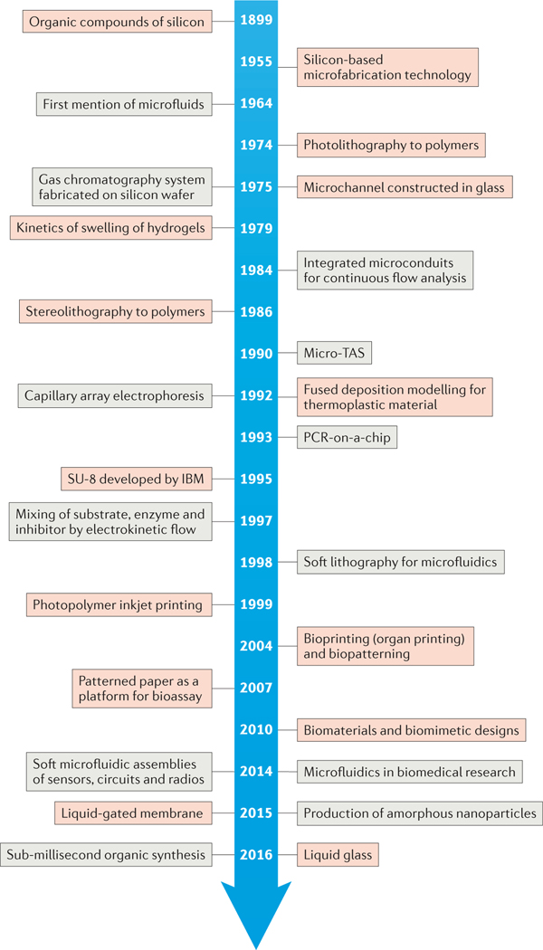
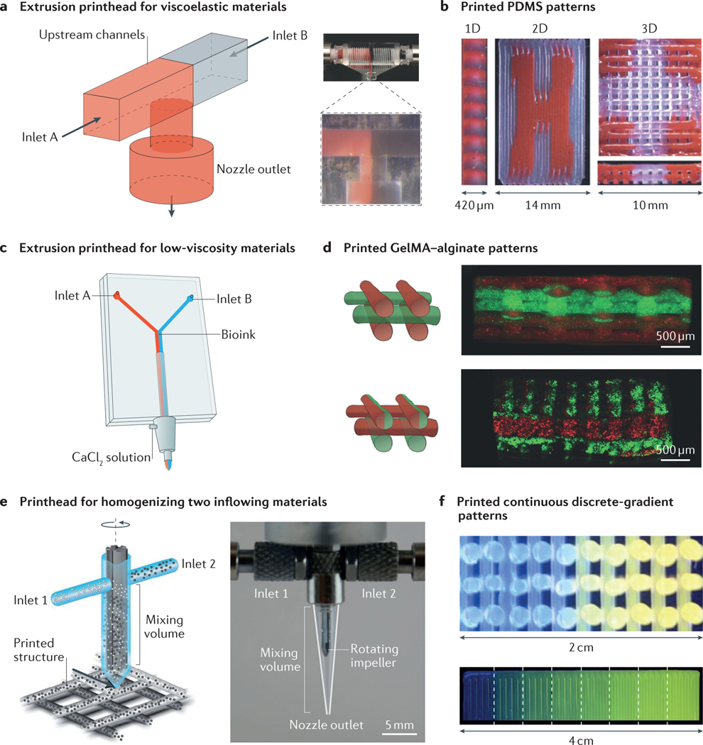
Developments in the field of microfluidics have triggered technological revolutions in many disciplines, including chemical synthesis, electronics, diagnostics, single-cell analysis, micro- and nanofabrication, and pharmaceutics. In many of these areas, rapid growth is driven by the increasing synergy between fundamental materials development and new microfluidic capabilities. In this Review, we critically evaluate both how recent advances in materials fabrication have expanded the frontiers of microfluidic platforms and how the improved microfluidic capabilities are, in turn, furthering materials design. We discuss how various inorganic and organic materials enable the fabrication of systems with advanced mechanical, optical, chemical, electrical and biointerfacial properties - in particular, when these materials are combined into new hybrids and modular configurations. The increasing sophistication of microfluidic techniques has also expanded the range of resources available for the fabrication of new materials, including particles and fibres with specific functionalities, 3D (bio)printed composites and organoids. Together, these advances lead to complex, multifunctional systems, which have many interesting potential applications, especially in the biomedical and bioengineering domains. Future exploration of the interactions between materials science and microfluidics will continue to enrich the diversity of applications across engineering as well as the physical and biomedical sciences.


Conflict of interest statement

Competing interests statement The authors declare no competing interests.

Figures

Figure 1 |
Figure 1 |. Historical timeline of developments in materials and microfluidics.
Key advances in materials (red) and microfluidics (grey) have aided the interplay between the two complementary fields and their synergistic growth. PCR, polymerase chain reaction; TAS, total analysis system.
Figure 2 |
Figure 2 |. Elastomer-based stretchable microfluidics.
a | Schematic illustrations and optical micrographs of a stretchable antenna. The half-wave dipole antenna was made of EGaIn (eutectic gallium–indium, a liquid metal alloy) embedded in microfluidic channels composed of polydimethylsiloxane (PDMS) and Ecoflex (an elastomeric silicone polymer that is softer than PDMS). b | Photographs of a microfluidics-based solution for the fabrication of a stretchable radio-frequency electronic radiation sensor that can operate in an ordinary office environment. The insets are the LED indicators of the sensor, which show no difference in the signal with different degrees of stretching. From left to right: non-stretched; strained with 15% elongation along the y axis; manually applied strain in both x and y directions; and severe twisting. c | Schematic illustrations and images of a soft, stretchable electronic system that integrates strain-isolated device components and a free-floating interconnecting network in a thin elastomeric microfluidic enclosure. The inset in the far right is an optical micrograph of the device, which contains a pair of epidermal electrodes in a serpentine mesh layout. LED, light-emitting diode. Panel a is adapted with permission from REF. , Wiley-VCH. Panel b is adapted with permission from REF. , Royal Society of Chemistry. Panel c is adapted with permission from REF. , AAAS.
Figure 3 |
Figure 3 |. Representative materials for microfluidic devices.
a | A free-standing SU-8-based microfluidic sensor. The inset shows a cross-sectional scanning electron microscopy (SEM) image of a microchannel. b | A polytetrafluoroethylene (PTFE)-based microfluidic device with two-layer microchannels separated by a thin membrane. The inset shows a cross-sectional SEM image of the microchannel. c | Fabrication process of a two-layer silk hydrogel microfluidic device using gelatin moulding and layer-by-layer stacking methods. The inset shows optical micrographs of the minimal microchannel diameters achievable with the silk hydrogel microfluidic device. d | Paper-based 3D microfluidic device for multiple bioassays and sequential fluidic manipulation. The inset on the right shows a magnified view of the device before liquid injection, and that on the bottom shows the device after injection. Panel a is adapted with permission from REF. , Copernicus Publications. Panel b is adapted with permission from REF. , National Academy of Sciences. Panel c is adapted with permission from REF. , Elsevier. Panel d is adapted with permission from REF. , Elsevier.
Figure 4 |
Figure 4 |. Fabrication of micro- and nanoparticles in microfluidic systems.
a | Microfluidic fabrication of monodispersed microparticles. The two scanning electron microscopy (SEM) images show the formation of Janus particles (left) and microcapsule particles (right). b | Amorphous nanoparticles prepared using a microfluidic nebulator (the example shown is for CaCO3 nanoparticles, although this method can be applied for a wide range of materials). c | Synthesis of noble metal nanoparticles (for example, Ag nanocubes, Ag truncated octahedra and Au–Ag nanocups) in microfluidic droplet reactors with multistep adsorption and reactions. Transmission electron microscope images of the particles are shown on the right. d | In situ synthesis of ZnO nanowires in a microfluidic chip; comparison of global synthesis in the entire fluidic channel and local synthesis by microheaters in the fluidic channel. SEM images of the nanowires are shown below. AA, ascorbic acid; CA, citric acid; PDMS, polydimethylsiloxane. Panel a is adapted with permission from REF. , Royal Society of Chemistry. Panel b is adapted with permission from REF. , AAAS. Panel c is adapted with permission from REF. , American Chemical Society. Panel d is adapted with permission from REF. , Royal Society of Chemistry.
Figure 5 |
Figure 5 |. Crystallization in microfluidic systems.
a | Precipitation of protein crystals in a microfluidic reactor. By changing solvent ratios, the degree of mixing, the protein solution and other additives, different conditions can be established in each drop. b | Multichamber microfluidic system that enabled up to 144 different experimental conditions to be established, as indicated in the fluorescence images that show the gradient of fluorescein (left). In the right part, different conditions were used for investigating the optimal crystallization conditions of lysozymes (Lyz). The arrows indicate increasing concentrations of the denoted chemicals, and the coloured squares indicate the conditions under which crystals form. Panel a is adapted with permission from REFS , American Chemical Society. Panel b is adapted with permission from REF. , American Chemical Society.
Figure 6 |
Figure 6 |. Microfluidics-enabled 3D bioprinting.
a | Microfluidic printhead for the extrusion of two viscoelastic materials. b | Printed heterogeneous 1D, 2D and 3D polydimethylsiloxane (PDMS) patterns. c | Dual-layer microfluidic printhead for the extrusion of two materials with low viscosities. d | Printed heterogeneous gelatin methacryloyl (GelMA)–alginate patterns. e | Microfluidic printhead containing an active rotating impeller for homogenizing two inflowing materials before they are extruded from the printhead to deposit patterns. f | Printed continuous- and discrete-gradient patterns. Panels a and b are adapted with permission from REF. , Wiley-VCH. Panels c and d are adapted with permission from REF. , Wiley-VCH. Panels e and f are adapted with permission from REF. , National Academy of Sciences.

Similar articles

Cited by

References

    1. Whitesides GM The origins and the future of microfluidics. Nature 442, 368–373 (2006).

      A comprehensive review of the background, applications and future perspectives of microfluidics.

    1. Huh D, Hamilton GA & Ingber DE From 3D cell culture to organs-on-chips. Trends Cell Biol. 21, 745–754 (2011). - PMC - PubMed
    1. Bhatia SN & Ingber DE Microfluidic organs-on-chips. Nat. Biotechnol 32, 760–772 (2014). - PubMed
    1. Esch EW, Bahinski A. & Huh D. Organs-on-chips at the frontiers of drug discovery. Nat. Rev. Drug Discov. 14, 248–260 (2015). - PMC - PubMed
    1. Hughes AJ & Herr AE Microfluidic western blotting. Proc. Natl Acad. Sci. USA 109, 21450–21455 (2012). - PMC - PubMed