. 2024 Jun 27:15:1400700.
doi: 10.3389/fmicb.2024.1400700. eCollection 2024.

Characterization and genomic analysis of a lytic Stenotrophomonas maltophilia short-tailed phage A1432 revealed a new genus of the family Mesyanzhinovviridae

Affiliations

Characterization and genomic analysis of a lytic Stenotrophomonas maltophilia short-tailed phage A1432 revealed a new genus of the family Mesyanzhinovviridae

Shixia Li et al. Front Microbiol. .

Abstract

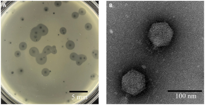
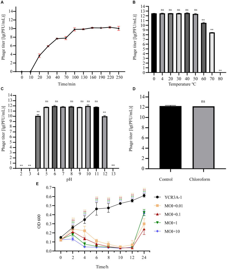
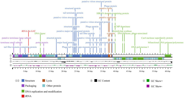
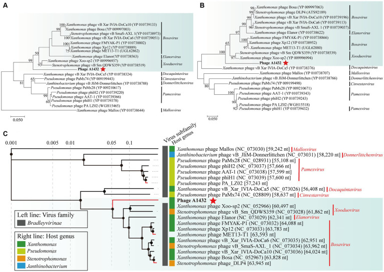
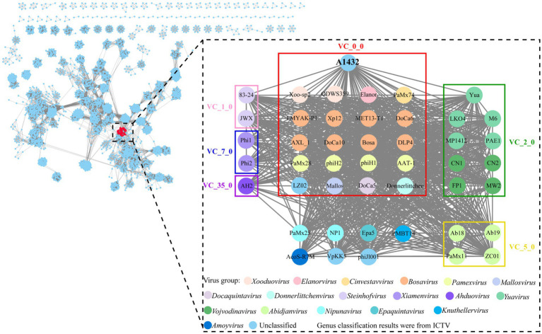
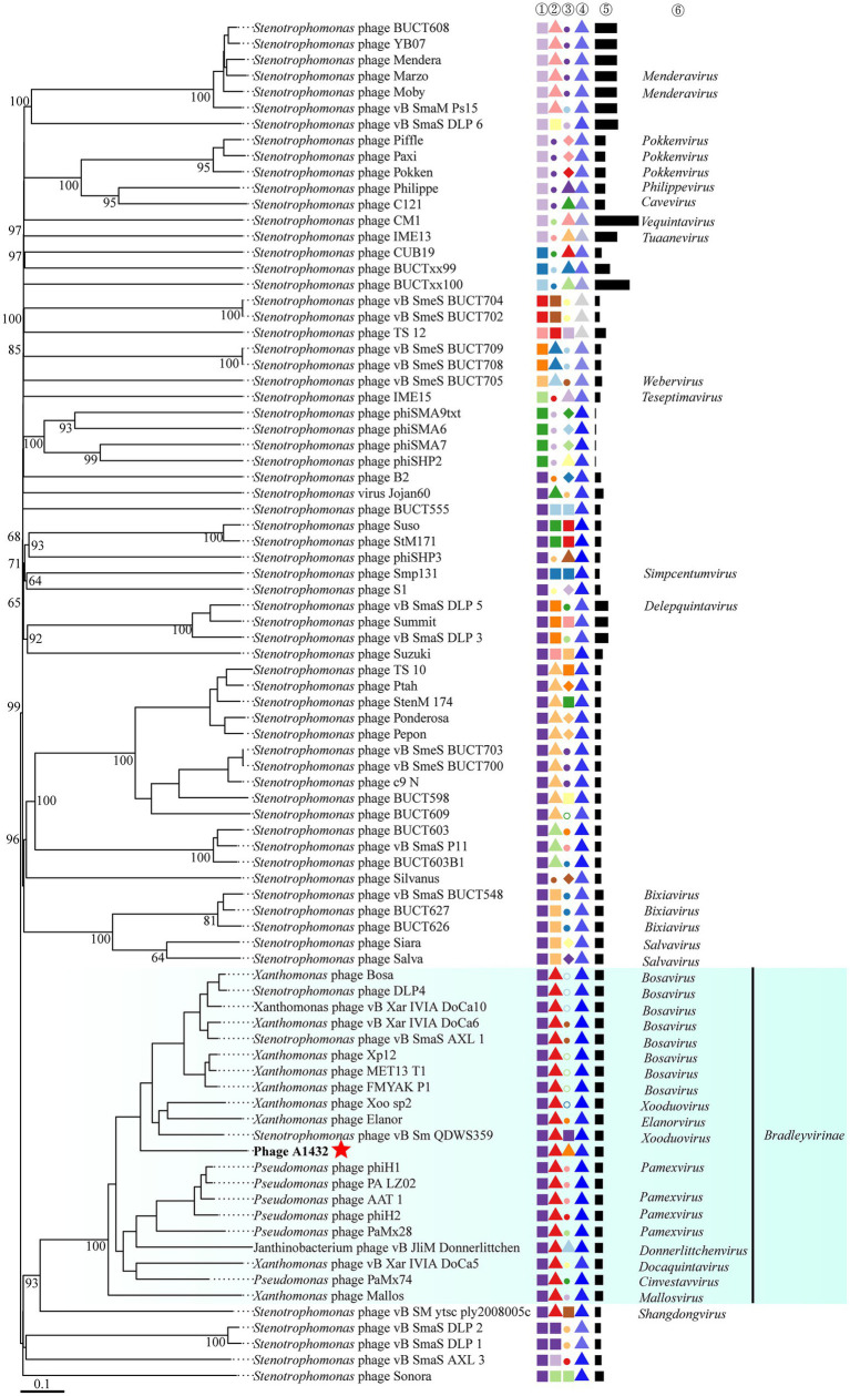
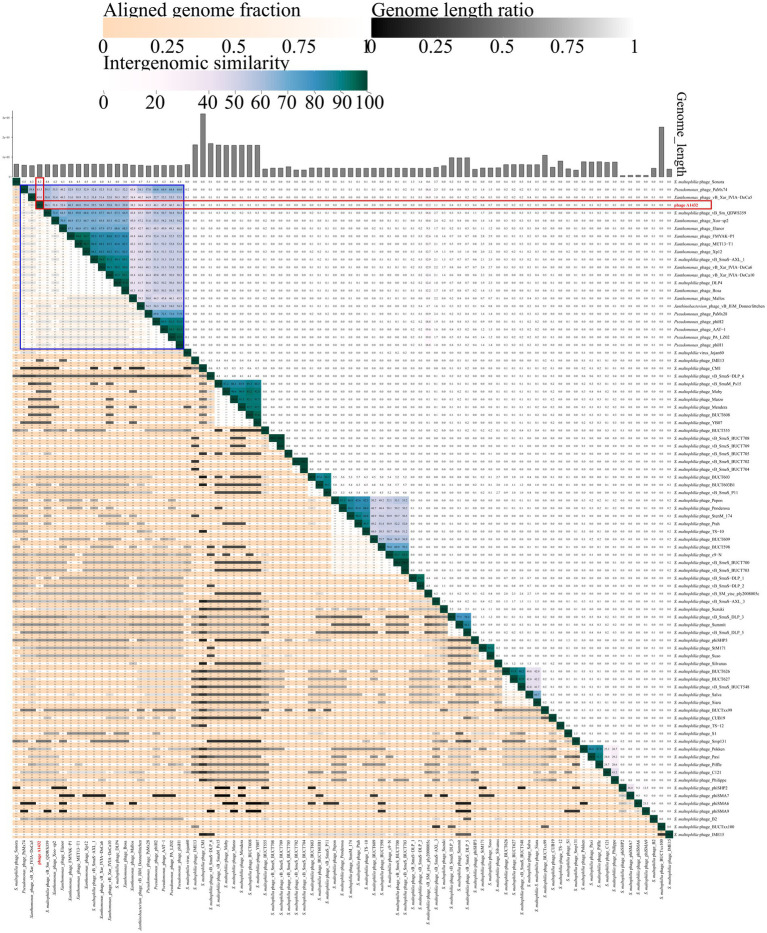
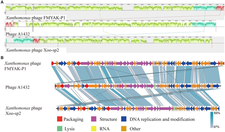
Stenotrophomonas maltophilia (S. maltophilia) is an emerging opportunistic pathogen that exhibits resistant to a majority of commonly used antibiotics. Phages have the potential to serve as an alternative treatment for S. maltophilia infections. In this study, a lytic phage, A1432, infecting S. maltophilia YCR3A-1, was isolated and characterized from a karst cave. Transmission electron microscopy revealed that phage A1432 possesses an icosahedral head and a shorter tail. Phage A1432 demonstrated a narrow host range, with an optimal multiplicity of infection of 0.1. The one-step growth curve indicated a latent time of 10 min, a lysis period of 90 min, a burst size of 43.2 plaque-forming units per cell. In vitro bacteriolytic activity test showed that phage A1432 was capable to inhibit the growth of S. maltophilia YCR3A-1 in an MOI-dependent manner after 2 h of co-culture. BLASTn analysis showed that phage A1432 genome shares the highest similarity (81.46%) with Xanthomonas phage Xoo-sp2 in the NCBI database, while the query coverage was only 37%. The phage contains double-stranded DNA with a genome length of 61,660 bp and a GC content of 61.92%. It is predicted to have 79 open reading frames and one tRNA, with no virulence or antibiotic resistance genes. Phylogenetic analysis using terminase large subunit and DNA polymerase indicated that phage A1432 clustered with members of the Bradleyvirinae subfamily but diverged into a distinct branch. Further phylogenetic comparison analysis using Average Nucleotide Identity, proteomic phylogenetic analysis, genomic network analysis confirmed that phage A1432 belongs to a novel genus within the Bradleyvirinae subfamily, Mesyanzhinovviridae family. Additionally, phylogenetic analysis of the so far isolated S. maltophilia phages revealed significant genetic diversity among these phages. The results of this research will contribute valuable information for further studies on their morphological and genetic diversity, will aid in elucidating the evolutionary mechanisms that give rise to them.

Keywords: Bradleyvirinae; Mesyanzhinovviridae; Stenotrophomonas maltophilia; karst cave; lytic phage; new genus; phage.


Conflict of interest statement

The authors declare that the research was conducted in the absence of any commercial or financial relationships that could be construed as a potential conflict of interest.

Figures

Figure 1
Figure 1
Morphology of phage A1432. (A) Phage plaques formed on the lawn of isolate YCR3A-1. (B) Transmission electron micrograph.
Figure 2
Figure 2
Biological characteristics of phage A1432. (A) One-step growth curve of phage A1432. (B) Temperature stability. (C) pH stability. (D) chloroform stability. (E) In vitro bacteriolytic activity of phage A1432 on host YCR3A-1. All assays were performed in triplicate, the error bars indicate the standard deviation (SD) of three replicates. One-way ANOVA and Dunnett’s post hoc test were used. **p < 0.01, ns: no significant difference (p > 0.05).
Figure 3
Figure 3
Genome map of phage A1432. ① and ② display the coding sequences (CDS) on the positive and negative strands, with different colors denoting distinct gene functions. ③ illustrates the GC content, while the innermost ④ represents the GC-Skew value.
Figure 4
Figure 4
Phylogenetic analysis of phage A1432. Phylogenetic trees of the terminase large subunit (A) and DNA polymerase I (B). The phylogenetic trees were constructed based on the amino acid sequences by MEGA 7.0 software. The evolutionary history was inferred by using the Maximum Likelihood method based on the JTT matrix-based model. All parameters are default except the bootstrap value is 1,000. (C) Proteomic tree of phage A1432 and its related phages by Viptree. The top 20 hit viruses in Genbank were manually added (red branches) to the Viptree database (last updated in November 17, 2023). Duplicate and unassociated sequences are removed. The classification status of viruses is derived from the ICTV (May 2024). Phage A1432 labeled with a red star. Branch lengths are shown on a logarithmic scale from the root of the tree. The inner nodes of the tree are shown as filled circles, each links to a genomic alignment of the sequences included in its subtree.
Figure 5
Figure 5
Genomic network map. Genomic network analysis was performed using vConTACT2, MCL, ClusterONE and Cytoscape. The reference database is Prokaryotic Viral RefSeq211-Merged (updated in June 2022), the top 20 similar sequences in Genbank were manually added. Duplicate and unassociated sequences are removed. Viruses are represented as circles (nodes) connected with each other (edges) based on a significant number of shared protein clusters. Circles in various colors indicate viruses of different genera and the classification status of viruses is derived from the ICTV (May 2024). Gray lines connect nodes in the network. Edges represents the strength between two genomes measured by significance score. Clusters of viruses are shown as boxes of different colors: VC_0_0 (red), VC_1_0 (pink), VC_2_0 (green), VC_5_0 (yellow), VC_7_0 (blue), VC_35_0 (purple).
Figure 6
Figure 6
Phylogenetic analysis of the genome sequences by VICTOR analysis. The analysis was performed on the genome sequences of 85 viruses, included the 20 best hit phages whose genome sequences closely related with phage A1432 by BLASTn analysis, 68 S. maltophilia phages (including A1432) with publicly-available genome sequences. Duplicate and unassociated sequences are removed. All the genome sequences of the phages were downloaded from NCBI database. The intergenomic distances were inferred using formula D6 with 53% support rate. ①②③④⑤ are derived from VICTOR analysis results. ①②③ represent the clustering information of the phages at the family, genus and species rank, different shapes and colors indicate different family/genus/species. ④ represent the G + C% content of the phages (35.38%/light gray - 67.41%/dark blue). ⑤ represent the genome length of the phages (Min: 5,819 bp; Max: 319,518 bp). ⑥ represent the genus name of the phage classified according to ICTV (February 2024), viruses without genus names indicate that they have not classified in ICTV.
Figure 7
Figure 7
VIRIDIC heatmap of phage A1432. Totally, 85 viruses included the 20 best hit phages whose genome sequences closely related with phage A1432 by BLASTn analysis, 68 S. maltophilia phages (including A1432) with publicly-available genome sequences. Duplicate and unassociated sequences are removed. The average nucleotide identity (ANI) of the pairwise intergenomic distances were calculated by VIRIDIC. The red box indicates the ANI value of A1432 compared to the 85 phages. The blue box indicates the ANI value of A1432 compared to the 20 best hit phages.
Figure 8
Figure 8
Genome comparison between A1432 and Xanthomonas phage Xoo-sp2 and FMYAK-P1. (A) Local blocks of colinearity between phages were analyzed using Mauve. The three colinear blocks between the three phages, indicated by different colors. The genome of phage A1432 was rearranged compared to the other two phages. (B) Comparison of gene levels of phages using Easyfig software. Arrows indicate predicted ORFs and are shown in different colors depending on the predicted function.

Similar articles

Cited by

References

    1. Ackermann H. W., Eisenstark A. (1974). The present state of phage taxonomy. Intervirology 3, 201–219. doi: 10.1159/000149758 - DOI - PubMed
    1. Aiewsakun P., Adriaenssens E. M., Lavigne R., Kropinski A. M., Simmonds P. (2018). Evaluation of the genomic diversity of viruses infecting bacteria, archaea and eukaryotes using a common bioinformatic platform: steps towards a unified taxonomy. J. Gen. Virol. 99, 1331–1343. doi: 10.1099/jgv.0.001110, PMID: - DOI - PMC - PubMed
    1. Alcock B. P., Huynh W., Chalil R., Smith K. W., Raphenya A. R., Wlodarski M. A., et al. (2023). CARD 2023: expanded curation, support for machine learning, resistome prediction at the comprehensive antibiotic resistance database. Nucleic Acids Res. 51, D690–d699. doi: 10.1093/nar/gkac920, PMID: - DOI - PMC - PubMed
    1. Arnaud C. A., Effantin G., Vivès C., Engilberge S., Bacia M., Boulanger P., et al. (2017). Bacteriophage T5 tail tube structure suggests a trigger mechanism for Siphoviridae DNA ejection. Nat. Commun. 8:1953. doi: 10.1038/s41467-017-02049-3, PMID: - DOI - PMC - PubMed
    1. Assefa M. (2022). Multi-drug resistant gram-negative bacterial pneumonia: etiology, risk factors, drug resistance patterns. Pneumonia (Nathan) 14:4. doi: 10.1186/s41479-022-00096-z, PMID: - DOI - PMC - PubMed