Review
. 2024 Jul 10;41(2):154-169.
doi: 10.1055/s-0044-1787165. eCollection 2024 Apr.

Recent Advances in Minimally Invasive Management of Osteolytic Periacetabular Skeletal Metastases

Affiliations
Review

Recent Advances in Minimally Invasive Management of Osteolytic Periacetabular Skeletal Metastases

Will Jiang et al. Semin Intervent Radiol. .

Abstract

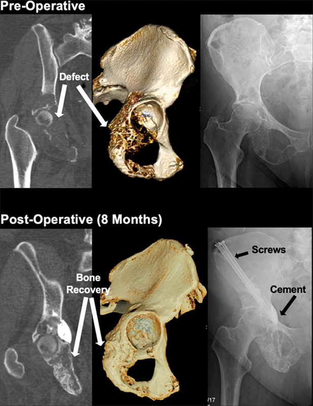
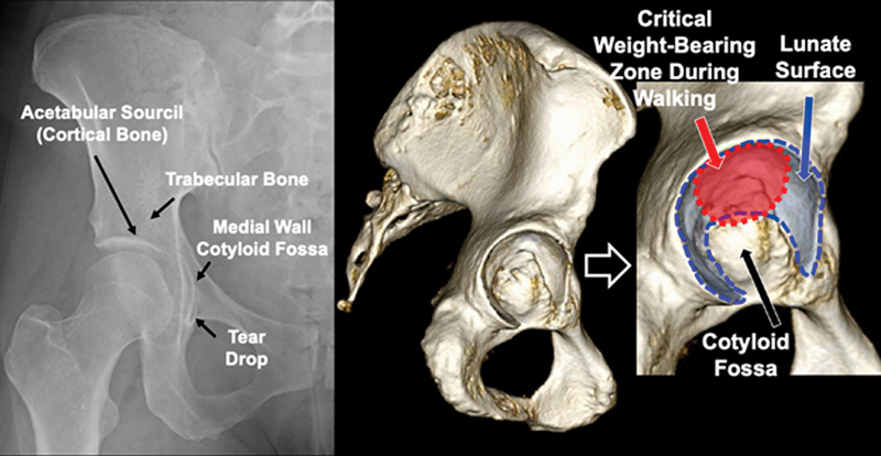
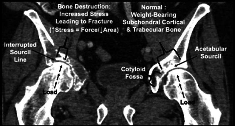
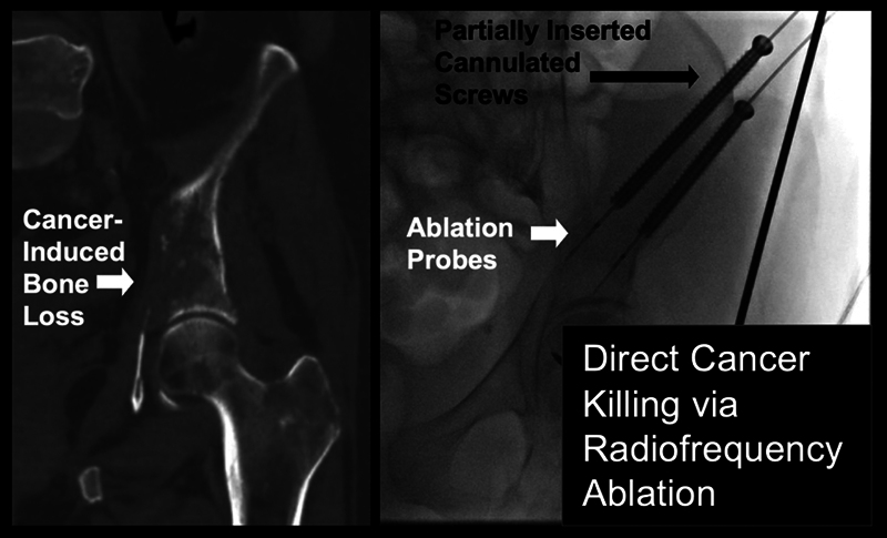
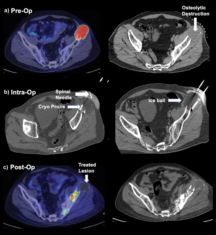
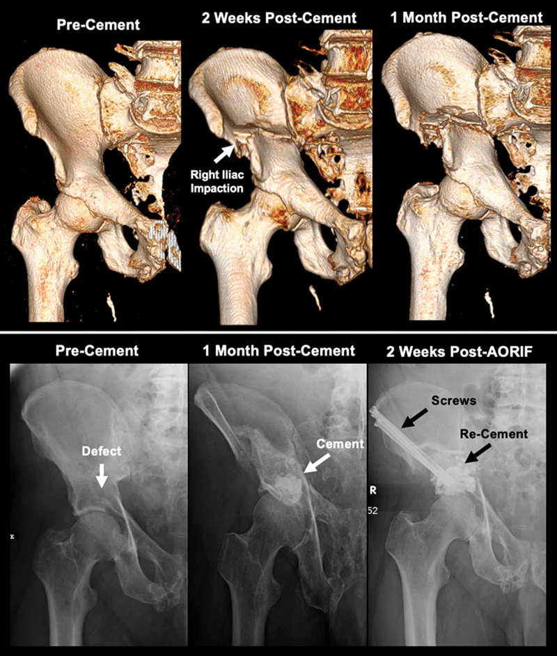
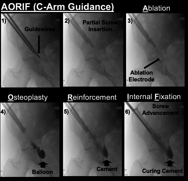
Painful skeletal osteolytic metastases, impending pathological fractures, and nondisplaced fractures present as a devastating clinical problem in advanced stage cancer patients. Open surgical approaches provide excellent mechanical stabilization but are often associated with high complication rates and slow recovery times. Percutaneous minimally invasive interventions have arisen as a pragmatic and logical treatment option for patients with late-stage cancer in whom open surgery may be contraindicated. These percutaneous interventions minimize soft tissue dissection, allow for the immediate initiation or resumption of chemotherapies, and present with fewer complications. This review provides the most up-to-date technical and conceptual framework for the minimally invasive management of osseous metastases with particular focus on periacetabular lesions. Fundamental topics discussed are as follows: (1) pathogenesis of cancer-induced bone loss and the importance of local cytoreduction to restore bone quality, (2) anatomy and biomechanics of the acetabulum as a weight-bearing zone, (3) overview of ablation options and cement/screw techniques, and (4) combinatorial approaches. Future studies should include additional studies with more long-term follow-up to better assess mechanical durability of minimally invasive interventions. An acetabulum-specific functional and pain scoring framework should be adopted to allow for better cross-study comparison.

Keywords: ablation; cement; metastases; percutaneous; periacetabular.


Figures

Fig. 1
Fig. 1
Pre- and postoperative bone mass improvement after radiofrequency ablation, cementoplasty, and screw fixation. A 61-year-old female with metastatic breast cancer with chemotherapy-resistant acetabular bone defect of the superior medial and posterior aspect of the right acetabulum. Preoperative imaging shows extensive bone mass loss of the ischium, posterior aspect of the right acetabulum, and superior medial aspect of the right acetabulum. The patient underwent radiofrequency ablation, cementoplasty, and screw fixation. At 8 months of follow-up, there is significant bone reconstitution visualized on imaging. Patient is clinically ambulating without pain at follow-up.
Fig. 2
Fig. 2
Functional anatomy of the acetabulum for targeted interventional reinforcement of acetabular bone defects secondary to osteolytic metastases. Effective delivery of bone cement above the acetabular sourcil is meaningful for maintenance and restoration of ambulatory function.
Fig. 3
Fig. 3
A clinical example of normal acetabulum (left) and pathologic fracture (right). Osteolytic bone defects in the weight-bearing region increases contact stress at the deficient acetabulum by the femoral head in the weight-bearing region. Skeletal intervention is aimed to reinforce the bone defect that is most pertinent to ambulation.
Fig. 4
Fig. 4
Radiofrequency ablation. Delivery of radiofrequency ablation in a 66-year-old female with metastatic lung cancer and cancer-induced bone loss in the supra-acetabular region. Ablation probes were advanced through partially inserted cannulated screws for positioning in the center of the lesion. In this technique, screws are partially inserted to allow for direct lesion ablation and subsequent filling with cement before screws are advanced through the curing cement into the final position. Left is 3 days preoperative and right image is intraoperative fluoroscopy.
Fig. 5
Fig. 5
Demonstrative CT and PET scan of a metastatic lung cancer patient presenting with severe pain due to an expansile lesion in the supra-acetabular region. ( a ) Preoperative imaging shows strong uptake on PET scan and visible osteolytic site on CT. ( b ) Image showing spinal needle placed in the subcutaneous plane for hydrodissection to protect against thermal damage to the skin during cryoablation. A total of five cryoprobes (yellow arrow) were used for treatment. Adjacent to the right shows hypodense ice ball. CT visibility is due to Hounsfield unit change of unfrozen to frozen tissue. Warm saline-filled glove was placed over the skin after the procedure to prevent frost bite from the remaining ice ball should hydrodissection disperse before the ice melts. ( c ) Follow-up PET CT shows no activity in the cryoablation-treated ASIS metastasis. There are other new foci of metastatic deposits in the rest of the pelvis.
Fig. 6
Fig. 6
A failed case of cement augmentation without screw fixation. A 61-year-old male with metastatic hepatocellular carcinoma who initially received cementoplasty without screw fixation (not done at the authors' institution) for a pathologic fracture. Two weeks later, postoperative imaging demonstrated impaction of the right iliac bone with worsening pain. Patient received radiation and chemotherapy. The patient presented at preoperative visit with 8/10 pain and ambulated with a walker. Patient received radiofrequency ablation, cementoplasty, and screw fixation (AORIF procedure). At 2 weeks postoperative, patient was able to walk comfortably with a cane and is rapidly improving as muscle weakness dissipates.
Fig. 7
Fig. 7
Guidewire placement via 3-View technique shot on C-arm fluoroscopy using the anterior iliac crest view, iliac wing view, and lateral view. Guidewire insertion on anterior iliac crest view should be within the inner and outer cortex boundaries. Confirmation of desired placement can be done on iliac wing and lateral view. Guidewire will be then used for screw placement.
Fig. 8
Fig. 8
Intraoperative fluoroscopy of combined ablation, screw fixation, and cementoplasty. Procedure begins with guidewire insertion at the anterior iliac crest. Fully threaded, cannulated screws are partially advanced at the boundary of the lesion site. Radiofrequency ablation probes are passed through the cannulated screws into the lesion. After, balloon osteoplasty is applied to create a cleaner space for subsequent PMMA cement injection. Finally, screws are advanced into the curing cement to their final position for full anchorage.

Similar articles

References

    1. Walker R H. Pelvic reconstruction/total hip arthroplasty for metastatic acetabular insufficiency. Clin Orthop Relat Res. 1993;(294):170–175. - PubMed
    1. Enneking W F, Dunham W K. Resection and reconstruction for primary neoplasms involving the innominate bone. J Bone Joint Surg Am. 1978;60(06):731–746. - PubMed
    1. Harrington K D. The management of acetabular insufficiency secondary to metastatic malignant disease. J Bone Joint Surg Am. 1981;63(04):653–664. - PubMed
    1. Houdek M T, Ferguson P C, Abdel M P et al. Comparison of porous tantalum acetabular implants and Harrington reconstruction for metastatic disease of the acetabulum. J Bone Joint Surg Am. 2020;102(14):1239–1247. - PMC - PubMed
    1. Tillman R M, Myers G J, Abudu A T, Carter S R, Grimer R J. The three-pin modified ‘Harrington’ procedure for advanced metastatic destruction of the acetabulum. J Bone Joint Surg Br. 2008;90(01):84–87. - PubMed