. 2024 May 17;8(4):102443.
doi: 10.1016/j.rpth.2024.102443. eCollection 2024 May.

Salvianolic acid B inhibits thrombosis and directly blocks the thrombin catalytic site

Affiliations

Salvianolic acid B inhibits thrombosis and directly blocks the thrombin catalytic site

Miguel A D Neves et al. Res Pract Thromb Haemost. .

Abstract

Background: Salvianolic acid B (SAB) is a major component of Salvia miltiorrhiza root (Danshen), widely used in East/Southeast Asia for centuries to treat cardiovascular diseases. Danshen depside salt, 85% of which is made up of SAB, is approved in China to treat chronic angina. Although clinical observations suggest that Danshen extracts inhibited arterial and venous thrombosis, the exact mechanism has not been adequately elucidated.

Objective: To delineate the antithrombotic mechanisms of SAB.

Methods: We applied platelet aggregation and coagulation assays, perfusion chambers, and intravital microscopy models. The inhibition kinetics and binding affinity of SAB to thrombin are measured by thrombin enzymatic assays, intrinsic fluorescence spectrophotometry, and isothermal titration calorimetry. We used molecular in silico docking models to predict the interactions of SAB with thrombin.

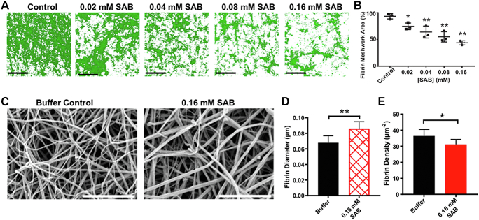
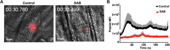
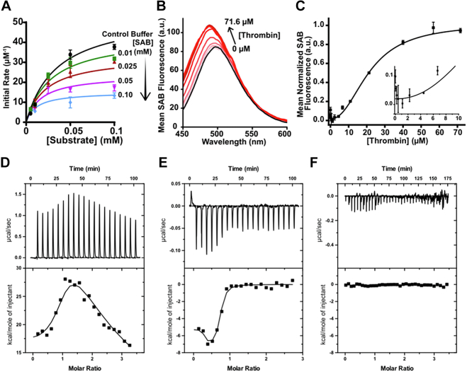
Results: SAB dose-dependently inhibited platelet activation and aggregation induced by thrombin. SAB also reduced platelet aggregation induced by adenosine diphosphate and collagen. SAB attenuated blood coagulation by modifying fibrin network structures and significantly decreased thrombus formation in mouse cremaster arterioles and perfusion chambers. The direct SAB-thrombin interaction was confirmed by enzymatic assays, intrinsic fluorescence spectrophotometry, and isothermal titration calorimetry. Interestingly, SAB shares key structural similarities with the trisubstituted benzimidazole class of thrombin inhibitors, such as dabigatran. Molecular docking models predicted the binding of SAB to the thrombin active site.

Conclusion: Our data established SAB as the first herb-derived direct thrombin catalytic site inhibitor, suppressing thrombosis through both thrombin-dependent and thrombin-independent pathways. Purified SAB may be a cost-effective agent for treating arterial and deep vein thrombosis.

Keywords: blood coagulation; platelet; salvianolic acid B (SAB); thrombin; thrombosis.


Figures

Figure 1
Figure 1
SAB inhibits thrombin-induced human platelet aggregation. Human gel-filtered platelet aggregation was induced with 0.02 U/mL thrombin (A, B). Panel A shows a representative single experiment, and panel B displays dose–response and statistical analyses of thrombin-induced aggregation as a function of SAB concentration. The EC50 dose is quantified (0.044 ± 0.01) mM SAB, R2 = 0.9974. (C) Human platelet activation analyses using flow cytometry. Platelets were labeled at room temperature for 30 minutes, and 10,000 events were captured on a Fortessa flow cytometer. The analyzed histograms and their corresponding activation markers are shown on top, and statistical analysis is displayed on bottom panels. The black, blue, and red lines indicate the groups of PBS treatment with no agonists, PBS treatment with thrombin, and SAB treatment with thrombin, respectively. ∗∗P value < .01, n = 3-4. EC50, effective dose concentration; PBS, phosphate-buffered saline; SAB, salvianolic acid B.
Figure 2
Figure 2
SAB inhibits human blood coagulation in vitro. (A) SAB decreased the dry weight of clots formed in whole blood. (B, C) SAB prolonged PT and aPTT at 0.6 mM. (D) Delay of blood coagulation in the clot turbidity assay. Superimposed turbidity plots as a function of SAB concentration. Turbidity at 405 nm was used as an indication of coagulation and monitored for 20 minutes at 37 °C. Differential turbidity values were analyzed using a Boltzmann kinetics model. SAB dosages from 0.08 to 0.32 mM were utilized. (E) The measured delay time of blood coagulation plots shown in panel D was analyzed as a function of SAB concentration, where an EC50 of 0.062 ± 0.07 mM SAB was quantified (dotted red line), R2 = 0.9844. ∗∗P < .01, ∗P < .05. aPTT, activated partial thromboplastin time; PT, prothrombin time; SAB, salvianolic acid B.
Figure 3
Figure 3
SAB reduces thrombin-induced fibrin network formation with rhFg. Human blood coagulation was induced by 0.02 U/mL bovine thrombin in rhFg. (A, B) Dose-dependent inhibition of surface fibrin clot coverage by SAB at dosages from 0.02 mM to 0.16 mM. Each black scale bar signifies 100 microns. (C, D, E) 0.16 mM SAB significantly increased the fibrin–fiber diameter and reduced fiber density. Each white scale bar measures 50 microns. ∗∗P < .01, ∗P < .05. rhFg, recombinant human fibrinogen; SAB, salvianolic acid B.
Figure 4
Figure 4
SAB inhibits thrombus formation in vivo. (A) Representative image of thrombus formation induced in a murine cremaster muscle laser injury model. The platelets were labeled in red. The scale bar denotes 10 microns. (B) SAB markedly reduced thrombus formation in vivo as shown by platelet MFI. n = 4. SAB was injected through the jugular vein cannula to a final in vivo concentration of 0.010 mM based on the weight of each mouse (80 μL/g total blood volume). MFI, mean fluorescence intensity; SAB, salvianolic acid B.
Figure 5
Figure 5
Direct binding of SAB to thrombin. (A) SAB directly inhibits the enzymatic activity of thrombin. Plot of initial rate vs substrate concentration at different SAB concentrations. Global Michaelis–Menten model (fit all curves to shared parameters). (B) Exhibits intrinsic fluorescence of 0.03 mM SAB (λex 382 nm) titrated with thrombin in PBS (pH 7.4) at 20 °C. (C) Data obtained from the fluorescence binding detection assay were fit to our previously published binding script. Intrinsic fluorescence of SAB shows direct binding to thrombin. The inset displays a zoomed view of 2 binding events, quenching and enhancement. Characterization of thrombin binding to (D) SAB and (E) dabigatran using ITC. (F) Shows the ITC titration of SAB into control lysozyme solution, where no binding was observed. ITC reveals direct binding of SAB to thrombin. For each ITC panel, on top is the raw titration data demonstrating the heat resulting from each injection of ligand into protein solution. The bottom shows the integrated heat plot after correcting for the heat of dilution. Binding experiments were performed in PBS buffer (pH 7.4) at 20 °C. n = 3 to 4. ITC, isothermal titration calorimetry; PBS, phosphate-buffered saline; SAB, salvianolic acid B.
Figure 6
Figure 6
SAB occupies the 2-site binding pocket of the trisubstituted benzimidazole thrombin inhibitors. Molecular structure of SAB (A) and dabigatran (B). Thrombin binding pocket interactions with (C, E) SAB in red and (D, F) the inhibitor dabigatran in fuchsia resolved using computational docking analysis. The blue ribbon displays the published X-ray crystal structure of thrombin (PDB: 1KTS). Purple side chains show interacting residues with the bound ligands. SAB, salvianolic acid B.

References

    1. Coller B.S. Historical perspective and future directions in platelet research. J Thromb Haemost. 2011;9:374–395. - PMC - PubMed
    1. Wang Y., Andrews M., Yang Y., Lang S., Jin J.W., Cameron-Vendrig A., et al. Platelets in thrombosis and hemostasis: old topic with new mechanisms. Cardiovasc Hematol Disord Drug Targets. 2012;12:126–132. - PubMed
    1. Xu X.R., Gallant R.C., Ni H. Platelets, immune-mediated thrombocytopenias, and fetal hemorrhage. Thromb Res. 2016;141:S76–S79. - PubMed
    1. Stroncek D.F., Rebulla P. Platelet transfusions. Lancet. 2007;370:427–438. - PubMed
    1. Ni H., Chen P., Spring C.M., Sayeh E., Semple J.W., Lazarus A.H., et al. A novel murine model of fetal and neonatal alloimmune thrombocytopenia: response to intravenous IgG therapy. Blood. 2006;107:2976–2983. - PMC - PubMed

LinkOut - more resources