. 2024 Jul 11;8(7):e110.
doi: 10.1002/hem3.110. eCollection 2024 Jul.

Clonal competition assays identify fitness signatures in cancer progression and resistance in multiple myeloma

Affiliations

Clonal competition assays identify fitness signatures in cancer progression and resistance in multiple myeloma

Larissa Haertle et al. Hemasphere. .

Abstract

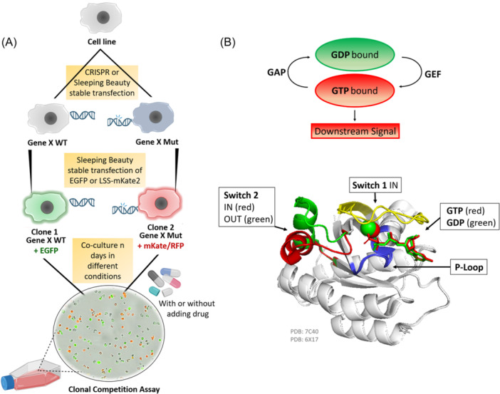
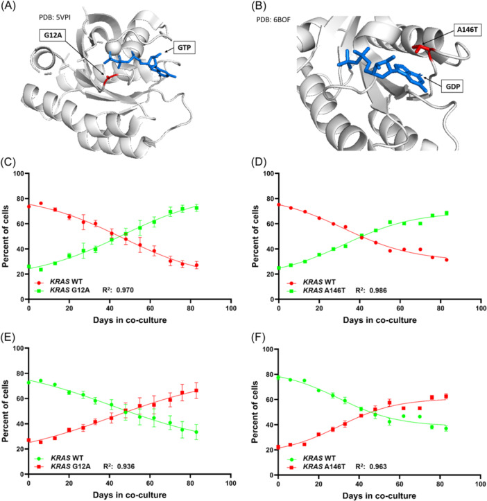
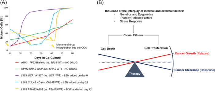
Multiple myeloma (MM) is a genetically heterogeneous disease and the management of relapses is one of the biggest clinical challenges. TP53 alterations are established high-risk markers and are included in the current disease staging criteria. KRAS is the most frequently mutated gene affecting around 20% of MM patients. Applying Clonal Competition Assays (CCA) by co-culturing color-labeled genetically modified cell models, we recently showed that mono- and biallelic alterations in TP53 transmit a fitness advantage to the cells. Here, we report a similar dynamic for two mutations in KRAS (G12A and A146T), providing a biological rationale for the high frequency of KRAS and TP53 alterations at MM relapse. Resistance mutations, on the other hand, did not endow MM cells with a general fitness advantage but rather presented a disadvantage compared to the wild-type. CUL4B KO and IKZF1 A152T transmit resistance against immunomodulatory agents, PSMB5 A20T to proteasome inhibition. However, MM cells harboring such lesions only outcompete the culture in the presence of the respective drug. To better prevent the selection of clones with the potential of inducing relapse, these results argue in favor of treatment-free breaks or a switch of the drug class given as maintenance therapy. In summary, the fitness benefit of TP53 and KRAS mutations was not treatment-related, unlike patient-derived drug resistance alterations that may only induce an advantage under treatment. CCAs are suitable models for the study of clonal evolution and competitive (dis)advantages conveyed by a specific genetic lesion of interest, and their dependence on external factors such as the treatment.


Conflict of interest statement

Joaquin Martinez‐Lopez and Santiago Barrio are equity shareholders of Altum Sequencing Co. The remaining authors declare no competing financial interests.

Figures

Figure 1
Figure 1
Clonal competition assay (CCA) for the study of the long‐term impact of genomic alterations on the overall cell fitness, and KRAS in its active and inactive conformational shape. (A) Schematic illustration of the general principle of the CCA. Initially, the alteration of interest (e.g., point mutation, deletion, etc.) is introduced into a cell line by gene editing approaches like CRISPR Cas9 or Sleeping Beauty (SB). Then, SB vectors for the stable expression of fluorescent proteins (e.g., EGFP or mKate/RFP) are integrated. Different colors are used to differentiate between the WT and the mutant or between different alterations of interest. Co‐cultures are set up by mixing cells of different colors. The culture is regularly split and drugs can be added. Every 4–5 days a sample is taken from the culture and the ratio of differently colored cells is measured by flow cytometry. (B) Schematic representation of RAS superfamily proteins activation and inactivation cycle by hydrolyzing GTP into GDP via the GTPase activating proteins (GAPs) and the Guanine exchange factor (GEF) proteins triggering or stopping downstream signaling. Superimposition of the cartoon diagram of KRAS displaying the conformational change in switch 2 induced by the hydrolysis of GTP changing from the signaling conformation (red, PDB: 6XI7, 20 ) to the nonsignaling conformation (green, PDB: 7C40, 21 ). The backbone is shown in white, the P‐loop Walker Motif A in blue, and switch 1 in yellow as it is in the IN conformation in both PDBs.
Figure 2
Figure 2
Clonal competition assays of OPM2 sublines bearing KRAS mutations G12A or A146T compared to OPM2 KRAS WT cells. (A) Structural representation of the oncogenic KRAS mutation G12A (PDB: 5VPI, 42 ) in complex with GTP and the magnesium ion cofactor. (B) KRAS A146T mutation in complex with GDP (PBD: 6BOF, 40 ), there is no magnesium ion coordination in this structure. The mutated residues are represented in red. GTP and GDP are displayed in blue; for clarity no different colors indicating charges are assigned to any atom. Clonal Competition Assays reveal a gradual increase of the mutants KRAS G12A (C, E) and KRAS A146T (D, F) outcompeting the OPM2 KRAS WT cells due to enhanced fitness. This effect was confirmed by technical replicates and by switching the color coding between WT and mutant cells. The error bars indicate the two‐paired 95% CI.
Figure 3
Figure 3
RNA‐seq transcriptome analysis and differentially expressed genes (DEGs) in the KRAS A146T and G12A mutation‐bearing cells. (A) Principal‐component analysis (PCA) results. The figure shows a scatter plot of the first two principal components (PC1 and PC2) depicting the distribution of the gene expression profile according to the mutational status. Percentages on each axis represent the percentages of variation explained by the principal components. (B) Bar plots showing expression levels in transcripts per million (TPM) in KRAS mutants and KRAS WT. (C) Volcano plots indicating gene expression differences between KRAS mutants and KRAS WT. DEGs with an adjusted p < 0.05 and a log2FC >│1│ are depicted in colors. Triangles indicate genes with a –Log10 adjusted p‐value higher than 300. (D) Heatmap of the 57 overlapping DEGs in both comparisons. Log2FC > 0 is indicated in red and log2FC < 0 in blue. Venn diagrams of all DEGs (upper panel), upregulated DEGs (middle panel), and downregulated DEGs (lower panel) found in the two comparisons.
Figure 4
Figure 4
Gene Set Enrichment Analysis (GSEA) results of the G12A mutation‐bearing cells. (A) Barplot with GSEA results depicting positively and negatively enriched gene sets in KRAS G12A altered cells related to proliferation, cell division, growth, or cell death. A false discovery rate (FDR) of <0.25 was chosen as the cut‐off. (B) Enrichment plots of the PI3K‐Akt, MAPK, and JAK‐STAT signaling pathways. The enrichment profile is indicated by the red line. (C) Heatmap of log2FC on overlapping genes shared by the MAPK, JAK‐STAT, and PI3K‐Akt pathways in the GSEA analysis. Each row represents one of the three evaluated pathways and the columns represent those 28 genes that were shared at least by two of the three pathways and had a substantial impact on the GSEA analysis.
Figure 5
Figure 5
Clonal competition assays (CCA) examples of different genetic alterations and models of the cell death/cell growth equilibrium with regard to clonal fitness and therapy. (A) Alterations in drug resistance‐associated genes (CUL4B and PSMB5) only confer a proliferative fitness in the presence of the respective drug (Lenalidomide or Bortezomib). In the absence of selection pressure, the WT cells outcompete the mutant‐bearing sublines in the CCAs. On the contrary, KRAS and TP53 alterations intrinsically confer a growth advantage to the affected cells. Shown here is a meta‐analysis of CCAs made for previously published studies, and the OPM2 KRAS WT vs KRAS G12A comparison. (B) The complex interplay between clonal fitness and antitumor therapy determines if the cancer gets cleared as a response to the treatment or if it grows, provoking a relapse. The CCA is a suitable tool to study the interaction and outcome by modulating internal and external factors of the system.

References

    1. John L, Poos AM, Brobeil A, et al. Resolving the spatial architecture of myeloma and its microenvironment at the single‐cell level. Nat Commun. 2023;14(1):5011. 10.1038/s41467-023-40584-4 - DOI - PMC - PubMed
    1. Barrio S, Munawar U, Zhu YX, et al. IKZF1/3 and CRL4(CRBN) E3 ubiquitin ligase mutations and resistance to immunomodulatory drugs in multiple myeloma. Haematologica. 2020;105(5):e237‐e241. 10.3324/haematol.2019.217943 - DOI - PMC - PubMed
    1. Morgan GJ, Walker BA, Davies FE. The genetic architecture of multiple myeloma. Nat Rev Cancer. 2012;12(5):335‐348. 10.1038/nrc3257 - DOI - PubMed
    1. Manier S, Salem KZ, Park J, Landau DA, Getz G, Ghobrial IM. Genomic complexity of multiple myeloma and its clinical implications. Nat Rev Clin Oncol. 2017;14(2):100‐113. 10.1038/nrclinonc.2016.122 - DOI - PubMed
    1. Bolli N, Avet‐Loiseau H, Wedge DC, et al. Heterogeneity of genomic evolution and mutational profiles in multiple myeloma. Nat Commun. 2014;5:2997. 10.1038/ncomms3997 - DOI - PMC - PubMed