. 2024 Jun 12:5:uqae014.
doi: 10.1093/femsml/uqae014. eCollection 2024.

Coordinated regulation of osmotic imbalance by c-di-AMP shapes ß-lactam tolerance in Group B Streptococcus

Affiliations

Coordinated regulation of osmotic imbalance by c-di-AMP shapes ß-lactam tolerance in Group B Streptococcus

Terry Brissac et al. Microlife. .

Abstract

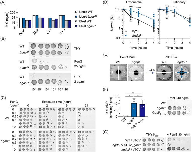
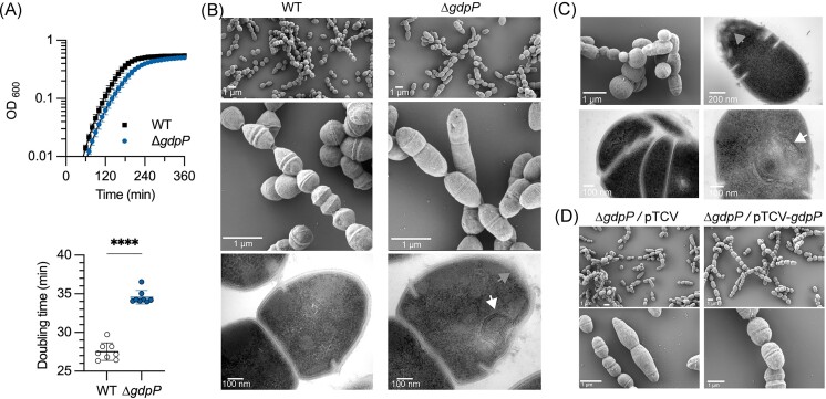
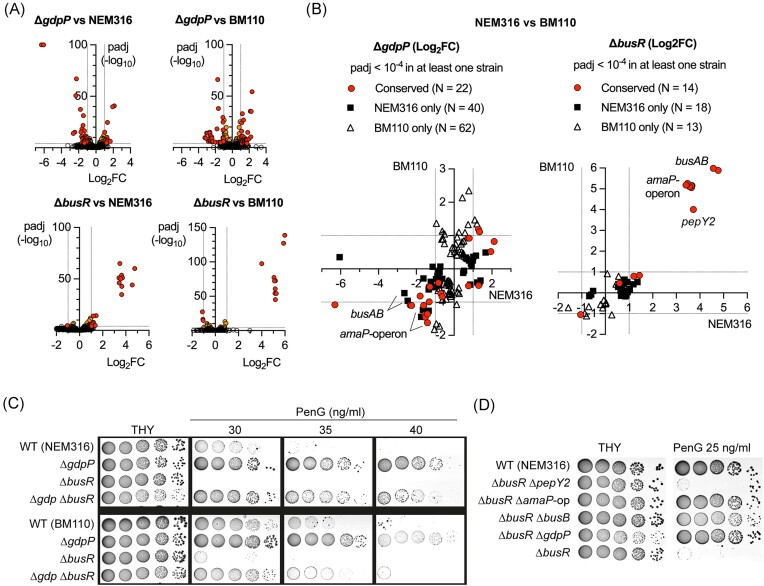
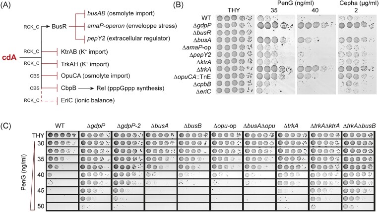
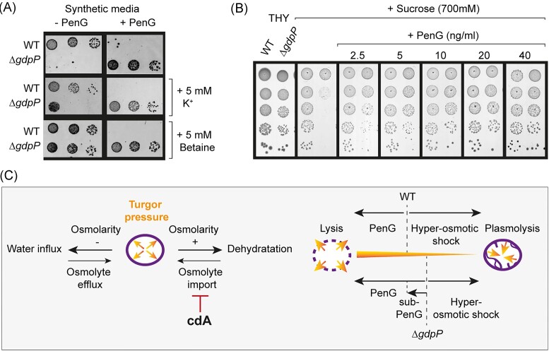
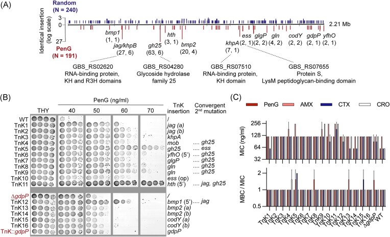
Streptococcus agalactiae is among the few pathogens that have not developed resistance to ß-lactam antibiotics despite decades of clinical use. The molecular basis of this long-lasting susceptibility has not been investigated, and it is not known whether specific mechanisms constrain the emergence of resistance. In this study, we first report ß-lactam tolerance due to the inactivation of the c-di-AMP phosphodiesterase GdpP. Mechanistically, tolerance depends on antagonistic regulation by the repressor BusR, which is activated by c-di-AMP and negatively regulates ß-lactam susceptibility through the BusAB osmolyte transporter and the AmaP/Asp23/GlsB cell envelope stress complex. The BusR transcriptional response is synergistic with the simultaneous allosteric inhibition of potassium and osmolyte transporters by c-di-AMP, which individually contribute to low-level ß-lactam tolerance. Genome-wide transposon mutagenesis confirms the role of GdpP and highlights functional interactions between a lysozyme-like hydrolase, the KhpAB RNA chaperone and the protein S immunomodulator in the response of GBS to ß-lactam. Overall, we demonstrate that c-di-AMP acts as a turgor pressure rheostat, coordinating an integrated response at the transcriptional and post-translational levels to cell wall weakening caused by ß-lactam activity, and reveal additional mechanisms that could foster resistance.

Keywords: Streptococcus; antibiotic; cell wall; nucleotide signaling; osmolytes; turgor pressure.


Conflict of interest statement

The authors declare no competing interests.

Figures

Figure 1.
Figure 1.
Inactivation of the cyclic-di-AMP phosphodiesterase GdpP confers beta-lactam tolerance. (A) MIC of antibiotics: conventional tests in liquid (broth microdilution) and solid (gradient Etest strips) in MH-F media with the WT strain and the ∆gdpP c-di-AMP phosphodiesterase deletion mutant for penicillin G (PenG), amoxicillin (AMX), and the third-generation cephalosporin cefotaxime (CTX), and ceftriaxone (CRO). (B) Spotting assays of the WT strain and ∆gdpP mutant: overnight cultures were serially diluted (101–10−5) and spotted on the surface of THY agar supplemented with PenG or the first-generation cephalosporin cephalexin (CEX). Images were taken after 16–24 h incubation at 37°C in aerobic condition with 5% CO2. (C) Dose and time-dependent killing of the WT strain and ∆gdpP mutant. High PenG concentrations (0.1–10 µg/ml) are added to exponential growing cultures of the WT strain and ∆gdpP mutant. Aliquots were taken at the indicate time, serial diluted, and spotted on THY without antibiotic. (D) Time-killing of exponential and stationary phase cultures of the WT strain and ∆gdpP mutant in presence of 10 µg/ml PenG. Aliquots were taken at the indicate time and dilution were plated on THY for colony-forming unit numeration. Data are from seven and six independent experiments for exponential and stationary time, respectively. Unpaired t-test were used to compare the WT and the mutant at each time point (* P < .05; ****  P < .0001). (E) Adaptation of the TD-test for GBS antibiotic tolerance. Diluted cultures of the WT strain and ∆gdpP mutant are spread on MH media and PenG disks (10 µg) were added. After a 24-h incubation, PenG disks were removed and replaced by glucose (Glc) disks (10 mg). Crossed white arrows highlight inhibition zones after 24 h, and crossed blue arrows highlight the decrease in the inhibition zone of the ∆gdpP mutant after an additional 24 h incubation with the Glc disk. (F) Penicillin tolerance depends on the GdpP phosphodiesterase activity. Left panel: quantification of intracellular c-di-AMP by LC-MS in the WT strain, the ∆gdpP mutant, and the catalytic inactivated GdpPD419A mutant. Data represent means and SD calculated from biological triplicate (N = 3) and analyzed with unpaired t-test (* P < .05; ** P < .01). Right panel: spotting assays on PenG plate. (G) Genetic complementation of the ∆gdpP mutant. Spotting assay with the WT and the ∆gdpP mutant containing the empty (pTCV) or complementing (pTCV_gdpP) vectors. Kanamycin (K500) is added to maintain the selective pressure for the vectors.
Figure 2.
Figure 2.
Cyclic-di-AMP phosphodiesterase deficiency leads to morphological and cell envelope defects. (A) Growth curves (upper panel) and doubling time in exponential phase (bottom panel) of the WT strain and ∆gdpP mutant in THY at 37°C. Means and SD are calculated from biological replicates (N = 8) and analyzed using unpaired t-test (****  P < .0001). (B) SEM and TEM of the WT strain and ∆gdpP mutant at similar scales. The white and grey arrows highlight intracytoplasmic membrane structures and areas with cell envelopes of heterogeneous thickness, respectively. (C) Additional electronic microscopy illustrating the heterogeneity and atypical ultrastructure of ∆gdpP cells. (D) Representative images of the ∆gdpP mutant with the empty (pTCV) or complementing (pTCV_gdpP) vectors observed by SEM.
Figure 3.
Figure 3.
ß-lactam tolerance depends on the c-di-AMP-activated BusR repressor. (A) Transcriptome analysis by RNA-seq of ∆gdpP and ∆busR mutants in two WT backgrounds (NEM316 and BM110). Colored dots on volcano plots highlight significant differential expression (P-adj < 10−4) with fold changes |FC| > 2 (red dots) and |FC| < 2 (orange dots). (B) Comparative analysis of the ∆gdpP and ∆busR transcriptomes between the two WT backgrounds. Red dots highlight conserved statistical significance (P-adj < 10−4) in the two strains, and black squares and white triangles highlight statistical significance in NEM316 only or BM110 only, respectively. (C) Tolerance of ∆gdpP to ß-lactam depends on the cdA-activated BusR repressor. Spotting assays (10−1–10−5 serial dilution) of ∆gdpP, ∆busR, and the double ∆gdpPbusR mutants in the two WT backgrounds on THY supplemented with penicillin. (D) Susceptibility of ∆busR to ß-lactam depends on busB and amaP operon. Spotting assays with ∆busR, and the double ∆busRpepY2, ∆busRamaP-operon, ∆busRbusB, and ∆busRgdpP mutants in the NEM316 background.
Figure 4.
Figure 4.
Coordinated regulation of osmolyte transporters confers ß-lactam tolerance. (A) Diagram of the c-di-AMP (cdA)-signaling network in GBS. The cdA-binding domain (RCK_C or CBS) of each effector is indicated on the connecting lines, with arrows indicating activation and final bars indicating repression or inhibition. The dotted line denotes predicted cdA binding to the RCK_C domain of EriC that has not been demonstrated experimentally. (B) Individual effectors contribute to ß-lactam susceptibility. Spotting assays (10−1–10−5 serial dilution) of a collection of deletion (∆) or insertion (:: TnE) mutants for each individual cdA effector on ß-lactam-containing plates. (C) Additive effect of cdA effectors on penicillin susceptibility. Spotting assays of single and double deletion mutants on plates containing increasing concentrations of penicillin, in increments of 2.5 ng/ml. An independent ∆gdpP-2 mutant was included for validation.
Figure 5.
Figure 5.
C-di-AMP balances osmotic and ß-lactam susceptibilities. (A) Penicillin alleviates ∆gdpP osmolyte requirements on minimal media. Spotting assays of the WT strain and ∆gdpP mutant on synthetic media supplemented with penicillin, potassium, and betaine at the indicated concentration. (B) Subinhibitory ß-lactam concentrations counteract ∆gdpP osmo-susceptibility. Spotting assays of the WT strain and ∆gdpP mutant under hyperosmotic condition (THY supplemented with sucrose) with and without penicillin. (C) Osmotic regulation by cdA and adaptation to osmotic imbalances. Left panel: the cellular turgor pressure is mainly regulated through osmolytes exchanges to respond to change in osmolarity. The import of osmolytes is necessary to counteract cellular dehydration due to increased osmolarity and is inhibited by cdA. Right panel: the high cdA concentration in the ∆gdpP mutant results in a hyperosmotic stress physiology in a standard environment. The advantage against the bactericidal activity of ß-lactam is at the cost of sensitivity to hyper osmotic condition. Fluidification of the cell wall with ß-lactam subinhibitory concentration allows osmolyte exchange and re-establishes a WT-like turgor pressure.
Figure 6.
Figure 6.
Genome-wide screening for ß-lactam tolerance in GBS. (A) Distribution of transposon insertions on the GBS chromosome. Vertical bars represent identical transposon insertions along the 2.1-Mb genome in randomly isolated colonies on THY (N = 240, upper blue bars) or THY supplemented with 35–40 ng/ml PenG (N = 191, lower red bars). Gene names are specified for mutants selected for secondary screening. The numbers in brackets indicated the total number of transposon insertions followed by the number of independent integration sites. Gene IDs and predicted functions are shown for the four genes showing a convergent mutation pattern. (B) Spotting assay with isolated insertional mutants (TnK) on plates with increasing PenG concentration. Genes inactivated by transposon insertion are indicated on the right, with independent insertion in the same gene indicated by letters (a and b), and whether the insertion is in the promoter (5') or in nonmonocistronic transcripts (-op). Convergent secondary mutations identified after genome sequencing are indicated after the dotted lines. Deletion (∆) and insertional (Tnk::) gdpP mutants are highlighted in red. (C) MIC and MBC for ß-lactams. MIC are determined in liquid (MH-F media) by serial dilution for penicillin G (PenG) amoxicillin (AMX), cefotaxime (CTX), and ceftriaxone (CRO). MBC are determined by numeration of viable bacteria after 16 h of contact with the antibiotic in MH-F. Data are mean and SD of two independent experiments (N = 2).

Similar articles

References

    1. Albarracin Orio AG, Pinas GE, Cortes PR et al. Compensatory evolution of pbp mutations restores the fitness cost imposed by beta-lactam resistance in Streptococcus pneumoniae. PLoS Pathog. 2011;7:e1002000. - PMC - PubMed
    1. Argudin MA, Roisin S, Nienhaus L et al. Genetic diversity among Staphylococcus aureus isolates showing oxacillin and/or cefoxitin resistance not linked to the presence of mec genes. Antimicrob Agents Chemother. 2018;62:e00091–18. - PMC - PubMed
    1. Ba X, Kalmar L, Hadjirin NF et al. Truncation of GdpP mediates β-lactam resistance in clinical isolates of Staphylococcus aureus. J Antimicrob Chemother. 2019;74:1182–91. - PubMed
    1. Bandera AM, Bartho J, Lammens K et al. BusR senses bipartite DNA binding motifs by a unique molecular ruler architecture. Nucleic Acids Res. 2021;49:10166–77. - PMC - PubMed
    1. Barros EM, Martin MJ, Selleck EM et al. Daptomycin resistance and tolerance due to loss of function in Staphylococcus aureus dsp1 and asp23. Antimicrob Agents Chemother. 2019;63:e01542–01518. - PMC - PubMed

LinkOut - more resources