. 2023 May 19:2:1176384.
doi: 10.3389/frtra.2023.1176384. eCollection 2023.

Recipient TIM4 signaling regulates ischemia reperfusion-induced ER stress and metabolic responses in liver transplantation: from mouse-to-human

Affiliations

Recipient TIM4 signaling regulates ischemia reperfusion-induced ER stress and metabolic responses in liver transplantation: from mouse-to-human

Hirofumi Hirao et al. Front Transplant. .

Abstract

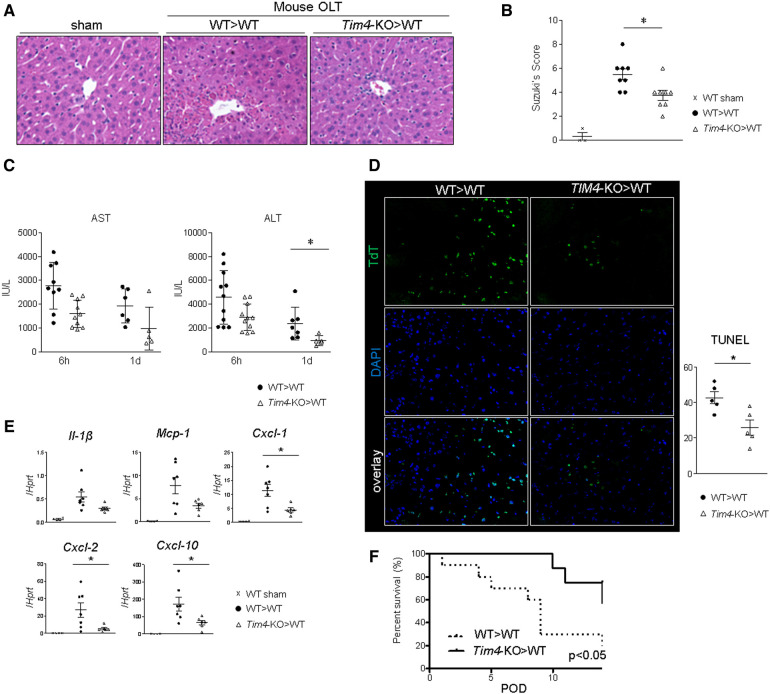
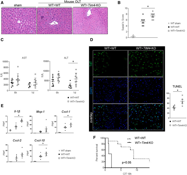
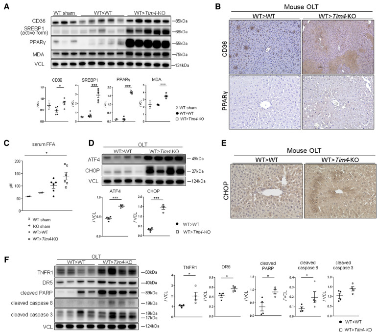
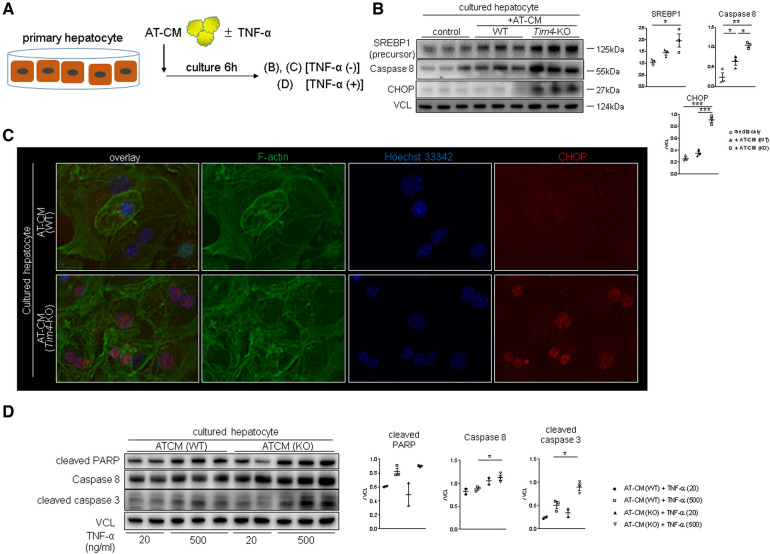
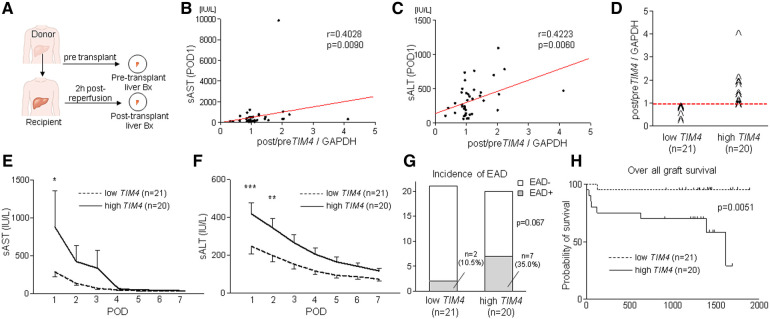
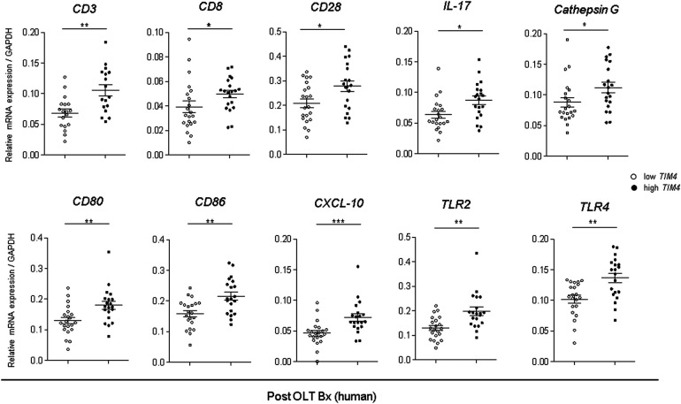
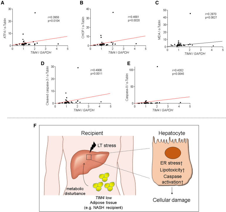
T-cell immunoglobulin and mucin (Tim)4 is expressed on APCs, including macrophages, as one of the main amplifiers in the mechanism of liver ischemia-reperfusion injury (IRI) following orthotopic liver transplantation (OLT). Though donor Tim4 selectively expressed on Kupffer cells serves as a checkpoint regulator of innate immune-driven IRI cascades, its role on cells outside the OLT remains unclear. To dissect the role of donor vs. recipient-specific Tim4 signaling in IR-induced stress and hepatocellular function, we employed a murine OLT model utilizing Tim4-knockout (KO) mice as either donor or recipient (WT → WT, WT → Tim4-KO, Tim4-KO → WT). In the experimental arm, disruption of donor Tim4 attenuated IRI-OLT damage, while recipient Tim4-null mutation aggravated hepatic IRI concomitant with disturbed lipid metabolism, enhanced endoplasmic reticulum stress, and activated pro-apoptotic signaling in the grafts. In the in vitro study, murine hepatocytes co-cultured with Tim4-null adipose tissue showed enhanced C/EBP homologous protein (CHOP) expression pattern and susceptibility to hepatocellular death accompanied by activated caspase cascade in response to TNF-α stimulation. In the clinical arm, liver grafts from forty-one transplant patients with enhanced TIM4 expression showed higher body mass index, augmented hepatic endoplasmic reticulum stress, enhanced pro-apoptotic markers, upregulated innate/adaptive immune responses, exacerbated hepatocellular damage, and inferior graft survival. In conclusion, although TIM4 is considered a principal villain in peri-transplant early tissue injury, recipient TIM4 signaling may serve as a savior of IR-triggered metabolic stress in mouse and human OLT recipients.

Keywords: endoplasmic reticulum stress; ischemia-reperfusion injury; liver transplantation; orthotopic liver tranplantation; t-cell immunoglobulin and mucin domain containing 4.


Conflict of interest statement

The authors declare that the research was conducted in the absence of any commercial or financial relationships that could be construed as a potential conflict of interest.

Figures

Figure 1
Figure 1
Donor Tim4-null mutation alleviates the hepatocellular damage and inflammatory response in IR-stressed mouse OLT: WT or Tim4-KO donor livers, stored in UW solution (4°C/18 h), were transplanted into WT recipients. OLT samples were collected at 6 h and 24 h post-reperfusion. (A) sAST and sALT levels (IU/L). (B) Representative H&E staining (original magnification, ×100) of sham livers and OLT (WT > WT, Tim4-KO > WT). (C) Suzuki's histological grading of liver IRI (n = 8/group). Data shown as mean ± SEM (*p < 0.05, 1-way ANOVA followed by Tukey's HSD test). (D) Representative TUNEL images and quantification of TUNEL + cells (n = 5/group). Data shown as mean ± SEM (*p < 0.05, Mann-Whitney U test). (E) qRT-PCR-assisted detection of mRNA coding for Il-1β, Mcp1, Cxcl1, Cxcl2 and Cxcl10. Data were normalized to Hprt gene expression. Sham: n = 4/group; OLT: n = 6–7/group. Data shown as mean ± SEM (*p < 0.05, Mann-Whitney U test). (F) Recipient mice after OLT were monitored for 14 days and cumulative survival was analyzed (Kaplan-Meier method). Dotted line: WT > WT; solid line Tim4-KO > WT (n = 10/group, *p < 0.05, log-rank test).
Figure 2
Figure 2
Recipient Tim4 disruption aggravates the hepatocellular damage and inflammatory response in IR-stressed mouse OLT: (A) sAST and sALT levels (IU/L). (B) Representative H&E staining (original magnification, ×100) of sham livers and OLT (WT > WT, Tim4-KO > WT). (C) Suzuki's histological grading of liver IRI (n = 8/group). Data shown as mean ± SEM (*p < 0.05, 1-way ANOVA followed by Tukey's HSD test). (D) Representative TUNEL images and quantification of TUNEL + cells (n = 5/group). Data shown as mean ± SEM (*p < 0.05, Mann–Whitney U-test). (E) qRT-PCR-assisted detection of mRNA coding for Il-1β, Mcp1, Cxcl1, Cxcl2 and Cxcl10. Data were normalized to Hprt gene expression. Sham: n = 3/group; OLT: n = 6–7/group. Data shown as mean ± SEM (*p < 0.05, Mann–Whitney U-test). (F) Recipient mice were monitored for 14 days and cumulative survival was analyzed (Kaplan–Meier method). Dotted line: WT > WT; solid line WT > Tim4-KO (n = 10/group, *p < 0.05, log-rank test).
Figure 3
Figure 3
Recipient Tim4-null mutation provokes hepatic lipid metabolism pathway and augments ER stress and apoptotic markers in OLT: (A) Western blot of CD36, SREBP1, PPARγ and MDA in sham and OLT livers (WT > WT, WT > Tim4-KO). The relative intensity to VCL was calculated. Data shown as mean ± SEM (*p < 0.05, ***p < 0.001, 1-way ANOVA followed by Tukey's HSD test). (B) Representative immunohistochemistry of CD36 and PPAR-γ in OLT livers (n = 2/group). (C) Serum FFA levels in sham and OLT mouse recipients. Data shown as mean ± SEM (*p < 0.05, 1-way ANOVA followed by Tukey's HSD test). (D) WB-assisted detection of ATF4, CHOP and VCL. The relative intensity to VCL was calculated. Data shown as mean ± SEM (***p < 0.001, Mann–Whitney U-test). (E) Representative staining for CHOP in OLT (n = 3/group). (F) WB of TNFR1, DR5, cleaved PARP, caspase 8, caspase 3 and VCL in OLT. The relative intensity to VCL was calculated. Data shown as mean ± SEM (*p < 0.05, Mann–Whitney U-test).
Figure 4
Figure 4
Tim4 deficient adipose tissue enhances hepatocyte SREBP1/CHOP and promotes TNF-α mediated cell death in vitro: (A) the scheme of the vitro experiment. (B) WB-assisted detection of SREBP1, caspase 8, CHOP and VCL. The relative intensity to VCL was calculated. Data shown as mean ± SEM (*p < 0.05, **p < 0.01, ***p < 0.001, 1-way ANOVA followed by Tukey's HSD test). (C) Representative immunocytochemistry of F-actin (green), nucleus (blue) and CHOP (red) in murine hepatocyte cultured with AT-CM from WT or Tim4-KO mice (n = 2). Original magnification, ×400. (D) WB of cleaved PARP, caspase 8, cleaved caspase 3 and VCL in cultured hepatocytes. The relative intensity to VCL was calculated. Data shown as mean ± SEM (*p < 0.05, 1-way ANOVA followed by Tukey's HSD test).
Figure 5
Figure 5
Peri-transplant TIM4 gene levels are associated with the hepatocellular function and OLT outcomes: (A) Pre-transplant (post–cold storage) and post-transplant (2 h after reperfusion) liver biopsies (Bx) were collected from forty-one OLT patients and analyzed for TIM4 by qRT-PCR with normalization to GAPDH. The relationship between TIM4 and sAST (B) and sALT (C) at POD1. r: Spearman correlation coefficient. (D) Human OLT Bx (2 h post-reperfusion) were classified into low (n = 21) and high (n = 20) TIM4 expression groups. (E,F) Serum AST and ALT levels at POD1-7 (*p < 0.05, **p < 0.01, ***p < 0.001, 2-way ANOVA, Bonferroni post hoc test; data shown as mean ± SEM. (G) Incidence of early allograft dysfunction (EAD) (Fisher's exact test). (H) The cumulative probability of overall graft survival. The solid line indicates TIM4-high, while the dotted line depicts TIM4-low OLT patient cohorts (Kaplan–Meier method, log-rank test).
Figure 6
Figure 6
Augmented innate and adaptive gene activation in human OLT is accompanied by high TIM4 levels: qRT-PCR-assisted detection of mRNA coding for CD3, CD8, CD28, IL-17, Cathepsin G, CD80, CD86, CXCL-10, TLR2, and TLR4. Data normalized to GAPDH gene expression are shown in dot plots and bars indicative mean ± SEM (*p < 0.05, ** p < 0.01, *** p < 0.001, Mann–Whitney U-test).
Figure 7
Figure 7
TIM4 expression is positively correlates with hepatic ATF4, CHOP, MDA, caspase 8 and cleaved caspase 3 levels in human OLT: the relationship between peri-transplant TIM4 levels and ATF4 (A) CHOP (B) MDA (C) cleaved caspase 3 (D) and caspase 8 (E) expression in OLT at 2 h after reperfusion. r: Spearman correlation coefficient. (F) A simplified scheme of how TIM4 signaling may affect IR-stress and hepatocellular damage in OLT recipients.

Similar articles

References

    1. Hirao H, Nakamura K, Kupiec-Weglinski JW. Liver ischaemia-reperfusion injury: a new understanding of the role of innate immunity. Nat Rev Gastroenterol Hepatol. (2022) 19(4):239–56. 10.1038/s41575-021-00549-8 - DOI - PubMed
    1. Kupiec-Weglinski JW. Grand challenges in organ transplantation. Front Transpl. (2022) 1:897679. 10.3389/frtra.2022.897679 - DOI
    1. Meyers JH, Chakravarti S, Schlesinger D, Illes Z, Waldner H, Umetsu SE, et al. TIM-4 is the ligand for TIM-1, and the TIM-1-TIM-4 interaction regulates T cell proliferation. Nat Immunol. (2005) 6(5):455–64. 10.1038/ni1185 - DOI - PubMed
    1. Liu W, Xu L, Liang X, Liu X, Zhao Y, Ma C, et al. Tim-4 in health and disease: friend or foe? Front Immunol. (2020) 11:537. 10.3389/fimmu.2020.00537 - DOI - PMC - PubMed
    1. Wong K, Valdez PA, Tan C, Yeh S, Hongo JA, Ouyang W. Phosphatidylserine receptor tim-4 is essential for the maintenance of the homeostatic state of resident peritoneal macrophages. Proc Natl Acad Sci U S A. (2010) 107(19):8712–7. 10.1073/pnas.0910929107 - DOI - PMC - PubMed

LinkOut - more resources