. 2023 Nov 9:2:1233322.
doi: 10.3389/frtra.2023.1233322. eCollection 2023.

Farnesyltransferase-inhibitors exert in vitro immunosuppressive capacity by inhibiting human B-cells

Affiliations

Farnesyltransferase-inhibitors exert in vitro immunosuppressive capacity by inhibiting human B-cells

Shilei Xu et al. Front Transplant. .

Abstract

Objectives: Farnesyltransferase inhibitors (FTI), which inhibit the prenylation of Ras GTPases, were developed as anti-cancer drugs. As additional target proteins for prenylation were identified in the past, it is likely that FTI have potential value for therapeutic purposes beyond cancer. The effect of FTI on B-cells remains unclear. To address this issue, we investigated the effects of in vitro FTI treatment on effector and regulatory B-cells in healthy controls and renal transplant patients.

Methods: For this purpose, B-cells were isolated from the peripheral blood of healthy controls and renal transplant patients. Purified B-cells were stimulated via Toll-like-receptor 9 (TLR-9) in the presence or absence of FTI. Regulatory functions, such as IL-10 and Granzyme B (GrB) secretion, were assessed by flow cytometry. In addition, effector B-cell functions, such as plasma cell formation and IgG secretion, were studied.

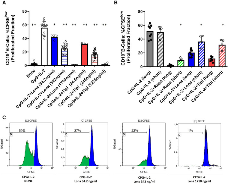
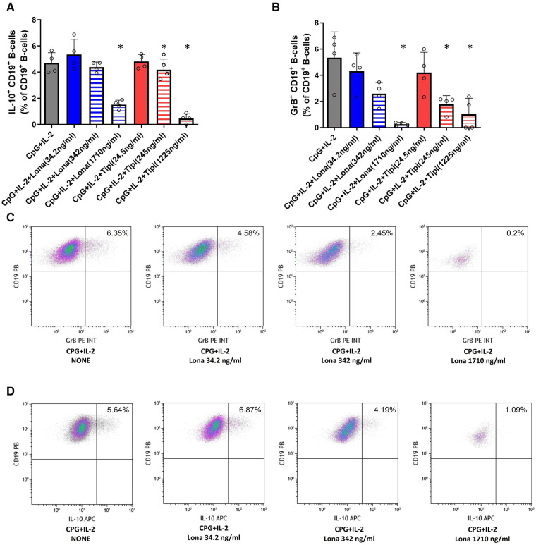
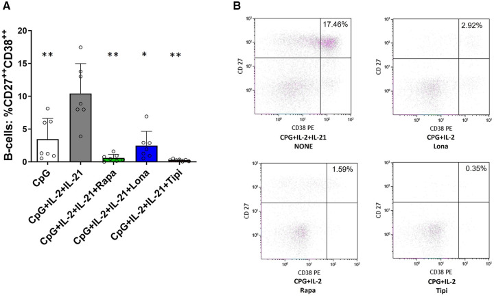
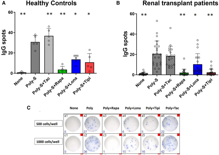
Results: The two FTI Lonafarnib and tipifarnib both suppressed TLR-9-induced B-cell proliferation. Maturation of IL-10 producing B-cells was suppressed by FTI at high concentrations as well as induction of GrB-secreting B-cells. Plasma blast formation and IgG secretion were potently suppressed by FTI. Moreover, purified B-cells from immunosuppressed renal transplant patients were also susceptible to FTI-induced suppression of effector functions, evidenced by diminished IgG secretion.

Conclusion: FTI suppress in vitro B-cell proliferation and plasma cell formation while partially preserving IL-10 as well as GrB production of B-cells. Thus, FTI may have immunosuppressive capacity encouraging further studies to investigate the potential immunomodulatory value of this agent.

Keywords: B-cells; farnesyltransferase inhibitors; humoral rejection; plasma cells; renal transplantation.


Conflict of interest statement

The authors declare that the research was conducted in the absence of any commercial or financial relationships that could be construed as a potential conflict of interest. The authors BW declared that they were an editorial board member of Frontiers, at the time of submission. This had no impact on the peer review process and the final decision.

Figures

Figure 1
Figure 1
Dose-dependent suppression of TLR9-induced B-cell proliferation by FTI. (A) Purified B-cells from healthy donors were labeled with CFSE and stimulated with CpG plus IL-2 in the presence of different concentrations of FTI (tipifarnib and lonafarnib). After 72 h, CD19+ B-cell proliferation was determined by CFSE dilution. Stimulation with CPG + IL-2 in absence of FTI served as control condition. Statistical significance was calculated by repeated-measures ANOVA and corrections for multiple comparisons were done by Dunnett's test (all conditions were compared vs. CPG + IL-2; *p: < 0.05, **p: < 0.005). CpG + IL-2 n = 16, Lona (34.2 ng/ml) n = 4, Lona (342 ng/ml) n = 16, Lona (1710 ng/ml) n = 4, Tipi (24.5 ng/ml) n = 4, Tipi (245 ng/ml) n = 16, Tipi (1,225 ng/ml) n = 4. (B) B-cells from healthy donors were exposed either for only 24 h to FITs (short) or over the whole culture period (long). B-cells were stimulated in all conditions with CPG + IL-2. For short exposure to FTI, B-cells were washed after the first 24 h of culture followed by culture with CPG + IL-2 in the absence of FTI/rapamycin for an additional period of 48 h. Tipi and lona were used at concentrations of 245 ng/ml and 342 ng/ml, respectively. Statistical significance was calculated by repeated-measures ANOVA and corrections for multiple comparisons were done by Dunnett's test (the respective conditions with short exposure were compared to the matching conditions with long exposure. *p: < 0.05). CpG + IL-2 (long) n = 8, CpG + IL-2 (short) n = 4, Rapa (long) n = 8, Rapa (short) n = 4, Lona (long) n = 8, Lona (short) n = 4, Tipi (long) n = 8, Tipi (short) n = 4. (C) Representative flow cytometric data. Plots are gated on viable CD19+ B-cells. Proliferation data was analyzed by gating on the cells that have undergone at least one round of division and is given as “Proliferated fraction”.
Figure 2
Figure 2
Effect of FTI on the expression of costimulatory molecules and CD25 on activated B-cells. CD19+ B-cells from healthy donors were stimulated with CpG in the presence of FTI for 72 h. Rapamycin was used as a comparative immunosuppressive agent. Stimulation with CPG in absence of FTI served as control condition. Tipi and lona were used at concentrations of 245 ng/ml and 342 ng/ml, respectively.The expression of (A) PDL-1, (B) CD27 and (C) CD25 was analyzed by flow cytometry after 72 h of culture. For the determination of the MFI, cells were gated on viable CD19+ B-cells (all conditions n = 5). Statistical significance was calculated by repeated-measures ANOVA and corrections for multiple comparisons were done by Dunnett's test (all conditions were compared vs. CPG; *p < 0.05, **p: < 0.005). (D) Representative flow cytometric data. Plots are gated on viable CD19+ B-cells. B-cells with detectable expression of the respective costimulators or CD25 are depicted in the upper right quadrant.
Figure 3
Figure 3
Impact of FTI on IL-10+ B-cells and GrB+ regulatory B-cells. B-cells from healthy donors were incubated for 72 h with FTI at different concentrations in the presence of CpG plus IL-2. After three days, the production of IL-10 and GrB by CD19+ B-cells was determined by flow cytometry. Stimulation with CPG + IL-2 in absence of FTI served as control condition. Tipi and lona were used at concentrations of 245 ng/ml and 342 ng/ml, respectively. (A) Impact of FTI on IL-10+ CD19+ B-cells upon CpG plus IL-2 stimulation (all conditions n = 4). (B) Impact of FTI on GrB+ CD19+ B-cells upon CpG plus IL-2 stimulation (all conditions n = 4). Statistical significance was calculated by repeated-measures ANOVA and corrections for multiple comparisons were done by Dunnett's test (all conditions were compared vs. CpG + IL-2; *p: < 0.05). (C) and (D) Representative flow cytometric data of GrB and IL-10 expressing B-cells. Plots are gated on viable CD19+ B-cells. B-cells with detectable IL-10 or GrB expression are depicted in the upper right quadrant.
Figure 4
Figure 4
Impact of FTI on plasma cell formation. (A) Plasma cell formation is efficiently inhibited by FTI. B-cells from healthy donors were stimulated for 6 days in absence or presence of FTI. Differentiation into plasma cells was determined by flow cytometry (all conditions n = 7). Stimulation with CPG + IL-2 + IL-21 in absence of FTI served as control condition.Tipi and lona were used at concentrations of 245 ng/ml and 342 ng/ml, respectively. Statistical significance was calculated by repeated-measures ANOVA and corrections for multiple comparisons were done by Dunnett's test (all conditions were compared vs. CPG + IL-2 + IL-21, *p: < 0.05, **p: < 0.005). (B) Representative flow cytometric data. Plots are gated on viable CD19+ B-cells. Plasma cells were defined as B-cells with high of expression of CD27 and CD38 (CD27++CD38++, upper right quadrant).
Figure 5
Figure 5
Effect of FTI on IgG secretion by plasma cells. Purified CD19+ B-cells were initially cultured at 5 × 104 cells/well under Poly-S (10 μg/ml) stimulation in the presence of FTI for 4 days and then transferred to ELISpot plates at a density of 1,000 cells/well for another 24 h. Rapamycin and tacrolimus were used as controls. IgG secretion was detected by EliSpot. Tipifarnib (Tipi) and lonafarnib (Iona) were used at concentrations of 245 ng/ml and 342 ng/ml, respectively. (A) Impact of FTI on IgG secretion by plasma cells from HC (n = 5). (B) Impact of FTI on IgG secretion by plasma cells from renal transplant patients (n = 26; none n = 20, Poly-S n = 23, Poly-S + Rapa n = 22, Poly-S + Lona n = 19, Poly-S + Tipi n = 25, Poly-S + Tax n = 18). (C) Representative photos of IgG-specific ELISpot plates. Statistical significance was calculated by repeated-measures ANOVA and corrections for multiple comparisons were done by Dunnett's test (all conditions were compared vs. Poly-S, *p: < 0.05, **p: < 0.005).

Similar articles

Cited by

References

    1. Abe T, Ishii D, Gorbacheva V, Kohei N, Tsuda H, Tanaka T, et al. Anti-Hucd20 antibody therapy for antibody-mediated rejection of renal allografts in a mouse model. Am J Transplant. (2015) 15(5):1192–204. 10.1111/ajt.13150 - DOI - PMC - PubMed
    1. Rodriguez-Ramirez S, Al Jurdi A, Konvalinka A, Riella LV. Antibody-mediated rejection: prevention, monitoring and treatment dilemmas. Curr Opin Organ Transplant. (2022) 27(5):405–14. 10.1097/MOT.0000000000001011 - DOI - PMC - PubMed
    1. Karp JE, Lancet JE, Kaufmann SH, End DW, Wright JJ, Bol K, et al. Clinical and biologic activity of the farnesyltransferase inhibitor R115777 in adults with refractory and relapsed acute leukemias: a phase 1 clinical-laboratory correlative trial. Blood. (2001) 97(11):3361–9. 10.1182/blood.v97.11.3361 - DOI - PubMed
    1. Si MS, Ji P, Tromberg BJ, Lee M, Kwok J, Ng SC, et al. Farnesyltransferase inhibition: a novel method of immunomodulation. Int Immunopharmacol. (2003) 3(4):475–83. 10.1016/S1567-5769(02)00278-3 - DOI - PubMed
    1. Marchwicka A, Kamińska D, Monirialamdari M, Błażewska KM, Gendaszewska-Darmach E. Protein prenyltransferases and their inhibitors: structural and functional characterization. Int J Mol Sci. (2022) 23(10):5424. 10.3390/ijms23105424 - DOI - PMC - PubMed

LinkOut - more resources