

The emerging role of Piezo1 in the musculoskeletal system and disease
- PMID: 38994033
- PMCID: PMC11234281
- DOI: 10.7150/thno.96959
The emerging role of Piezo1 in the musculoskeletal system and disease
Abstract
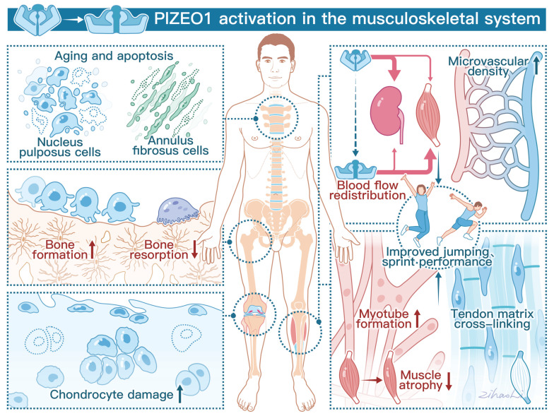
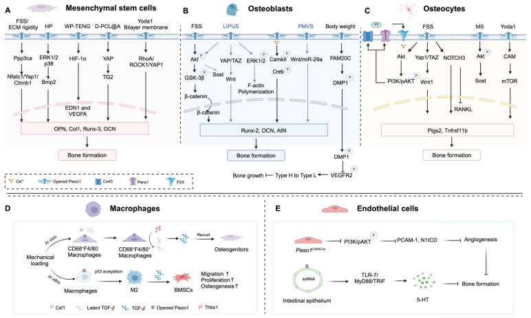
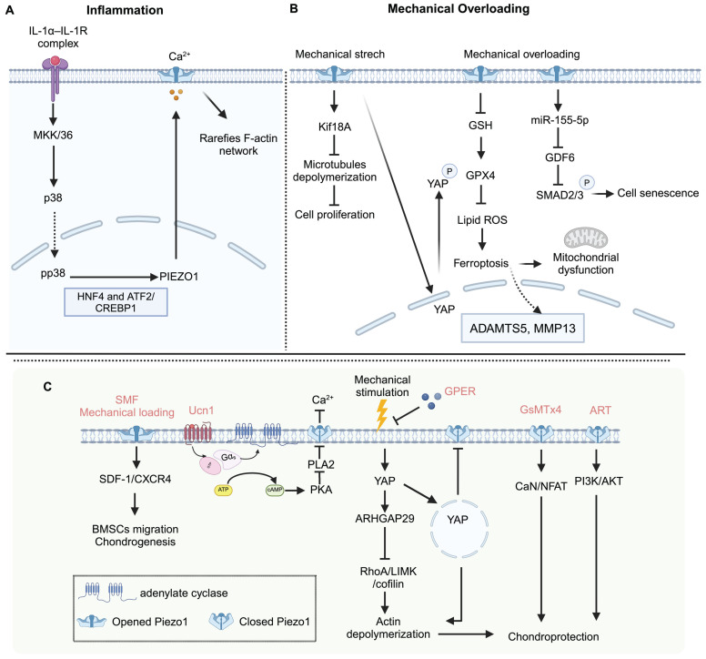
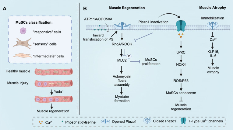
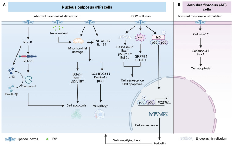
Piezo1, a mechanosensitive ion channel, has emerged as a key player in translating mechanical stimuli into biological signaling. Its involvement extends beyond physiological and pathological processes such as lymphatic vessel development, axon growth, vascular development, immunoregulation, and blood pressure regulation. The musculoskeletal system, responsible for structural support, movement, and homeostasis, has recently attracted attention regarding the significance of Piezo1. This review aims to provide a comprehensive summary of the current research on Piezo1 in the musculoskeletal system, highlighting its impact on bone formation, myogenesis, chondrogenesis, intervertebral disc homeostasis, tendon matrix cross-linking, and physical activity. Additionally, we explore the potential of targeting Piezo1 as a therapeutic approach for musculoskeletal disorders, including osteoporosis, muscle atrophy, intervertebral disc degeneration, and osteoarthritis.
Keywords: Piezo1; bone; cartilage; intervertebral disc; muscle.
© The author(s).
Conflict of interest statement
Competing Interests: The authors have declared that no competing interest exists.
Figures
Similar articles
-
The role of PIEZO ion channels in the musculoskeletal system.Am J Physiol Cell Physiol. 2023 Mar 1;324(3):C728-C740. doi: 10.1152/ajpcell.00544.2022. Epub 2023 Jan 30. Am J Physiol Cell Physiol. 2023. PMID: 36717101 Free PMC article. Review.
-
Mechanosensitive Ion Channel Piezo1 Activated by Matrix Stiffness Regulates Oxidative Stress-Induced Senescence and Apoptosis in Human Intervertebral Disc Degeneration.Oxid Med Cell Longev. 2021 Feb 10;2021:8884922. doi: 10.1155/2021/8884922. eCollection 2021. Oxid Med Cell Longev. 2021. PMID: 33628392 Free PMC article.
-
The mechanosensitive Piezo1 channel is required for bone formation.Elife. 2019 Jul 10;8:e47454. doi: 10.7554/eLife.47454. Elife. 2019. PMID: 31290742 Free PMC article.
-
The State of the Art of Piezo1 Channels in Skeletal Muscle Regeneration.Int J Mol Sci. 2022 Jun 14;23(12):6616. doi: 10.3390/ijms23126616. Int J Mol Sci. 2022. PMID: 35743058 Free PMC article. Review.
-
Piezo-Type Mechanosensitive Ion Channel Component 1 (Piezo1): A Promising Therapeutic Target and Its Modulators.J Med Chem. 2022 May 12;65(9):6441-6453. doi: 10.1021/acs.jmedchem.2c00085. Epub 2022 Apr 24. J Med Chem. 2022. PMID: 35466678 Review.
Cited by
-
Tensile force promotes osteogenic differentiation via ephrinB2-EphB4 signaling pathway in orthodontic tooth movement.BMC Oral Health. 2025 Jan 22;25(1):118. doi: 10.1186/s12903-025-05491-8. BMC Oral Health. 2025. PMID: 39844202 Free PMC article.
-
Mechanosensitive Piezo1 channel: an emerging target in demyelination disease.Front Cell Neurosci. 2025 Jul 9;19:1556892. doi: 10.3389/fncel.2025.1556892. eCollection 2025. Front Cell Neurosci. 2025. PMID: 40703567 Free PMC article. Review.
-
PIEZO1-Mediated Calcium Signaling and Podocyte Injury in Diabetic Kidney Disease.J Am Soc Nephrol. 2025 Feb 11;36(7):1310-1326. doi: 10.1681/ASN.0000000634. J Am Soc Nephrol. 2025. PMID: 39932793
-
Mechanosensitive miRNAs in Cartilage and Subchondral Bone Remodeling: Emerging Targets for Osteoarthritis Therapy.J Inflamm Res. 2025 Jul 19;18:9609-9625. doi: 10.2147/JIR.S529149. eCollection 2025. J Inflamm Res. 2025. PMID: 40703641 Free PMC article. Review.
-
Pathophysiologic Mechanisms of Severe Spinal Cord Injury and Neuroplasticity Following Decompressive Laminectomy and Expansive Duraplasty: A Systematic Review.Neurol Int. 2025 Apr 16;17(4):57. doi: 10.3390/neurolint17040057. Neurol Int. 2025. PMID: 40278428 Free PMC article. Review.
References
-
- Douguet D, Honoré E. Mammalian Mechanoelectrical Transduction: Structure and Function of Force-Gated Ion Channels. Cell. 2019;179:340–54. - PubMed
-
- Martinac B. Mechanosensitive ion channels: molecules of mechanotransduction. J Cell Sci. 2004;117:2449–60. - PubMed
-
- Honoré E, Martins JR, Penton D, Patel A, Demolombe S. The Piezo Mechanosensitive Ion Channels: May the Force Be with You! Rev Physiol Biochem Pharmacol. 2015;169:25–41. - PubMed
Publication types
MeSH terms
Substances
LinkOut - more resources
Full Text Sources
