

Fabrication and evaluation of a bi-layered electrospun PCL/PVA patch for wound healing: Release of vitamins and silver nanoparticle
- PMID: 38994056
- PMCID: PMC11238126
- DOI: 10.1016/j.heliyon.2024.e33178
Fabrication and evaluation of a bi-layered electrospun PCL/PVA patch for wound healing: Release of vitamins and silver nanoparticle
Abstract
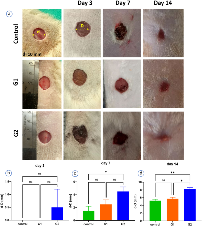
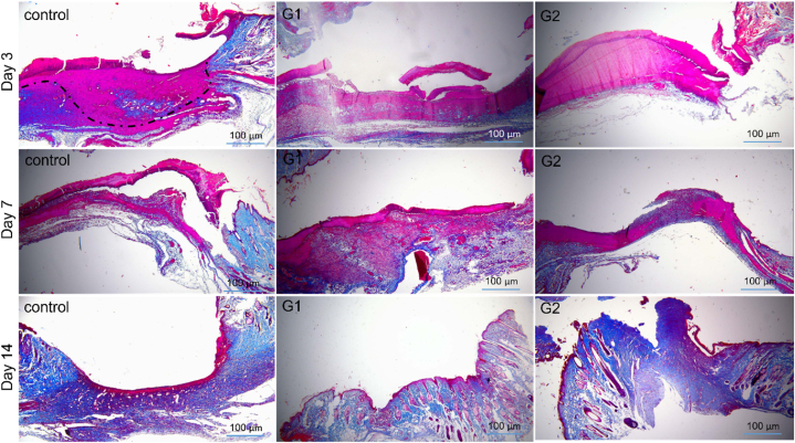
There is still little research on the co-delivery of vitamins and AgNPs to accelerate wound healing. In this study, a bi-layered electrospun PCL/PVA patch loaded with Vitamin C, Vitamin B12, and AgNPs was fabricated using a co-spinning technique. SEM, FTIR, degradation, swelling, tensile strength, disk diffusion, and MTT assay were studied. Nine rats were placed in three groups (control: no treatment, G1: without agents, and G2: with agents) for 14 days in an in-vivo study. H&E and Masson Trichrome staining were employed for histological analysis. Results showed that the final electrospun wound dressings depicted nanofibers with diameters ranging from 100 to 500 nm. The presence of AgNP enhanced the mechanical strength (40-50 MPs). An appropriate swelling (100 %) and degradation (50 %) rate was observed for groups with no significant difference (P > 0.05). G1 and G2 did not show a significant difference in terms of porosity (65 % vs. 69 %). Regarding WVTR, G2 demonstrated higher WVTR (88 vs. 95 g/m2. h). G2 showed a vitamin release of more than 90 % after 48 h. Compared to G1, G2 demonstrated good antibacterial activity (>3 cm) against E. Coli and S. aureous (P < 0.01), with cell viability of more than 93 % (P > 0.05). Furthermore, the in-vivo study approved that G2 accelerated wound healing in full-thickness wounds, compared to the control groups, with notable wound size reduction (8 mm), epithelialization, and collagen formation. The findings support the use of this simple but potent electrospun wound dressing for the healing of full-thickness wounds.
Keywords: Bi-layered; Co-delivery; Electrospinning; Nanoparticle; Vitamin; Wound dressing.
© 2024 The Authors.
Conflict of interest statement
The authors declare that they have no known competing financial interests or personal relationships that could have appeared to influence the work reported in this paper.
Figures


Similar articles
-
A comparative study of wound dressings loaded with silver sulfadiazine and silver nanoparticles: In vitro and in vivo evaluation.Int J Pharm. 2019 Jun 10;564:350-358. doi: 10.1016/j.ijpharm.2019.04.068. Epub 2019 Apr 24. Int J Pharm. 2019. PMID: 31028800
-
Wound dressing based on electrospun PVA/chitosan/starch nanofibrous mats: Fabrication, antibacterial and cytocompatibility evaluation and in vitro healing assay.Int J Biol Macromol. 2019 Feb 1;122:238-254. doi: 10.1016/j.ijbiomac.2018.10.115. Epub 2018 Oct 18. Int J Biol Macromol. 2019. PMID: 30342125
-
Physical and biological properties of blend-electrospun polycaprolactone/chitosan-based wound dressings loaded with N-decyl-N, N-dimethyl-1-decanaminium chloride: An in vitro and in vivo study.J Biomed Mater Res B Appl Biomater. 2020 Nov;108(8):3084-3098. doi: 10.1002/jbm.b.34636. Epub 2020 May 27. J Biomed Mater Res B Appl Biomater. 2020. PMID: 32459395
-
Silver/Snail Mucous PVA Nanofibers: Electrospun Synthesis and Antibacterial and Wound Healing Activities.Membranes (Basel). 2022 May 20;12(5):536. doi: 10.3390/membranes12050536. Membranes (Basel). 2022. PMID: 35629862 Free PMC article.
-
Co-electrospun nanofibrous mats loaded with bitter gourd (Momordica charantia) extract as the wound dressing materials: in vitro and in vivo study.BMC Complement Med Ther. 2021 Apr 7;21(1):111. doi: 10.1186/s12906-021-03284-4. BMC Complement Med Ther. 2021. PMID: 33827547 Free PMC article.
Cited by
-
Copper Nanoparticle Loaded Electrospun Patches for Infected Wound Treatment: From Development to In-Vivo Application.Polymers (Basel). 2024 Sep 27;16(19):2733. doi: 10.3390/polym16192733. Polymers (Basel). 2024. PMID: 39408444 Free PMC article.
-
Mechanical Properties of Biodegradable Fibers and Fibrous Mats: A Comprehensive Review.Molecules. 2025 Aug 5;30(15):3276. doi: 10.3390/molecules30153276. Molecules. 2025. PMID: 40807450 Free PMC article. Review.
-
Formulation and Evaluation of Licorice-Extract-Enhanced Chitosan, PVA, and Gelatin-Derived Hydrogels for Wound Dressing.Bioengineering (Basel). 2025 Apr 23;12(5):439. doi: 10.3390/bioengineering12050439. Bioengineering (Basel). 2025. PMID: 40428058 Free PMC article.
-
Research Progress on the Application of Novel Wound Healing Dressings in Different Stages of Wound Healing.Pharmaceutics. 2025 Jul 28;17(8):976. doi: 10.3390/pharmaceutics17080976. Pharmaceutics. 2025. PMID: 40870999 Free PMC article. Review.
References
-
- Li Y., Zhao W., Chen S., Zhai H., Wu S. Bioactive electrospun nanoyarn-constructed textile dressing patches delivering Chinese herbal compound for accelerated diabetic wound healing. Mater. Des. 2024;237
-
- Li H., Wang M., Williams G.R., Wu J., Sun X., Lv Y., Zhu L.-M. Electrospun gelatin nanofibers loaded with vitamins A and E as antibacterial wound dressing materials. RSC Adv. 2016;6(55):50267–50277.
-
- Barjasteh M., Dehnavi S.M., Seyedkhani S.A., Akrami M., Rahimi M. Hybrid nanocomposite wound dressings by a novel nanorod vitamin-B3-Ag metal-organic framework and bacterial cellulose nanofibers. J. Drug Deliv. Sci. Technol. 2024
-
- Demir G.C., Erdemli Ö., Keskin D., Tezcaner A. Xanthan-gelatin and xanthan-gelatin-keratin wound dressings for local delivery of Vitamin C. Int. J. Pharm. 2022;614 - PubMed
LinkOut - more resources
Full Text Sources
Miscellaneous
