. 2024 Jun 14;10(12):e33146.
doi: 10.1016/j.heliyon.2024.e33146. eCollection 2024 Jun 30.

Infliximab inhibits TNF-α-dependent activation of the NLRP3/IL-1β pathway in acne inversa

Affiliations

Infliximab inhibits TNF-α-dependent activation of the NLRP3/IL-1β pathway in acne inversa

Yanyan He et al. Heliyon. .

Abstract

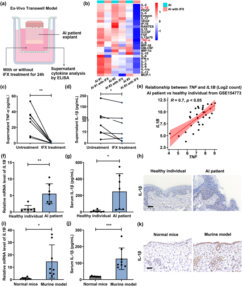
Background: Acne inversa (AI) is a refractory inflammatory skin disease, and TNF-α plays an important role in the pathogenesis of AI. By blocking TNF-α, infliximab (IFX) has been proven to be a promising method.

Objectives: To explore the underlying mechanisms of IFX treatment in AI patients.

Methods: In this research, we integrated transcriptome sequencing data from the samples of our patients with AI and the GEO database. Ex vivo skin culture of AI patients was conducted to evaluate the efficacy of IFX treatment. Animal studies and cell experiments were used to explore the therapeutic effect and mechanism of IFX treatment.

Results: Both TNF-α and NLRP3 inflammasome-related pathways were enriched in skin lesions of AI patients and murine AI models. After IFX treatment, the NLRP3 inflammasome-related pathway was effectively blocked, and the IL-1β level was normalized in ex vivo AI skin explants and murine AI models. Mechanistically, IFX suppressed the NF-κB signaling pathway to lower the expression of NLRP3 and IL-1β in keratinocytes.

Conclusions: IFX treatment alleviated skin lesions in murine AI models and downregulated NLRP3 and IL-1β expression levels by inhibiting the NF-κB signaling pathway, which was helpful for understanding the mechanism of IFX therapy.

Keywords: Acne inversa; IL-1β; Infliximab; NLRP3; TNF-α.


Conflict of interest statement

The authors declare that they have no known competing financial interests or personal relationships that could have appeared to influence the work reported in this paper.

Figures

Fig. 1
Fig. 1
The TNF signaling pathway was significantly enriched in the skin lesions of AI patients and murine AI models. (a) The top 22 inflammation-related KEGG pathways enriched from transcriptome sequencing in the skin of AI patients versus healthy individuals (GSE148027). n = 18 AI patients and n = 8 healthy individuals. (b) Skin sections from AI patients and healthy individuals were examined following H&E and TNF-α staining by IHC. Scale bar = 100 μm for H&E staining, Scale bar = 25 μm for IHC. (c) TNF-α levels in the serum of AI patients and healthy individuals were detected using ELISA analysis. AI patients (n = 7) and healthy individuals (n = 7) for panels (b–c). (d) Schematic diagram of murine AI model construction and representative images of normal mice and murine AI models harvested on day 48 after the initial tamoxifen treatment (20 mg/kg). (e) The top 18 enriched KEGG pathways identified from genes significantly increased in the skin lesions of murine AI models compared to normal mice. n = 4 mice per group. (f) Skin sections from normal mice and murine AI models were examined following TNF-α expression level by IHC analysis. Scale bar = 100 μm. (g–h) TNF-α expression level in the skin or serum of normal mice and murine AI models by qRT‒PCR (g) and ELISA (h). n = 8 mice per group for panels (d, f-h). *P < 0.05, **P < 0.01.
Fig. 2
Fig. 2
Infliximab treatment decreased TNF-α and IL-1β expression levels. (a) Schematic diagram of the ex vivo Transwell model. (b) Cluster analysis of cytokines in the culture supernatant of AI patient lesions via Luminex multifactor detection technology. n = 3 patients. (c) TNF-α expression levels were detected in the culture supernatant of AI patient lesions treated with IFX (20 μg/mL) or PBS by ELISA. (d) ELISA was applied to detect IL-1β expression levels in the culture supernatant of AI patient lesions treated with IFX (20 μg/mL) or PBS. n = 7 AI patients for panels (c–d). (e) Correlation analysis of TNF and IL1B levels in the skin of AI patients and healthy individuals (data from GSE154773). n = 22 AI patients and n = 10 healthy individuals. (f–h) The mRNA and protein levels of IL-1β in the skin or serum of AI patients and healthy individuals by qRT‒PCR (f), ELISA (g), and IHC (h). Scale bar = 100 μm. AI patients (n = 7) and healthy individuals (n = 7) for panels (c–d). (i–k) IL-1β expression level in the skin or serum of normal mice and murine AI models using qRT‒PCR 7 (i), ELISA 7 (j) and IHC analysis (k). Scale bar = 100 μm. n = 8 mice per group for panels (i–k). *P < 0.05, **P < 0.01.
Fig. 3
Fig. 3
Infliximab treatment selectively downregulated NLRP3 expression levels. (a) qRT‒PCR technology was used to measure the level of inflammasome sensors in the skin lesions of AI patients compared to healthy individuals. (b) The level of inflammasomes in the skin lesions of AI patients with IFX (20 μg/mL) or PBS treatment using qRT‒PCR. (c) Correlation analysis of TNF and NLRP3 levels in the skin of AI patients and healthy subjects (data from GSE154773). n = 22 AI patients and n = 10 healthy individuals. (d) NLRP3 staining was performed in the skin of AI patients and healthy individuals via IF. Scale bar = 100 μm. (e) Immunostainings showed NLRP3 expression levels in the skin lesions of AI patients treated with IFX (20 μg/mL) or PBS. Scale bar = 100 μm. (f–g) NLRP3 expression level in the skin of normal mice and murine AI models by qRT‒PCR (f) and immunostainings (g). AI patients (n = 7) and healthy individuals (n = 7) for panels (a-b, d). n = 7 AI patients for panels (b, e). n = 8 mice per group for panels (f–g). Scale bar = 50 μm *P < 0.05, **P < 0.01, ***P < 0.001, ****P < 0.0001.
Fig. 4
Fig. 4
TNF-α promoted NLRP3IL-1β pathways by activating NF-κB signaling pathways in keratinocytes. (a) The level of inflammasomes and IL-1β expression in HaCaT cells with or without TNF-α (20 ng/mL) treatment using qRT‒PCR. n = 3 biologically independent samples. (b) Western blot was performed to semiquantify NLRP3 and IL-1β expression in HaCaT cells with or without TNF-α (20 ng/mL) treatment, and the grayscale analysis of NLRP3 and IL-1β was normalized to the unstimulated group. (c) GSEA plot showing the enrichment of genes upregulated in the NF-κB signaling pathway and cytokine‒cytokine receptor interaction. (d) P-IκB and IκB expression in HaCaT cells with or without TNF-α (20 ng/mL) treatment by Western blot, and the gray levels of P-IκB and IκB expression were normalized to those in the unstimulated group. (e) HaCaT cells were pre-treated with 10 μM BAY11-7082 for 1 h and TNF-α (20 ng/mL) was added into the media containing BAY11-7082 for another 24 h. NLRP3 and IL-1β expression was determined by Western blot. The grayscale analysis of NLRP3 and IL-1β expression was normalized to the unstimulated group. (f) HaCaT cells were incubated with 20 μg/mL IFX for 1 h and TNF-α (20 ng/mL) was added into the media containing IFX for another 24 h. P-IκB, IκB, NLRP3 and IL-1β expression was determined by Western blot. The grayscale analysis of p-IκB, IκB, NLRP3 and IL-1β expression was normalized to the unstimulated group. *P < 0.05, **P < 0.01, ***P < 0.001, ****P < 0.0001.
Fig. 5
Fig. 5
Infliximab treatment reduced TNF-α, IL-1β and NLRP3 expression levels in murine AI models. (a) Schematic diagram of murine AI models with or without IFX (5 mg/kg) treatment. (b) Gross appearance of skin lesions in murine AI models with or without IFX (5 mg/kg) treatment. (c) H&E staining of skin sections from normal mice, lesional skin of murine AI models, and lesional skin of murine AI models with IFX (5 mg/kg) therapy. Scale bar = 100 μm. (d) Representative images showing the protein levels of TNF-α and IL-1β in the skin of normal mice, murine AI models, and murine AI models with IFX (5 mg/kg) therapy via IHC analysis. Scale bar = 100 μm. (e) NLRP3 expression level in the skin of normal mice and murine AI models treated with IFX (5 mg/kg) or IgG by immunostainings. Scale bar = 50 μm. n = 8 mice per group for panels (a–e).

Similar articles

Cited by

References

    1. Saunte D.M.L., Jemec G.B.E. Hidradenitis suppurativa: advances in diagnosis and treatment. JAMA. 2017;318(20):2019–2032. doi: 10.1001/jama.2017.16691. - DOI - PubMed
    1. Goldburg S.R., Strober B.E., Payette M.J. Hidradenitis suppurativa: epidemiology, clinical presentation, and pathogenesis. J. Am. Acad. Dermatol. 2020;82(5):1045–1058. doi: 10.1016/j.jaad.2019.08.090. - DOI - PubMed
    1. Matusiak L., Bieniek A., Szepietowski J.C. Increased serum tumour necrosis factor-alpha in hidradenitis suppurativa patients: is there a basis for treatment with anti-tumour necrosis factor-alpha agents? Acta Derm. Venereol. 2009;89(6):601–603. doi: 10.2340/00015555-0749. - DOI - PubMed
    1. Hotz C., Boniotto M., Guguin A., Surenaud M., Jean-Louis F., Tisserand P., et al. Intrinsic defect in keratinocyte function leads to inflammation in hidradenitis suppurativa. J. Invest. Dermatol. 2016;136(9):1768–1780. doi: 10.1016/j.jid.2016.04.036. - DOI - PubMed
    1. Moran B., Sweeney C.M., Hughes R., Malara A., Kirthi S., Tobin A.M., et al. Hidradenitis suppurativa is characterized by dysregulation of the Th17:treg cell Axis, which is corrected by anti-TNF therapy. J. Invest. Dermatol. 2017;137(11):2389–2395. doi: 10.1016/j.jid.2017.05.033. - DOI - PubMed

LinkOut - more resources