. 2024 Jun 15;10(12):e32528.
doi: 10.1016/j.heliyon.2024.e32528. eCollection 2024 Jun 30.

Epigenetic silencing of miR-125a-3p promotes the progress of human cholangiocarcinoma via increasing CAC1 expression

Affiliations

Epigenetic silencing of miR-125a-3p promotes the progress of human cholangiocarcinoma via increasing CAC1 expression

Xiaojuan Guo et al. Heliyon. .

Abstract

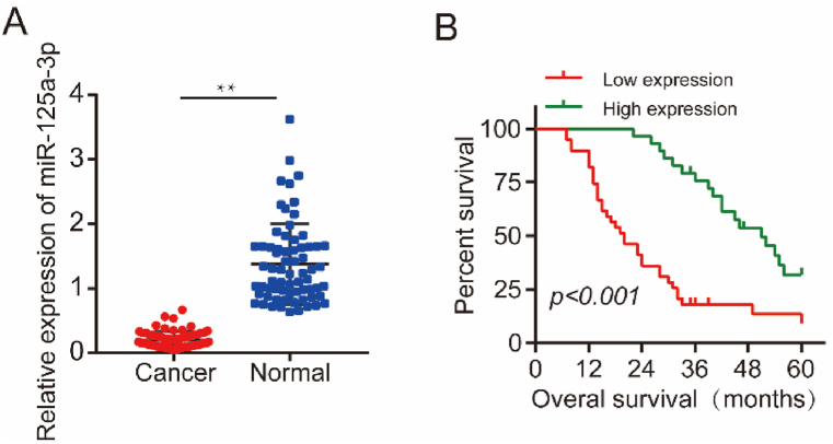
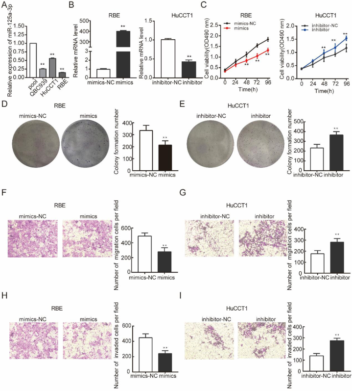
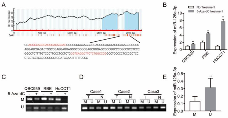
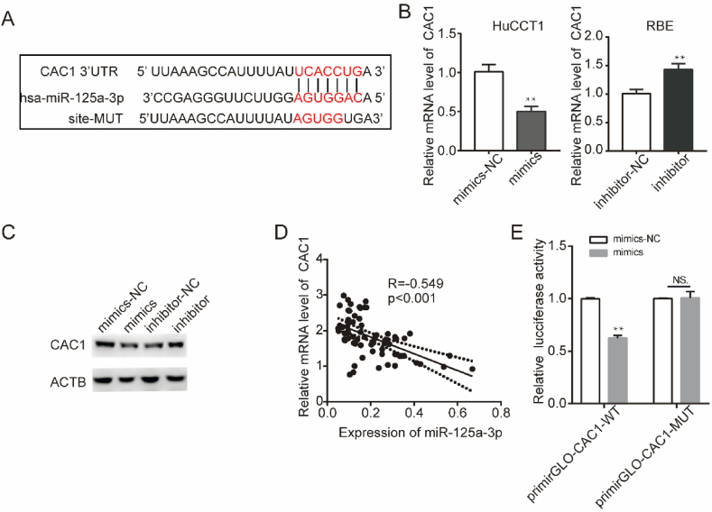
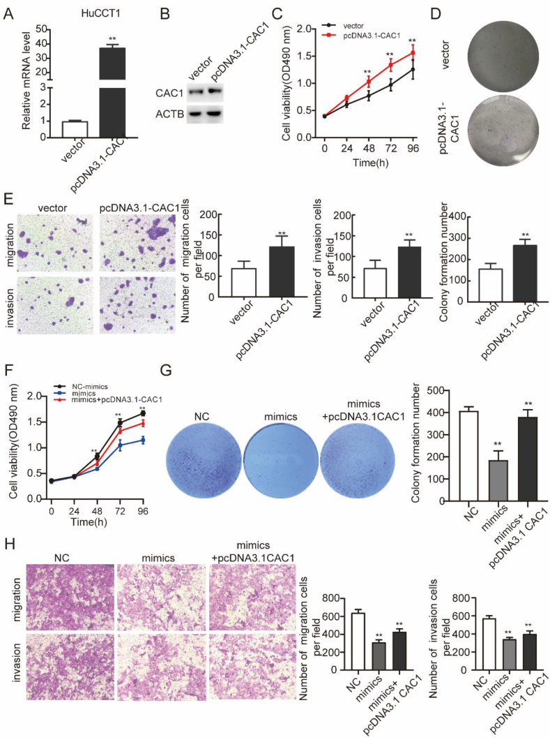
We aimed to investigate the dysregulation of the microRNAs(miRNAs) in cholangiocarcinoma (CCA), including its impact on the homeostasis of the transcriptome and cellular behavior. MiRNAs serve as potent epigenetic regulators of transcriptional output, targeting various signaling pathways. This study aimed to investigate the expression level, epigenetic mechanism and function of miR-125a-3 in CCA. The study data showed that the expression level of miR125a-3p was decreased in CCA tissue samples and cell lines, and it was closely related to lymph node metastasis, tissue differentiation and TNM stage. The data demonstrate a strong association between decreased miR-125a-3p expression and poorer prognosis in cholangiocarcinoma patients. miR-125a-3p acts as a tumor suppressor by inhibiting the viability, migration and invasion of CCA cells. There are CpG islands in the promoter region of miR-125a-3p gene, and the methylation of the promoter region of miR-125a-3p gene leads to the transcriptional repression of miR-125a-3p. In addition, miR125a-3p can target and regulate CAC1 mRNA and protein expression in the downstream mechanism, and the high expression of CAC1 can promote the proliferation, migration and invasion of cholangiocarcinoma cells. These data demonstrate that miR-125a-3p promoter methylation leads to silencing of its expression. Mechanically, miR-125a-3p acts as a tumor suppressor and participates in the occurrence and development of CCA through targeting CAC1 gene expression. Therefore, miR-125a-3p may serve as a new target for the diagnosis, prognostic assessment or molecular therapy of CCA.

Keywords: CAC1; Cholangiocarcinoma; Malignant progression; Methylation; miR-125a-3p; microRNA.


Conflict of interest statement

The authors declare that they have no known competing financial interests or personal relationships that could have appeared to influence the work reported in this paper.

Figures

Fig. 1
Fig. 1
miR-125a-3p is upregulated in CCA and associated with the prognostic of patients. a. miR-125a-3p expression levels in paired cholangiocarcinoma tissues and corresponding para-cancerous normal bile duct tissues detected by qRT-PCR (n = 68). b. Kaplan-Meier curves show that CCA patients with very high miR-125a-3p expression levels show significantly prolonged overall survival (green Kaplan-Meier curve) compared to patients with lower miR-125a-3p expression levels (red Kaplan-Meier curve). **P < 0.01.
Fig. 2
Fig. 2
miR-125a-3p represses CCA cell proliferation, migration and invasion in vitro. a. Expression levels in cholangiocarcinoma cell lines relative to normal bile duct cells analyzed by qRT-PCR. The data are shown as the mean ± S.D. **p < 0.01. b. Using miR-125a-3p mimics or miR-125a-3p inhibitor were transfected into REB and HuCCT1 cells, to overexpress or downregulate the expression of miR-125a-3p. c, d and e. MTS and colony formation assays were performed to evaluate RBE and HuCCT1 cells proliferation after miR-125a-3p overexpression or knockdown. f-i. Cell migration and invasion ability were analyzed by Transwell assays (Magnification: × 100).
Fig. 3
Fig. 3
Methylation of the promoter region of miR-125a-3p gene in CCA leads to silencing of its expression. a. Distribution of CpG islands in the promoter region of miR-125a-3p genome. The two MSP regions analyzed are indicated. b. Relative miR-125a-3p expression in CCA cells treated with 10 mmol/L 5-Aza-deoxycytidine (5-Aza-dC) or vehicle for 72 h, as determined by quantitative RT-PCR (RT-qPCR) analysis. c. Methylation-specific PCR (MSP) of genomic DNA from RBE, HuccT-1 and QBC939 cells treated with or without 10 mmol/L 5-Aza-dC treatment. (The original image is provided in the Supplementary file). d. MSP is performed to analyze in CCA tissues. (The original image is provided in the Supplementary file). Case 1–3: CCA case representative. e. RT-qPCR was used to detect the relative expression of miR-125a-3p in tissues with miR-125a-3p gene methylation in CCA tissues. T: Tumor, N: Normal, M: Methylation, U: Un-methylation. **P < 0.01.
Fig. 4
Fig. 4
miR-125a-3p regulates CAC1 expression. a. The sequences of the potential binding sites between 3′ UTR of CAC1 and miR-125a-3p. CAC1-MUT is the binding sites-mutated segments. b-c. CAC1 mRNA and protein expression levels in the indicated cholangiocarcinoma cells transfected with miR-125a-3p mimics or inhibitor. ACTB was used as an internal control for Western blot. (The original image is provided in the Supplementary file). d. Correlation between CAC1 expression and miR-125a-3p expression in cholangiocarcinoma tissues detected by RT-qPCR. e. Luciferase activities were measured 24 h after the indicated cells transfected with miR-125a-3p mimics and reporter plasmid containing wild type 3′ UTR regions of CAC1 or mutated segments. **P < 0.01.
Fig. 5
Fig. 5
miR-125a-3p suppresses the proliferation, migration and invasion of cholangiocarcinoma cells by targeting CAC1 in vitro. a. CAC1 expressions were examined using qRT-PCR after pcDNA3.1-CAC1 transfection. b. CAC1 protein expression was examined using western bolt analysis after pcDNA3.1-CAC1 transfection. (The original image is provided in the Supplementary file). c and d. MTS and colony formation assays were performed to evaluate RBE cells proliferation after CAC1 overexpression. e. Cell migration and invasion ability were analyzed by transwell assays (Magnification: × 100). f and g. MTS and colony formation assays were performed to examine the effect of miR-125a-3p/CAC1 axis on cell proliferation ability of CCA cells. h. Transwell assay was performed for evaluating the effect of miR-125a-3p/CAC1 axis on cell invasion and metastatic ability of CCA cells in each group (Magnification: × 100). **P < 0.01.

Similar articles

References

    1. Lamarca A., Santos-Laso A., Utpatel K., La Casta A., Stock S., Forner A., Adeva J., Folseraas T., Fabris L., Macias R.I.R., Krawczyk M., Krawczyk M., Cardinale V., Braconi C., Alvaro D., Evert M., Banales J.M., Valle J.W., Group: on behalf of the European Network for the Study of C Liver metastases of intrahepatic cholangiocarcinoma: implications for an updated staging system. Hepatology. 2021;73:2311–2325. - PMC - PubMed
    1. Nara S., Esaki M., Ban D., Takamoto T., Shimada K., Ioka T., Okusaka T., Ishii H., Furuse J. Adjuvant and neoadjuvant therapy for biliary tract cancer: a review of clinical trials. Jpn. J. Clin. Oncol. 2020;50:1353–1363. - PubMed
    1. Tang Y.Y., Zhao Y.N., Zhang T., Chen Z.Y., Ma X.L. Comprehensive radiomics nomogram for predicting survival of patients with combined hepatocellular carcinoma and cholangiocarcinoma. World J. Gastroenterol. 2021;27:7173–7189. - PMC - PubMed
    1. Tummanatsakun D., Proungvitaya T., Roytrakul S., Limpaiboon T., Wongkham S., Wongkham C., Silsirivanit A., Somintara O., Sangkhamanon S., Proungvitaya S. Serum apurinic/apyrimidinic endodeoxyribonuclease 1 (APEX1) level as a potential biomarker of cholangiocarcinoma. Biomolecules. 2019;9 - PMC - PubMed
    1. Xu G., Sun L., Li Y., Xie F., Zhou X., Yang H., Du S., Xu H., Mao Y. The clinicopathological and prognostic value of PD-L1 expression in cholangiocarcinoma: a meta-analysis. Front. Oncol. 2019;9:897. - PMC - PubMed

LinkOut - more resources