. 2024 Jun 14;10(12):e33060.
doi: 10.1016/j.heliyon.2024.e33060. eCollection 2024 Jun 30.

Differential peptide-dependent regulation of growth hormone (GH): A comparative analysis in pituitary cultures of reptiles, birds, and mammals

Affiliations

Differential peptide-dependent regulation of growth hormone (GH): A comparative analysis in pituitary cultures of reptiles, birds, and mammals

Valeria A Urban-Sosa et al. Heliyon. .

Abstract

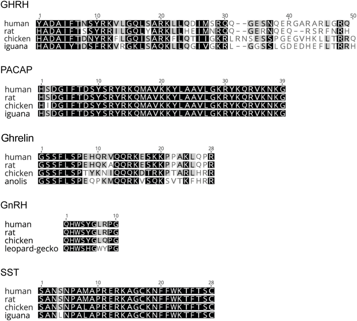
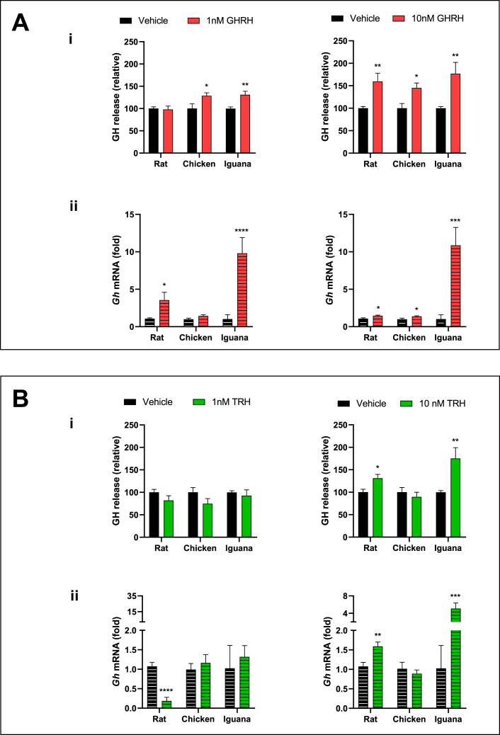
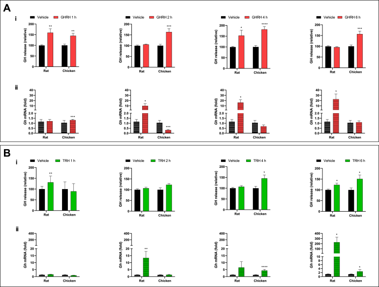
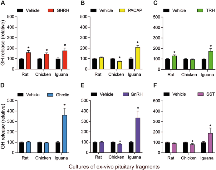
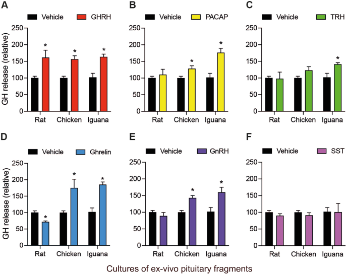
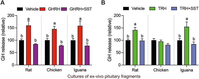
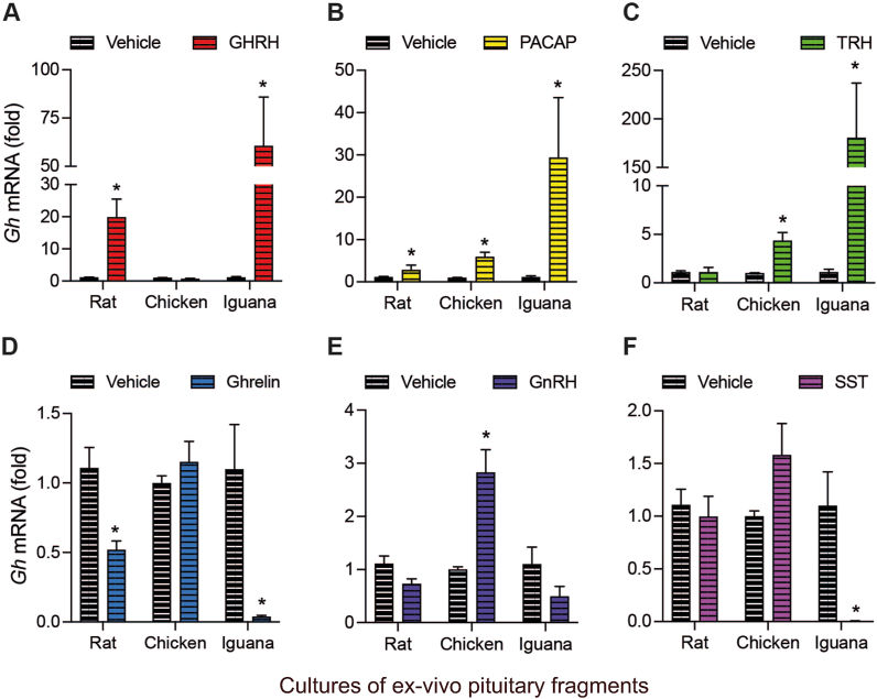
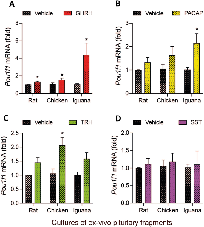
Growth hormone (GH) is a pituitary protein that exerts pleiotropic roles in vertebrates. The mechanisms regulating GH synthesis and secretion are finely controlled by hypothalamic neuropeptides and other factors. These processes have been considerably studied in mammals but are still poorly understood in other groups. To better understand the pituitary GH regulation during vertebrate phylogeny, we compared the effects of incubating several peptides on cultures of ex-vivo pituitary fragments obtained from representative specimens of reptiles (iguana), birds (chicken) and mammals (rat). The peptides used were: growth hormone-releasing hormone (GHRH), thyrotropin-releasing hormone (TRH), pituitary adenylate cyclase-activating polypeptide (PACAP), ghrelin, gonadotropin-releasing hormone (GnRH), and somatostatin (SST). In rat pituitary cultures, GH secretion was stimulated by GHRH and TRH, while gh mRNA expression was increased by GHRH and PACAP. In the case of chicken pituitaries, GH release was promoted by GHRH, ghrelin, PACAP, and GnRH, although the latter two had a dual effect since at a shorter incubation time they decreased GH secretion; in turn, gh mRNA expression was significantly stimulated by TRH, PACAP, and GnRH. The most intense effects were observed in iguana pituitary cultures, where GH secretion was significantly augmented by GHRH, PACAP, TRH, ghrelin, and GnRH; while gh mRNA expression was stimulated by GHRH, TRH, and PACAP, but inhibited by ghrelin and SST. Also, in the three species, SST was able to block the GHRH-stimulated GH release. Furthermore, it was found that the expression of Pou1f1 mRNA was increased with greater potency by GHRH and PACAP in the iguana, than in chicken or rat pituitary cultures. Additionally, in-silico analysis of the gh gene promoter structures in the three species showed that the reptilian promoter has more Pit-1 consensus binding sites than their avian and mammalian counterparts. Taken together, results demonstrate that pituitary peptide-mediated GH regulatory mechanisms are differentially controlled along vertebrate evolution.

Keywords: GH-Regulatory peptides; Growth hormone; Iguana; Pit-1; Pituitary.


Conflict of interest statement

The authors declare that they have no known competing financial interests or personal relationships that could have appeared to influence the work reported in this paper.

Figures

Fig. 1
Fig. 1
Multiple alignments of hypothalamic peptides of human, rat, chicken and reptiles. Similar residues are colored according to BLOSUM62 as follows: black = 100 % similar; dark gray = 80–99 % similar; light gray = 60–79 % similar; white = less than 60 % similar. Since there are no reported sequences for iguana ghrelin and GnRH, those of anole and leopard-gecko, lizards belonging to the order Squamata, were employed for comparison.
Fig. 2
Fig. 2
Effects of GHRH or TRH upon GH secretion and gh mRNA expression as a function of dose.Ex-vivo cultures of pituitary fragments from rat, chicken or iguana were incubated with 1 or 10 nM of either GHRH (Panel A) or TRH (Panel B) for 1 h, and their effects were evaluated on i) GH secretion, determined by ELISA, and ii)gh mRNA expression determined by qPCR. The bars represent the mean ± SEM, n = 4. *p < 0.05, **p < 0.01, ***p < 0.005, ****p < 0.001 in Student's independent t-test. Illustration of results are given as relative proportion (%) of change in the treatment groups in comparison with the controls (100 %) for GH release; while for gh mRNA expression they are shown as -fold change as compared to the corresponding controls (1-fold).
Fig. 3
Fig. 3
Effects of GHRH or TRH upon secretion and gh mRNA expression as a function of incubation time.Ex-vivo cultures of pituitary fragments from rat or chicken were incubated with 10 nM of either GHRH (Panel A) or TRH (Panel B) for 1, 2, 4, and 6 h, and their effects were evaluated on i) GH secretion, determined by ELISA, and ii)gh mRNA expression determined by qPCR. The bars represent the mean ± SEM, n = 4. *p < 0.05, **p < 0.01, ***p < 0.005, ****p < 0.001 in Student's independent t-test. Illustration of results are given as relative proportion (%) of change in the treatment groups in comparison with the controls (100 %) for GH release; while for gh mRNA expression they are shown as -fold change as compared to the corresponding controls (1-fold).
Fig. 4
Fig. 4
Effects of peptide-dependent GH release in rat, chicken and iguana pituitary cultures after 1 h of treatment.Ex-vivo cultures of pituitary fragments were stabilized by 30 min and then exposed to GHRH, PACAP, TRH, ghrelin, GnRH or SST (10 nM) for 1 h. The GH release was quantified in the culture media using a homologous (rat and chicken) or heterologous (iguana, using an antibody against chicken GH) ELISA. A. GHRH induced GH release in rat (p = 0.0004), chicken (p = 0.0004) and iguana (p = 0.0045) pituitary cultures. B. PACAP treatment decreased GH release in chicken (p = 0.0008), while induced it in iguana (p < 0.0001) pituitaries. C. TRH caused GH release in rat (p = 0.0008) and iguana (p = 0.0023) pituitary cultures. D. ghrelin induced GH release only in iguana pituitaries (p < 0.0001). E. GnRH decreased GH release in chicken pituitary cultures (p = 0.0137), while in iguana it had a stimulatory effect (p < 0.0001). F. The SST treatment decreased GH concentration in chicken pituitary culture media (p = 0.0053), while it increased in iguana cultures (p = 0.0005). Bars represent the mean ± SEM. Asterisks indicate statistically significant differences (Student's independent t-test, p < 0.05, n = 4) comparing vehicle vs. treatment within rat, chicken or iguana pituitary cultures.
Fig. 5
Fig. 5
Effects of peptide-dependent GH release in rat, chicken and iguana pituitary cultures after 4 h of treatment.Ex-vivo cultures of pituitary fragments of rat, chicken and iguana were treated as described in Fig. 4, and GH release was quantified by ELISA after 4 h of treatment. A. GHRH induced GH release in rat (p = 0.0018), chicken (p < 0.0001) and iguana (p = 0.0023) pituitary cultures. B. PACAP treatment increased GH release in chicken (p = 0.01) and iguana (p = 0.0015) pituitaries. C. TRH stimulated GH release in chicken (p = 0.0036) and iguana (p = 0.018) pituitary cultures. D. ghrelin treatment decreased GH release in rat (p = 0.024), while induced it in chicken (p = 0.0037) and iguana (p = 0.0002) pituitaries. E. GnRH induced GH release in chicken (p = 0.0001) and iguana (p = 0.017) pituitary cultures. F. SST had no effect on GH release in any species. Bars represent the mean ± SEM. Asterisks indicate statistically significant differences (Student's independent t-test, p < 0.05, n = 4) comparing vehicle vs. treatment within rat, chicken or iguana pituitary cultures.
Fig. 6
Fig. 6
Effects of SST upon GHRH-stimulated or TRH-stimulated GH release in rat, chicken and iguana pituitary cultures.Ex-vivo cultures of pituitary fragments were stabilized for 30 min and then co-incubated with 10 nM GHRH/SST or TRH/SST for 1 h. GH concentration in the culture media was quantified by ELISA, as described in Fig. 4. A. Somatostatin inhibited GHRH-stimulated GH secretion in pituitary explants of the three studied species (rat: F(2,21) = 6.36, p = 0.0069; chicken; F(2,27) = 8.34, p = 0.0015; iguana F(2,21) = 6.36, p = 0.0037; one-way ANOVA followed by Tukey's post-hoc test; n = 4). B. Somatostatin inhibited TRH-stimulated GH release in rat and iguana pituitary cultures (rat: F(2,17) = 8.073, p = 0.0034; iguana F(2,26) = 6.66, p = 0.0046; one-way ANOVA followed by Tukey's post-hoc test; n = 4). Bars represent the mean ± SEM. Letters indicate statistically significant differences (p < 0.05) between treatments within pituitary cultures of each species.
Fig. 7
Fig. 7
Effects of peptide treatments on gh mRNA expression in rat, chicken and iguana pituitary cultures after 4 h of treatment.Ex-vivo cultures of pituitary fragments were stabilized for 30 min, exposed to 10 nM GHRH, PACAP, TRH, ghrelin, GnRH, or SST for 4 h; tissues were then harvested and RNA isolated. The gh mRNA expression was quantified by RT-qPCR. A. GHRH induced gh mRNA synthesis in rat (p < 0.0001) and iguana (p = 0.015) pituitary cultures. B. PACAP induced the rat (p = 0.042), chicken (p < 0.0001) and iguana (p = 0.0005) gh mRNAs expression. C. Treatment with TRH was effective in inducing gh mRNA expression in chicken (p < 0.0001) and iguana (p < 0.0001) pituitaries. D. ghrelin caused a decrease in levels of rat (p = 0.019) and iguana (p < 0.0001) gh mRNA expression. E. GnRH treatment induced gh mRNA expression only in chicken pituitary cultures (p = 0.0005). F. SST treatment strongly reduced the gh mRNA expression levels in iguana pituitaries (p = 0.0012). Bars represent the mean ± SEM. Asterisks indicate statistically significant differences (Student's independent t-test, p < 0.05, n = 4) comparing vehicle vs. treatment within rat, chicken or iguana pituitary cultures.
Fig. 8
Fig. 8
Effects of GHRH and ghrelin upon GH secretion and gh mRNA expression in rat pituitary dispersed cell cultures. Primary rat pituitary cell cultures were incubated with 10 nM GHRH or ghrelin for 1 h and 4 h. a) GHRH (p = 0.0001) and ghrelin (p = 0.0002) induced GH release after 1 h of incubation. b) GHRH (p = 0.0002) and ghrelin (p = 0.0043) induced GH release after 4 h of incubation. c) GHRH (p = 0.0175) and ghrelin (p = 0.0576) induced gh mRNA expression after 4 h of incubation. Bars represent the mean ± SEM. Asterisks indicate statistically significant differences (Student's independent t-test, p < 0.05, n = 5).
Fig. 9
Fig. 9
Effects of the peptides on Pou1f1 mRNA expression in rat, chicken and iguana pituitary cultures.Ex-vivo cultures of pituitary fragments were stabilized for 30 min, and then exposed to either GHRH, PACAP, TRH, or SST for another 30 min. Tissues where then harvested and RNA isolated. The Pou1f1 mRNA expression was quantified by RT-qPCR. A. GHRH stimulated Pou1f1 mRNA synthesis in rat (p = 0.012), chicken (p = 0.045) and iguana (p = 0.024) pituitary cultures. B. PACAP increased Pou1f1 mRNA levels only in iguana (p = 0.02) pituitaries. C. TRH only induced the chicken Pou1f1 mRNA expression (p = 0.015). D. Treatment with SST did not affect the Pou1f1 mRNA expression in any species, in comparison with the controls. Bars represent the mean ± SEM. Asterisks indicate statistically significant differences (Student's independent t-test, p < 0.05, n = 4) comparing vehicle vs. treatment within rat, chicken or iguana pituitary cultures.
Fig. 10
Fig. 10
Comparison of Pit-1 primary structure from mammalian, bird and reptile species, and analysis of Pit-1 consensus binding sites in gh gene promoters. A. Multiple alignment of rat (NP_037140.2), chicken (NP_001383031.1) and anole (XP_016848011.1) Pit-1 amino acid sequences. Similar residues are colored according to BLOSUM62 as follows: black = identical; gray = similar; white = no similarity. Red boxes highlight residues of the DNA-binding domain, which are identical in all three species. B. Diagram showing the location of predicted Pit-1 consensus binding sites (blue boxes) in the gh gene promoters of rat, chicken and anolis. The red line indicates the transcription start site (TSS). (For interpretation of the references to color in this figure legend, the reader is referred to the Web version of this article.)

Similar articles

Cited by

References

    1. Dehkhoda F., Lee C., Medina J., Brooks A. The growth hormone receptor: mechanism of receptor activation, cell signaling, and physiological aspects. Review Front Endocrinol (Lausanne) 2018;13:9–35. https://doi:10.3389/fendo.2018.00035 - DOI - PMC - PubMed
    1. Harvey S. In: Growth Hormone. Harvey S., Scanes C.G., Daughaday W.H., editors. CRC Press, Inc.; 1994. Growth hormone synthesis; pp. 55–78. ISBN 0-8493-8697-7.
    1. Harvey S., Martínez-Moreno C.G., Luna M., Arámburo C. Autocrine/paracrine roles of extrapituitary growth hormone and prolactin in health and disease: an overview. Gen. Comp. Endocrinol. 2015;220:103–111. doi: 10.1016/j.ygcen.2014.11.04. - DOI - PubMed
    1. Devesa J., Almengló C., Devesa P. Multiple effects of growth hormone in the body: is it really the hormone for growth? Clin. Med. Insights Endocrinol. Diabetes. 2016;9:CMED–S38201. doi: 10.4137/CMED.S38201. - DOI - PMC - PubMed
    1. Otteson D., Cirenza P., Hitchcock P. Persistent neurogenesis in the teleost retina: evidence for regulation by the growth hormone/insulin-like growth factor-I axis. Mech. Dev. 2002;117:137–149. doi: 10.1016/s0925-4773(02)00188-0. - DOI - PubMed

LinkOut - more resources