

Precise maxillofacial soft tissue reconstruction: A combination of cone beam computed tomography and 3dMD photogrammetry system
- PMID: 38994088
- PMCID: PMC11237927
- DOI: 10.1016/j.heliyon.2024.e32513
Precise maxillofacial soft tissue reconstruction: A combination of cone beam computed tomography and 3dMD photogrammetry system
Abstract
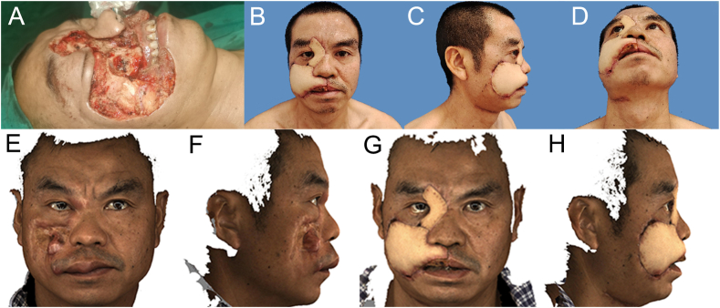
Introduction: The reconstruction of both extra- and intra-oral soft tissue defects, particularly in restoring the morphology of the lip and the corners of the mouth, has posed a significant challenge for surgeons. Inappropriate methods often lead to maxillofacial deformity which then causes psychological and functional problems. This study aimed to address the challenge of reconstructing extensive and complex maxillofacial soft tissue defects, mainly focusing on the lip, the corners of the mouth, and the surrounding areas.
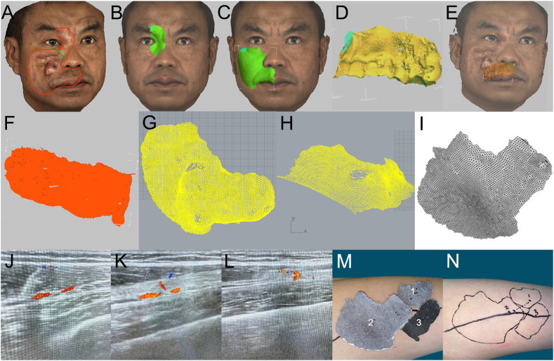
Materials and methods: We developed a reconstruction approach by combining the 3dMDface System (3dMD) with the cone beam computed tomography (CBCT). Firstly, with the extra-oral incision line, we evaluated the shape and the size of the extra-oral defect with 3dMD digitally. Then we used the corresponding maxillary and mandible tooth positions to record the intra-oral defect, which was then converted to digital images by combining 3dMD and CBCT. The islands of the anterolateral thigh perforator flap were then designed after the locations of the perforators were detected with Doppler ultrasonography.
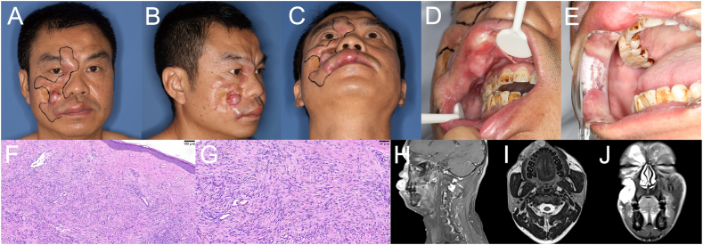
Results: A clinical case diagnosed as dermatofibrosarcoma protuberans was presented to illustrate the approach. The patient's tumor resection and the size of multiple defects were measured and simulated via the virtual surgery system. A three-island perforator flap from the descending branch of the lateral femoral circumflex artery was designed accurately. Two weeks postoperatively, the flap was healed as anticipated and the patient was satisfied with the profile.
Conclusion: The combination of the 3dMD and CBCT technologies improves the accuracy and fitness of extra- and intra-oral soft tissue reconstruction.
Keywords: 3dMD; CBCT; Digital planning; Maxillofacial surgery; Soft tissue reconstruction.
© 2024 The Authors.
Conflict of interest statement
The authors declare that they have no known competing financial interests or personal relationships that could have appeared to influence the work reported in this paper.
Figures
Similar articles
-
Reconstruction of an Extensive Maxillofacial Avulsion Injury Caused by Bear Attack With a Double-Island Anterolateral Thigh Free Flap.J Craniofac Surg. 2022 Jun 1;33(4):1122-1125. doi: 10.1097/SCS.0000000000008213. Epub 2021 Sep 22. J Craniofac Surg. 2022. PMID: 34560751
-
[Application value of CTA combined with digital technology in the design of anterolateral thigh flap in repairing operative defect of head, neck and maxillofacial tumor resection].Lin Chuang Er Bi Yan Hou Tou Jing Wai Ke Za Zhi. 2021 Nov;35(11):992-997. doi: 10.13201/j.issn.2096-7993.2021.11.007. Lin Chuang Er Bi Yan Hou Tou Jing Wai Ke Za Zhi. 2021. PMID: 34886602 Free PMC article. Chinese.
-
Three-dimensional prediction of nose morphology in Chinese young adults: a pilot study combining cone-beam computed tomography and 3dMD photogrammetry system.Int J Legal Med. 2020 Sep;134(5):1803-1816. doi: 10.1007/s00414-020-02351-8. Epub 2020 Jul 9. Int J Legal Med. 2020. PMID: 32647961
-
Reconstruction of complex soft-tissue defects in the extremities with chimeric anterolateral thigh perforator flap.Int J Surg. 2016 Feb;26:25-31. doi: 10.1016/j.ijsu.2015.12.035. Epub 2015 Dec 29. Int J Surg. 2016. PMID: 26739595
-
Combined use of lower medial thigh perforator (LMTP) flap and pedicled medial sural artery perforator flap (MSAP) for lateral knee defects coverage after sarcoma resection: A case report and literature review of soft tissue defect around knee reconstruction.Microsurgery. 2024 Jan;44(1):e31125. doi: 10.1002/micr.31125. Epub 2023 Oct 13. Microsurgery. 2024. PMID: 37830398 Review.
Cited by
-
The relationship between facial directional asymmetry, handedness, chewing side preference, and eyedness.Sci Rep. 2024 Oct 4;14(1):23131. doi: 10.1038/s41598-024-73077-5. Sci Rep. 2024. PMID: 39366983 Free PMC article.
References
-
- Vranckx J., Desmet O., Bila M., Wittesaele W., Wilssens N., Poorten V. Maxillomandibular reconstruction using insourced virtual surgical planning and homemade CAD/CAM: a single-center evolution in 75 patients. Plast. Reconstr. Surg. 2023;152(1):143e–154e. doi: 10.1097/prs.0000000000010142. - DOI - PubMed
-
- Scaglioni M., Meroni M., Fritsche E., Rajan G. Superficial circumflex iliac artery perforator flap in advanced head and neck reconstruction: from simple to its chimeric patterns and clinical experience with 22 cases. Plast. Reconstr. Surg. 2022;149(3):721–730. doi: 10.1097/prs.0000000000008878. - DOI - PubMed
LinkOut - more resources
Full Text Sources
