How does ferrocene correlate with ferroptosis? Multiple approaches to explore ferrocene-appended GPX4 inhibitors as anticancer agents
- PMID: 38994406
- PMCID: PMC11234876
- DOI: 10.1039/d4sc02002b
How does ferrocene correlate with ferroptosis? Multiple approaches to explore ferrocene-appended GPX4 inhibitors as anticancer agents
Abstract
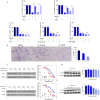
Ferroptosis has emerged as a form of programmed cell death and exhibits remarkable promise for anticancer therapy. However, it is challenging to discover ferroptosis inducers with new chemotypes and high ferroptosis-inducing potency. Herein, we report a new series of ferrocenyl-appended GPX4 inhibitors rationally designed in a "one stone kills two birds" strategy. Ferroptosis selectivity assays, GPX4 inhibitory activity and CETSA experiments validated the inhibition of novel compounds on GPX4. In particular, the ROS-related bioactivity assays highlighted the ROS-inducing ability of 17 at the molecular level and their ferroptosis enhancement at the cellular level. These data confirmed the dual role of ferrocene as both the bioisostere motif maintaining the inhibition capacity of certain molecules with GPX4 and also as the ROS producer to enhance the vulnerability to ferroptosis of cancer cells, thereby attenuating tumor growth in vivo. This proof-of-concept study of ferrocenyl-appended ferroptosis inducers via rational design may not only advance the development of ferroptosis-based anticancer treatment, but also illuminate the multiple roles of the ferrocenyl component, thus opening the way to novel bioorganometallics for potential disease therapies.
This journal is © The Royal Society of Chemistry.
Conflict of interest statement
Y. W., J. L., J. W., W. L., H. W. and X. Z. are inventors on a patent application related to this work.
Figures


Similar articles
-
ROS-responsive fluorinated polyethyleneimine vector to co-deliver shMTHFD2 and shGPX4 plasmids induces ferroptosis and apoptosis for cancer therapy.Acta Biomater. 2022 Mar 1;140:492-505. doi: 10.1016/j.actbio.2021.11.042. Epub 2021 Dec 5. Acta Biomater. 2022. PMID: 34879292
-
New Ferrocene Compounds as Selective Cyclooxygenase (COX-2) Inhibitors: Design, Synthesis, Cytotoxicity and Enzyme-inhibitory Activity.Anticancer Agents Med Chem. 2018;18(2):295-301. doi: 10.2174/1871520617666171003145533. Anticancer Agents Med Chem. 2018. PMID: 28971779
-
Dual degradation mechanism of GPX4 degrader in induction of ferroptosis exerting anti-resistant tumor effect.Eur J Med Chem. 2023 Feb 5;247:115072. doi: 10.1016/j.ejmech.2022.115072. Epub 2022 Dec 30. Eur J Med Chem. 2023. PMID: 36603510
-
GPX4-independent ferroptosis-a new strategy in disease's therapy.Cell Death Discov. 2022 Oct 30;8(1):434. doi: 10.1038/s41420-022-01212-0. Cell Death Discov. 2022. PMID: 36309489 Free PMC article. Review.
-
Targeting GPX4 in human cancer: Implications of ferroptosis induction for tackling cancer resilience.Cancer Lett. 2023 Apr 10;559:216119. doi: 10.1016/j.canlet.2023.216119. Epub 2023 Mar 8. Cancer Lett. 2023. PMID: 36893895 Review.
Cited by
-
Synthesis, Structure, Electrochemistry, and In Vitro Anticancer and Anti-Migratory Activities of (Z)- and (E)-2-Substituted-3-Ferrocene-Acrylonitrile Hybrids and Their Derivatives.Molecules. 2025 Jul 2;30(13):2835. doi: 10.3390/molecules30132835. Molecules. 2025. PMID: 40649349 Free PMC article.
-
Advances in Ferroptosis Research: A Comprehensive Review of Mechanism Exploration, Drug Development, and Disease Treatment.Pharmaceuticals (Basel). 2025 Feb 26;18(3):334. doi: 10.3390/ph18030334. Pharmaceuticals (Basel). 2025. PMID: 40143112 Free PMC article. Review.
-
Targeting Ferroptosis in Tumors: Novel Marine-Derived Compounds as Regulators of Lipid Peroxidation and GPX4 Signaling.Mar Drugs. 2025 Jun 19;23(6):258. doi: 10.3390/md23060258. Mar Drugs. 2025. PMID: 40559667 Free PMC article. Review.
-
Graphene Nanocomposites in the Targeting Tumor Microenvironment: Recent Advances in TME Reprogramming.Int J Mol Sci. 2025 May 9;26(10):4525. doi: 10.3390/ijms26104525. Int J Mol Sci. 2025. PMID: 40429669 Free PMC article. Review.
-
A potent and selective anti-glutathione peroxidase 4 nanobody as a ferroptosis inducer.Chem Sci. 2024 Oct 29;15(46):19420-19431. doi: 10.1039/d4sc05448b. eCollection 2024 Nov 27. Chem Sci. 2024. PMID: 39568902 Free PMC article.
References
-
- Dixon S. J. Lemberg K. M. Lamprecht M. R. Skouta R. Zaitsev E. M. Gleason C. E. Patel D. N. Bauer A. J. Cantley A. M. Yang W. S. Morrison III B. Stockwell B. R. Ferroptosis: An Iron-Dependent Form of Nonapoptotic Cell Death. Cell. 2012;149(5):1060–1072. doi: 10.1016/j.cell.2012.03.042. - DOI - PMC - PubMed
-
- Fang X. Wang H. Han D. Xie E. Yang X. Wei J. Gu S. Gao F. Zhu N. Yin X. Cheng Q. Zhang P. Dai W. Chen J. Yang F. Yang H.-T. Linkermann A. Gu W. Min J. Wang F. Ferroptosis as a target for protection against cardiomyopathy. Proc. Natl. Acad. Sci. U. S. A. 2019;116(7):2672–2680. doi: 10.1073/pnas.1821022116. - DOI - PMC - PubMed
-
- Sun W.-Y. Tyurin V. A. Mikulska-Ruminska K. Shrivastava I. H. Anthonymuthu T. S. Zhai Y.-J. Pan M.-H. Gong H.-B. Lu D.-H. Sun J. Duan W.-J. Korolev S. Abramov A. Y. Angelova P. R. Miller I. Beharier O. Mao G.-W. Dar H. H. Kapralov A. A. Amoscato A. A. Hastings T. G. Greenamyre T. J. Chu C. T. Sadovsky Y. Bahar I. Bayır H. Tyurina Y. Y. He R.-R. Kagan V. E. Phospholipase iPLA2β averts ferroptosis by eliminating a redox lipid death signal. Nat. Chem. Biol. 2021;17(4):465–476. doi: 10.1038/s41589-020-00734-x. - DOI - PMC - PubMed
LinkOut - more resources
Full Text Sources

