. 2024 Jun 10;15(27):10508-10518.
doi: 10.1039/d4sc00267a. eCollection 2024 Jul 10.

Hsp90α forms condensate engaging client proteins with RG motif repeats

Affiliations

Hsp90α forms condensate engaging client proteins with RG motif repeats

Jiaojiao Hu et al. Chem Sci. .

Abstract

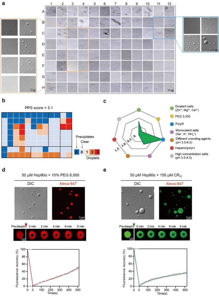
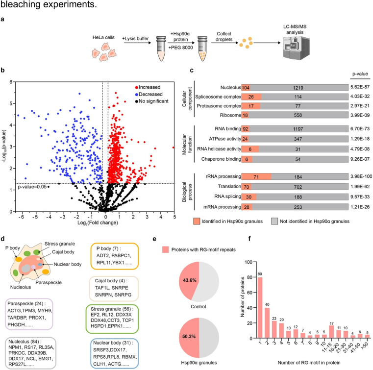
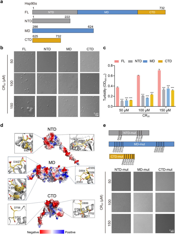
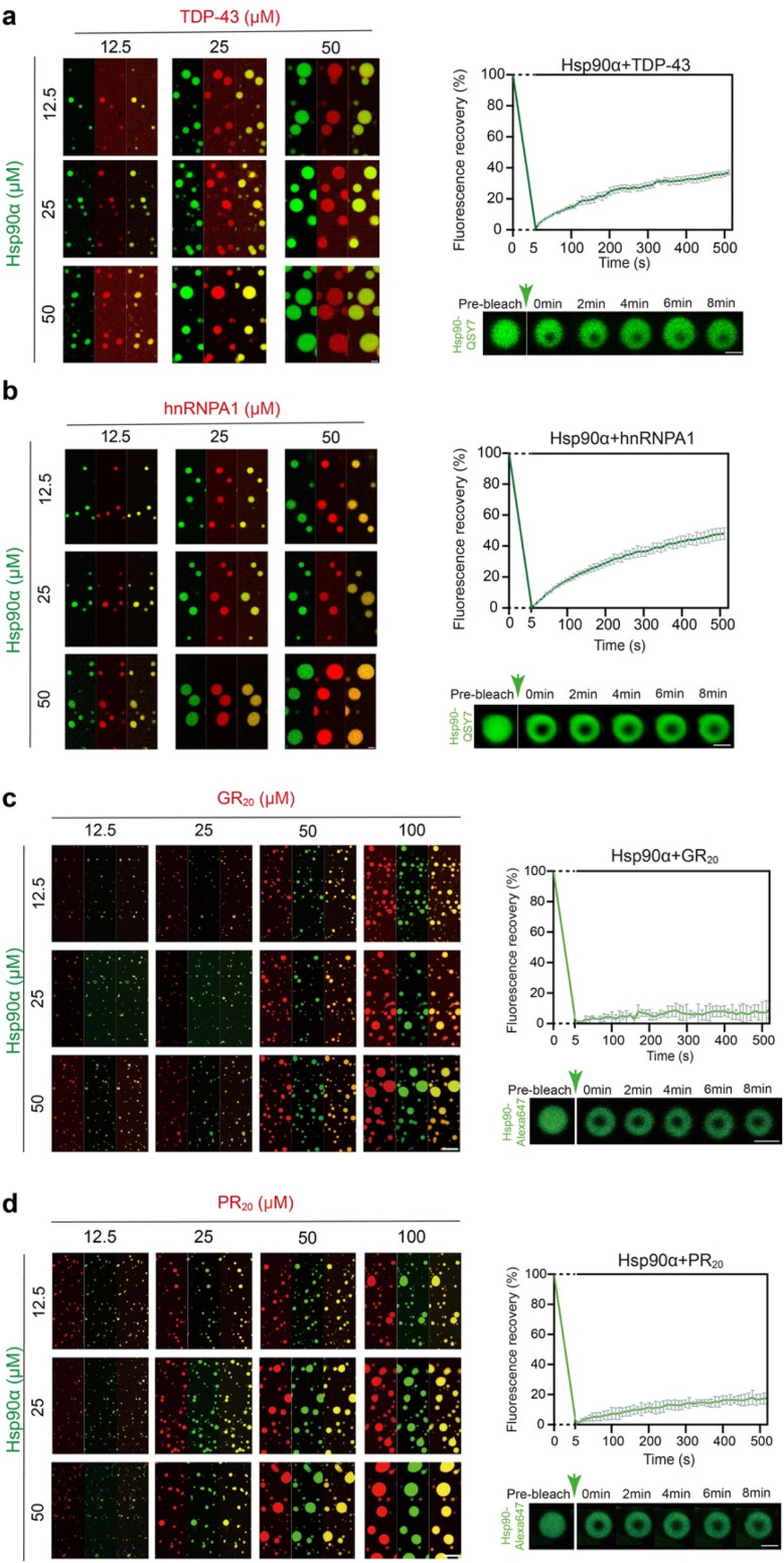
Hsp90α, a pivotal canonical chaperone, is renowned for its broad interaction with numerous protein clients to maintain protein homeostasis, chromatin remodeling, and cell growth. Recent studies indicate its role in modifying various components of membraneless organelles (MLOs) such as stress granules and processing bodies, suggesting its participation in the regulation of protein condensates. In this study, we found that Hsp90α possesses an inherent ability to form dynamic condensates in vitro. Utilizing LC-MS/MS, we further pinpointed proteins in cell lysates that preferentially integrate into Hsp90α condensates. Significantly, we observed a prevalence of RG motif repeats in client proteins of Hsp90α condensates, many of which are linked to various MLOs. Moreover, each of the three domains of Hsp90α was found to undergo phase separation, with numerous solvent-exposed negatively charged residues on these domains being crucial for driving Hsp90α condensation through multivalent weak electrostatic interactions. Additionally, various clients like TDP-43 and hnRNPA1, along with poly-GR and PR dipeptide repeats, exhibit varied impacts on the dynamic behavior of Hsp90α condensates. Our study spotlights various client proteins associated with Hsp90α condensates, illustrating its intricate adaptive nature in interacting with diverse clients and its functional adaptability across multiple MLOs.


Conflict of interest statement

The authors declare no conflict of interest.

Figures

Fig. 1
Fig. 1. Profiling in vitro phase separation of Hsp90α by HiPPS. (a) Microscopic images of HiPPS profiling for 25 μMHsp90α in the bright-field mode. Zoom in, DIC images for wells C3–F4 and B11–C12. (b) The HiPPS profile of 25 μM Hsp90α. The wells are graded and colored based on their microscopic images according to the grading criteria described previously. (c) Values of the average grades of each color zone in the HiPPS profiles. (d) DIC and fluorescence images of Hsp90α condensation under indicated conditions (top). FRAP measurement of Hsp90α droplets in 50 μM Hsp90α, pH 7.5, 100 mM NaCl, and 10% PEG 8000. Data shown are mean ± SEM, n = 3. Data are from three independent bleaching experiments. (e) DIC and fluorescence images of Hsp90α condensation with the addition of 150 μM CR20 (top). FRAP measurement of Hsp90α droplets in 50 μM Hsp90α, 150 μM CR20, pH 7.5, 100 mM NaCl. Data shown are mean ± SEM, n = 3. Data are from three independent bleaching experiments.
Fig. 2
Fig. 2. Proteomic analysis showing Hsp90α condensate containing proteins with RG motifs. (a) Schematic illustrating the generation and proteomic analysis of Hsp90α granules from cell lysates. (b) Volcano plot displaying protein enrichment in Hsp90α granules versus control, with significant changes highlighted in red (increased) and blue (decreased). (c) Gene ontology analysis of enriched proteins in Hsp90α granules. (d) Schematic of a cell with membrane less organelles related to enriched proteins in Hsp90α granules. (e) Percentage of RG motif-containing proteins within the Hsp90α granules compared to percentage of RG motif-containing proteins in the control. (f) The number of RG motifs in the client proteins of Hsp90α granules.
Fig. 3
Fig. 3. Characterization of phase separation by Hsp90α and its variants. (a) Domain organization of Hsp90α WT and truncated variants. (b) DIC and fluorescence images of 50 μM Hsp90α WT and variant condensate with the addition of varied concentrations of CR20 in the buffer containing 50 mM Tris, pH 7.5, 100 mM NaCl. (c) Turbidity measurement of Hsp90α FL and truncations in the addition of varying concentrations of CR20. Data shown are mean ± SD, n = 3. ***, p < 0.001. (d) Electrostatic surface representation of Hsp90α (structure generated by AlphaFold 2). The key negatively charged residues are shown in zoomed-in view. (e) DIC images for the condensation of 50 μM Hsp90α mutations in the addition of varied concentrations of CR20 in the buffer containing 50 mM Tris, pH 7.5, 100 mM NaCl.
Fig. 4
Fig. 4. The co-phase separation of Hsp90α and the four different RG motifs containing client proteins in vitro. (a) Fluorescence images of co-condensation of Hsp90α and TDP-43 (left). Scale bar, 5 μm. FRAP of droplets formed by 50 μM Hsp90α and 50 μM TDP-43 (right) in 50 mM Tris, pH 7.5, 100 mM NaCl, and 10% PEG 8000. Scale bar, 2 μm. (b) Fluorescence images of co-condensation of Hsp90α and hnRNPA1 (left). Scale bar, 5 μm. FRAP of droplets formed by 50 μM Hsp90α and 50 μM hnRNPA1 (right) in 50 mM Tris, pH 7.5, 100 mM NaCl, and 10% PEG 8000. Scale bar, 2 μm. (c) Fluorescence images of co-phase separation of Hsp90α and GR20 (left). Scale bar, 5 μm. FRAP of droplets formed by 50 μM Hsp90α and 50 μM GR20 (right) in 50 mM Tris, pH 7.5, 100 mM NaCl. Scale bar, 2 μm. (d) Fluorescence images of co-phase separation of Hsp90α and PR20 (left). Scale bar, 5 μm. FRAP of droplets formed by 50 μM Hsp90α and 50 μM PR20 (right) in 50 mM Tris, pH 7.5, 100 mM NaCl. Scale bar, 2 μm. The FRAP curves shown in (a)–(d) represents the recovery of Hsp90α fluorescence, with data presented as mean ± SEM, n = 3. Data are from three independent bleaching experiments.

Similar articles

References

    1. Kim Y. E. Hipp M. S. Bracher A. Hayer-Hartl M. Hartl F. U. Molecular chaperone functions in protein folding and proteostasis. Annu. Rev. Biochem. 2013;82:323–355. doi: 10.1146/annurev-biochem-060208-092442. - DOI - PubMed
    1. Saibil H. Chaperone machines for protein folding, unfolding and disaggregation. Nat. Rev. Mol. Cell Biol. 2013;14:630–642. doi: 10.1038/nrm3658. - DOI - PMC - PubMed
    1. Hartl F. U. Bracher A. Hayer-Hartl M. Molecular chaperones in protein folding and proteostasis. Nature. 2011;475:324–332. doi: 10.1038/nature10317. - DOI - PubMed
    1. Xu Q. Ma Y. Sun Y. Li D. Zhang X. Liu C. Protein amyloid aggregate: structure and function. Aggregate. 2023;4:e333. doi: 10.1002/agt2.333. - DOI
    1. Li D. Liu C. Molecular rules governing the structural polymorphism of amyloid fibrils in neurodegenerative diseases. Structure. 2023;31:1335–1347. doi: 10.1016/j.str.2023.08.006. - DOI - PubMed

LinkOut - more resources