

Effectiveness of human adipose tissue-derived mesenchymal stem cells expressing alpha-1 antitrypsin gene in liver fibrosis: a study in mice
- PMID: 38994502
- PMCID: PMC11234484
- DOI: 10.22037/ghfbb.v17i2.2923
Effectiveness of human adipose tissue-derived mesenchymal stem cells expressing alpha-1 antitrypsin gene in liver fibrosis: a study in mice
Abstract
Aim: The present study examined the protective potential of human adipose tissue-derived mesenchymal stem cells (hASCs) modified to overexpress alpha-1 antitrypsin (AAT), in a mouse model of the liver fibrosis.
Background: For the treatment of end-stage liver diseases, cell therapy has emerged as a promising noninvasive alternative to liver transplantation. Mesenchymal stem cells (MSCs) are being evaluated due to their dual capabilities of promoting liver regeneration and modulating the pathogenic inflammation of the immune system.
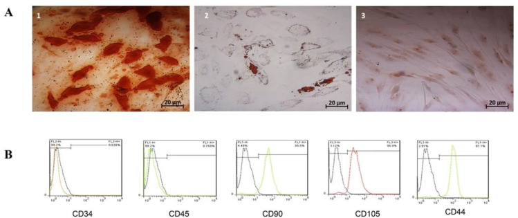
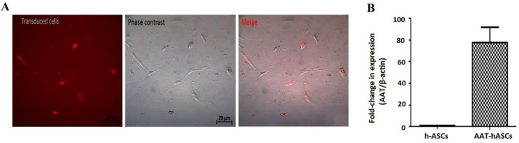
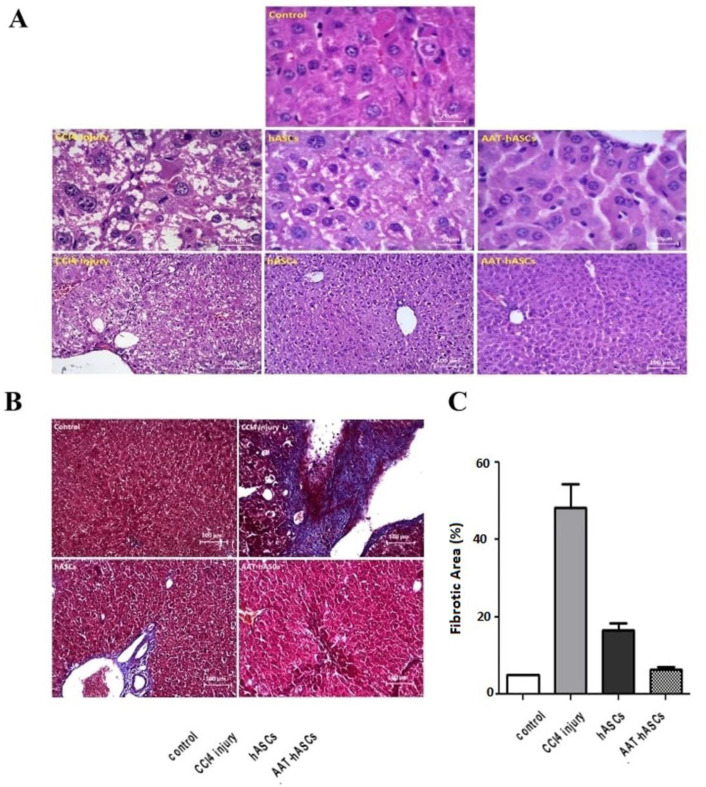
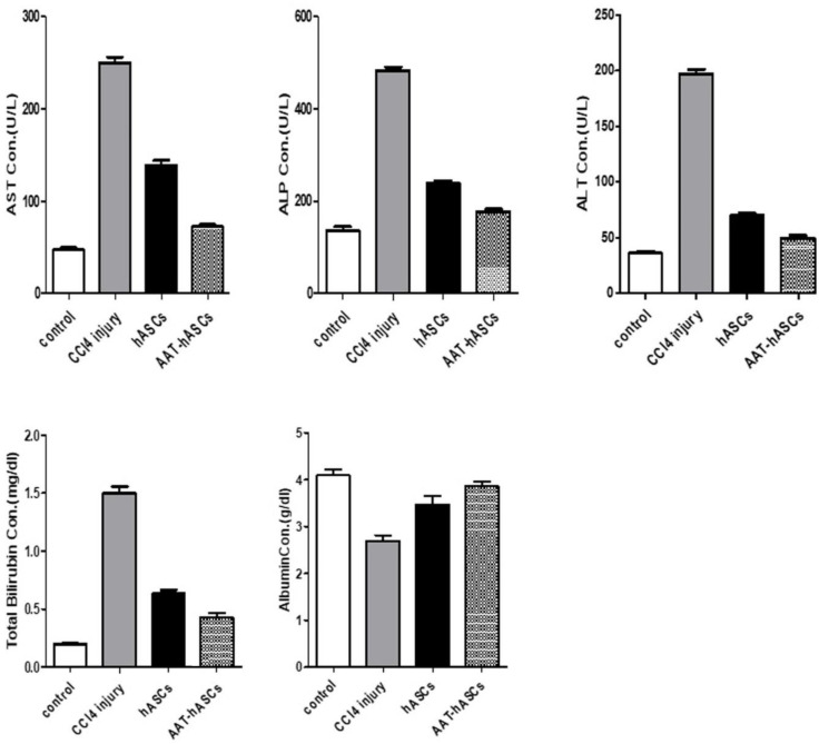
Methods: Liver fibrosis was induced in mice via the intraperitoneal injection of carbon tetrachloride (CCl4). MSCs were extracted from the human adipose tissue. After stemness confirmation, the cells were transduced with the lentiviruses containing the AAT gene, and then injected into the mice's tail vein. Fourteen days' post-transplantation, mice were sacrificed, and blood and tissue samples were collected for analysis. Important liver enzymes, including alanine transaminase (ALT), aspartate aminotransferase (AST), alkaline phosphatase (ALP), albumin, and total bilirubin (TB), were measured. Histological studies were carried out using the hematoxylin and eosin (H&E), as well as Masson's trichrome (MT) staining.
Results: Compared to hASCs, treatment with AAT-hASCs resulted in greater reductions in ALT, AST, ALP, and TB, as well as normalized albumin levels. AAT-hASCs promoted enhanced liver regeneration histologically, likely attributable to anti-inflammatory and anti-proteolytic properties of AAT.
Conclusion: These findings indicate AAT-engineered hASCs as a promising cell-gene therapy candidate for further study in liver cirrhosis models.
Keywords: Adipose tissue-derived mesenchymal stem cells; Alpha-1 antitrypsin; Carbon tetrachloride; Lentiviral vectors; Liver fibrosis.
© 2024, Gastroenterology and Hepatology From Bed to Bench (GHFBB).
Conflict of interest statement
The authors declare that they have no competing interests.
Figures
Similar articles
-
Effects of Bone Marrow-Derived Mesenchymal Stem Cells on Hypoxia and the Transforming Growth Factor beta 1 (TGFβ-1) and SMADs Pathway in a Mouse Model of Cirrhosis.Med Sci Monit. 2019 Sep 24;25:7182-7190. doi: 10.12659/MSM.916428. Med Sci Monit. 2019. PMID: 31550244 Free PMC article.
-
Comparison between the Regenerative and Therapeutic Impacts of Bone Marrow Mesenchymal Stem Cells and Adipose Mesenchymal Stem Cells Pre-Treated with Melatonin on Liver Fibrosis.Biomolecules. 2024 Mar 1;14(3):297. doi: 10.3390/biom14030297. Biomolecules. 2024. PMID: 38540717 Free PMC article.
-
[Effect of basic fibroblast growth factor treatment on efficacy of adipose-derived mesenchymal stem cells in liver cirrhosis].Zhonghua Gan Zang Bing Za Zhi. 2020 Dec 20;28(12):1030-1035. doi: 10.3760/cma.j.cn501113-20190407-00113. Zhonghua Gan Zang Bing Za Zhi. 2020. PMID: 34865351 Chinese.
-
Mesenchymal stem cells cultured under hypoxic conditions had a greater therapeutic effect on mice with liver cirrhosis compared to those cultured under normal oxygen conditions.Regen Ther. 2019 Sep 20;11:269-281. doi: 10.1016/j.reth.2019.08.005. eCollection 2019 Dec. Regen Ther. 2019. PMID: 31667206 Free PMC article.
-
Hepatoprotective effect of total flavonoids of Mallotus apelta (Lour.) Muell.Arg. leaf against carbon tetrachloride-induced liver fibrosis in rats via modulation of TGF-β1/Smad and NF-κB signaling pathways.J Ethnopharmacol. 2020 May 23;254:112714. doi: 10.1016/j.jep.2020.112714. Epub 2020 Feb 24. J Ethnopharmacol. 2020. PMID: 32105750
References
-
- Puche JE, Saiman Y, Friedman SL. Hepatic stellate cells and liver fibrosis. Compr Physiol. 2011;3:1473–1492. - PubMed
LinkOut - more resources
Full Text Sources
Research Materials
Miscellaneous