. 2024 Sep 9;52(16):9450-9462.
doi: 10.1093/nar/gkae600.

SMCHD1 activates the expression of genes required for the expansion of human myoblasts

Affiliations

SMCHD1 activates the expression of genes required for the expansion of human myoblasts

Matthew Man-Kin Wong et al. Nucleic Acids Res. .

Abstract

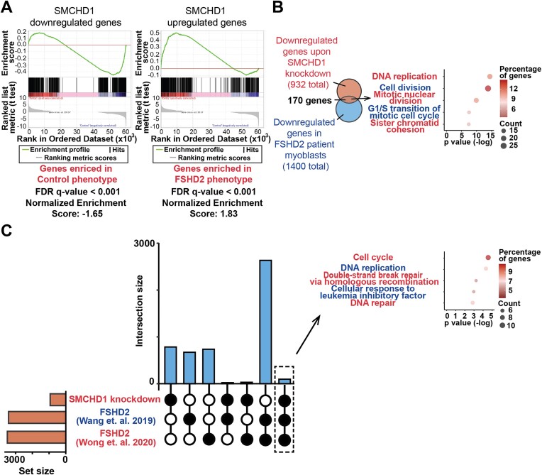
SMCHD1 is an epigenetic regulatory protein known to modulate the targeted repression of large chromatin domains. Diminished SMCHD1 function in muscle fibers causes Facioscapulohumeral Muscular Dystrophy (FSHD2) through derepression of the D4Z4 chromatin domain, an event which permits the aberrant expression of the disease-causing gene DUX4. Given that SMCHD1 plays a broader role in establishing the cellular epigenome, we examined whether loss of SMCHD1 function might affect muscle homeostasis through additional mechanisms. Here we show that acute depletion of SMCHD1 results in a DUX4-independent defect in myoblast proliferation. Genomic and transcriptomic experiments determined that SMCHD1 associates with enhancers of genes controlling cell cycle to activate their expression. Amongst these cell cycle regulatory genes, we identified LAP2 as a key target of SMCHD1 required for the expansion of myoblasts, where the ectopic expression of LAP2 rescues the proliferation defect of SMCHD1-depleted cells. Thus, the epigenetic regulator SMCHD1 can play the role of a transcriptional co-activator for maintaining the expression of genes required for muscle progenitor expansion. This DUX4-independent role for SMCHD1 in myoblasts suggests that the pathology of FSHD2 may be a consequence of defective muscle regeneration in addition to the muscle wasting caused by spurious DUX4 expression.


Figures

Graphical Abstract
Graphical Abstract
Figure 1.
Figure 1.
Loss of SMCHD1 leads to FSHD-like repression of PAX7 target genes in myoblasts without DUX4 target genes activation. (A) Nine days after lentiviral transduction, cells were observed under the fluorescence microscope. (White arrows) Transduced mCherry+SMCHD1 shRNA expressing cells. (Black arrows) Cells that do not express shRNA expression. (B) Volcano plot showing the DEGs after SMCHD1 knockdown. Genes with adjusted p-value <= 0.05 and log2 fold change > 0.5 were labeled red. (C) DUX4 score of samples transduced with non-silencing scrambled shRNA (Control) or SMCHD1 targeting shRNA (shSMCHD1) based on DUX4 targets identified by Choi et al. (9). (D) PAX7 scores of samples transduced with non-silencing scrambled shRNA (Control) or SMCHD1 targeting shRNA (shSMCHD1). (E) Venn diagram of the target genes activated by PAX7 and differentially expressed upon SMCHD1 depletion. (F) Number of PAX7-activated genes activated by PAX7 that were down-regulated (red) and up-regulated (blue) upon SMCHD1 depletion. (G) UCSC genome browser tracts showing RNA-Seq reads of samples transduced with non-silencing scrambled shRNA (Control) or SMCHD1 targeting shRNA (shSMCHD1) near the MYF5 gene. (H) Heatmap showing the expression of DUX4 target genes identified by Yao et. al. (1) upon SMCHD1 depletion. (I) UCSC genome browser tracts showing RNA-Seq reads of samples transduced with non-silencing scrambled shRNA (Control) or SMCHD1 targeting shRNA (shSMCHD1) near the DUX4 gene.
Figure 2.
Figure 2.
Loss of SMCHD1 in myoblasts leads to a decrease in proliferation. (A) GO terms of up-regulated genes after SMCHD1 depletion. (B) GO terms of down-regulated genes after SMCHD1 depletion. (C) Five days after lentiviral transduction, an EdU cell proliferation assay was performed, and the percentage of EdU+ cells was quantified. Error bars represent standard deviation from three independent experiments. (D) An equal number of myoblasts was seeded 3 days after lentiviral infection, and cell number was counted from day 5 to day 8 after lentiviral transduction. Error bars represent standard deviation from 3 independent experiments.
Figure 3.
Figure 3.
Comparison of changes in gene expression profiles upon SMCHD1 depletion and under FSHD2 condition. (A) Gene set enrichment analysis (GSEA) of DEGs in FSHD2 myoblasts compared to down-regulated (left panel) or up-regulated (right panel) genes upon SMCHD1 depletion. (B) (Left panel) Venn diagram of commonly down-regulated genes and (Right panel) dot plot of the top GO terms of the genes commonly down-regulated in SMCHD1-depleted myoblasts and in FSHD2 myoblasts. (C) (Left panel) Upset plot showing the commonly down-regulated genes upon SMCHD1 depletion and in FSHD2 biopsies from Wang et al. (45) and Wong et al. (43) studies. (Right panel) GO terms of commonly down-regulated genes upon SMCHD1 depletion and in FSHD2 biopsies.
Figure 4.
Figure 4.
Binding regions of SMCHD1 in the human myoblast genome. (A) The enrichment of H3K9me3, H3K4me1, H3K4me3, H3K27me3 and H3K27ac around SMCHD1 peak centers at SMCHD1 co-activated genes. (B) Percentage of direct targets-associated peaks that were located at promoters and enhancers. Z score and P-value were calculated by permutation test using regioneR. (C) UCSC genome browser tracts showing ENCODE cis-regulatory elements, GeneHancer annotated enhancer/promoter regions (¶) and their regulatory interactions, as well as ChIP-Seq (SMCHD1), CUT&Tag (H3K4me3 and H3K27ac) and RNA-Seq reads (control: proliferating myoblasts expressing non-silencing scrambled shRNA, shSMCHD1: proliferating myoblasts expressing shRNA targeting SMCHD1) near the LAP2 gene.
Figure 5.
Figure 5.
LAP2 depletion slows myoblast proliferation. (A) Heatmap showing the RNA expression of LAP2 in FSHD2 and control non-FSHD myoblasts (GEO accession number GSE143493). (B) Different LAP2 isoforms and target regions of different LAP2 shRNAs. (C) Nine days after LAP2 shRNA transduction, cells were observed under a fluorescence microscope for GFP+ shRNA-expressing cells. (D) Three days after LAP2 depletion, an equal number of myoblasts were seeded on 6 well plates. Cell number was counted on day 4, 6, 8 and 10 after lentiviral transduction. Error bars represent standard deviation from 3 independent experiments. (E) Lentivirus was added to the culture medium of proliferating myoblasts as indicated (control: non-silencing scrambled shRNA, shSMCHD1: shRNA targeting SMCHD1, vector: plenti-GIII vector alone, LAP2β: plenti-GIII vector expressing LAP2β transcript). Three days after lentiviral transduction, equal number of myoblasts were seeded on six-well plates. Cell number was counted on days 4, 6 and 10 after lentiviral transduction. Error bars represent standard deviation from three independent experiments.
Figure 6.
Figure 6.
Working model of how the loss of SMCHD1 function results in FSHD2-associated defects. (Left panel) Loss of SMCHD1 binding on the promoter and enhancer regions of its target genes in myoblasts may result in additional problems to myoblasts growth, on top of those caused by spurious DUX4 expression. (Right panel) Heterozygous SMCHD1 mutations in FSHD2 patients lead to a reduced SMCHD1 binding on the D4Z4 locus and the expression of DUX4 in muscle, which results in the degeneration of myofiber.

References

    1. Blewitt M.E., Vickaryous N.K., Hemley S.J., Ashe A., Bruxner T.J., Preis J.I., Arkell R., Whitelaw E.. An N-ethyl-N-nitrosourea screen for genes involved in variegation in the mouse. Proc. Natl. Acad. Sci. U.S.A. 2005; 102:7629–7634. - PMC - PubMed
    1. Blewitt M.E., Gendrel A.V., Pang Z., Sparrow D.B., Whitelaw N., Craig J., Apedaile A., Hilton D., Dunwoodie S., Brockdorff N.et al. .. SMCHD1, containing a structural-maintenance-of-chromosomes hinge domain, has a critical role in X inactivation. Nat. Genet. 2008; 40:663–669. - PubMed
    1. Wang C.Y., Jégu T., Chu H.P., Oh H.J., Lee J.T.. SMCHD1 merges chromosome compartments and assists formation of super-structures on the inactive X. Cell. 2018; 174:406–421. - PMC - PubMed
    1. Gendrel A.V., Apedaile A., Coker H., Termanis A., Zvetkova I.. SMCHD1-dependent and -independent pathways determine developmental dynamics of CpG island methylation on the inactive X chromosome. Dev. Cell. 2012; 23:265–279. - PMC - PubMed
    1. Huang Z., Yu J., Cui W., Johnson B.K., Kim K., Pfeifer G.P.. The chromosomal protein SMCHD1 regulates DNA methylation and the 2c-like state of embryonic stem cells by antagonizing TET proteins. Sci. Adv. 2021; 7:eabb9149. - PMC - PubMed

MeSH terms

Substances

LinkOut - more resources